Rotifer Genera Listed By Family
Table of Contents
1. Family Dicranophoridae: Dicranophorus
2. Family Floscularidae: Lacinularia; Limnias; Pyturga, Floscularia
3. Family Habrotrochidae :Habrotrocha
4. Family Lecanidae: Lecane
5. Family Lepadellidae: Lepadella, Colurella,Squatinella
6. Family Mytilinidae: Mytilina
7. Family Notommatidae: Cephalodella Type 1; Cephalodella Type 2; Eothinia; Monommata; Notommata; Taphrocampa
8. Family Philodinidae: Philodina; Rotaria
9. Family Synchaetidae: Synchaeta
10.Family Testudinellidae: Testudinella
11.Family Trichoceridae: Trichocera spp. Types 1 and 2
12.Family Trichotriidae: Macrochaetus, Trichotria
1.Family Dicranophoridae
Dicranophorus
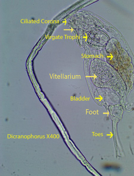
Dicranophorus, about 325 microns long, has a frontal hood that drapes over the anterior end, partly covering the ciliated ventral corona. The strong coronal cilia draw the animal smoothly forward.Long, wide toes allow the rotifer to quickly turn in any direction. During a turning maneuver, the toes first push against the substratum and allow the rotifer to pivot in the desired direction. The mastax has unique chitinous forcipate/virgate trophi with slender, pointed anterior jaws designed to penetrate and hold prey. Although I was unable to observe feeding,the structure of the mastax suggests that the rotifer is a predator. Two eye spots are visible on most specimens. Prey are passed by the mastax through a short oesophagus into the large stomach where digestion occurs. The indigestible remains are pushed into the intestine and then out of the anus. A clear, rounded, contractile Bladder is visible at the end of the body not far from the posterior part of the intestine. A relatively narrow foot is followed by two stout, relatively long toes.
http://vimeo.com/116342625 (Zottoli) X600
http://vimeo.com/116342624 (Zottoli) X400 . Fecal matter is ejected from the intestine through the anal opening near the beginning of the video.
2. Family Floscularidae
a. Lacinularia

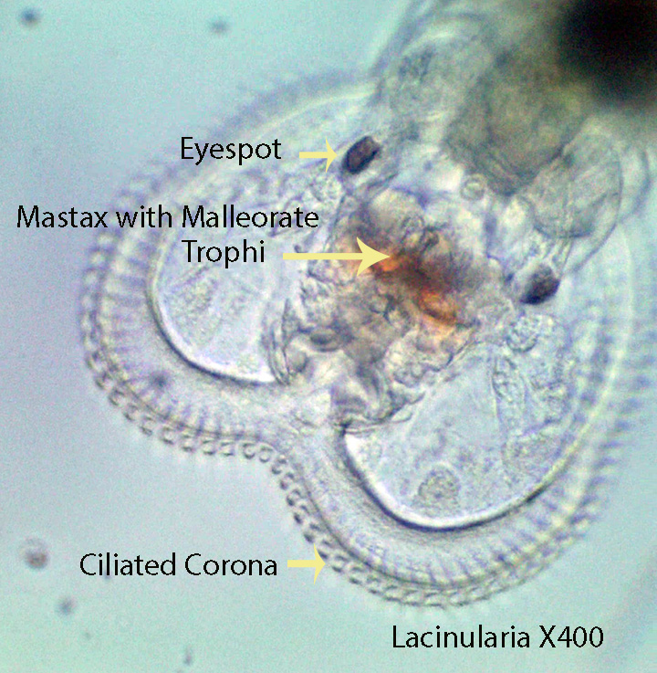
Lacinularia, about 1.7 mm long, lacks distinct ear-like coronal lobes. Instead the ciliated corona is heart-shaped. Each cilium on the corona beats followed by the next and the next, etc. giving the illusion that the entire corona is rotating. The colonial rotifers ,embedded in a gelatinous matrix, have a mastax with chitinous malleorate trophi. The first video shows a low power view of a rotifer colony. Note how particulate matter is moved around by the coronal cilia. The trophi are actively moving, breaking apart small organisms such as bacteria, unicellular algae and protozoans. From here the processed food passes into a short oesophagus and enters the stomach where digestion takes place.
http://vimeo.com/116291053 (Zottoli)X100
http://vimeo.com/116291050 (Zottoli) X400
b. Limnias


Limnias, about 1 mm long, has malleorate trophi that function in the manner described above for Lacinularia. The rotifer lives in a pipe-like tube of hardened secretions with a flange at the base. The ciliated corona is separated into two lateral lobes (Bi-lobed). .Each cilium on the corona beats followed by the next and the next, etc. giving the illusion that the entire corona is rotating. The animal is a suspension feeder consuming detritus, bacteria,unicellular algae and small protozoans.
http://vimeo.com/116202736 (Zottoli) X100.
http://vimeo.com/115734172 (Zottoli) X100
http://vimeo.com/115729978 (Zottoli) X400
c. Pyturga
Pyturga, about 1.5 mm long, is a sessile, filter feeding, solitary rotifer with an elongate body, that lives in a gelatinous tube adorned with round fecal pellets. Distinct ear-like coronal lobes are absent. The circular ciliated corona creates water currents that move food to the center of corona where it is passed to the mastax. Here the malleorate trophi process and push food through the short oesophagus and into the stomach. Each cilium on the corona beats followed by the next and the next, etc. giving the illusion that the entire corona is rotating.
http://vimeo.com/116272310 (Zottoli) X100 X400
d. Floscularia

Floscularia , about 2 mm long lives in a pipe-like tube composed of round pellets. A round, ciliated cup (Modulus) , visible in the photograph at the beginning of the second video, lies just behind the elliptical mouth. Reddish colored detritus (visible in the modulus) is mixed with mucus and shaped by cilia into a round ball (Pseudo-feces) ; eventually it is placed by the animal at the leading edge of the tube. As the animal grows it lengthens the tube through this process. A relatively short , finger-shaped, sensory antenna is also visible in the second video. The large corona is divided into two pairs of ciliated lobes. Each cilium beats followed by the next and the next, etc. giving the illusion that the entire lobe is rotating. The trophi are malleorate . As one might guess, the animal is a suspension feeder feeding for the most part on bacteria, detritus and small protozoans.
http://vimeo.com/115729976 (Zottoli) X400
http://vimeo.com/115729975 (Zottoli) X400. The photograph at the beginning of the video shows the modulus with its reddish brown contents and the sensory antenna.
3. Family Habrotrochidae
Habrotrocha spp.
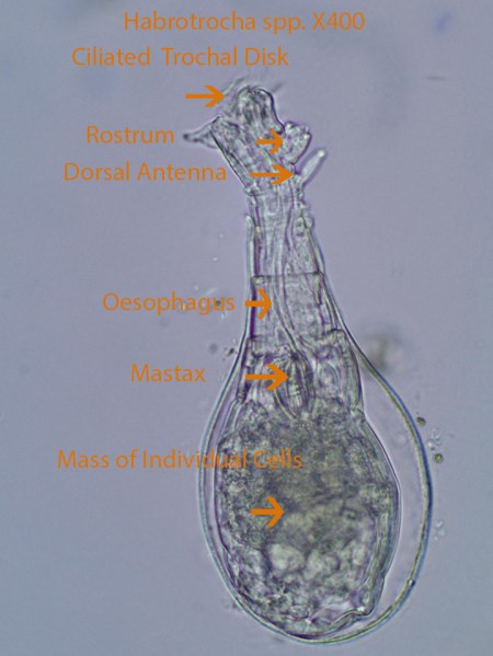
Habrotrocha, about 300 microns long, is often found in a mucus burrow secreted in layers. The rotifer is characterized by its lack of a stomach lumen (Cavity). Instead the stomach is formed by individual round cells that most likely consume food much like an amoeba. Small, ciliated trochal discs create a circular water flow from the sides of the lobes to above the head and back down again. Detritus, bacteria and small protozoans drop downward towards the mouth and then into a relatively wide ciliated canal that leads into the beating mastax. The mastax has ramate trophi that grind up the food and push it through the short oesophagus into the stomach where digestion takes place within cells.
http://vimeo.com/116342623 (Zottoli)X400
http://vimeo.com/116342622 (Zottoli) X400
4. Family Lecanidae
Lecane spp.

Lecane, about 120 microns long, is covered by a thick, inflexible lorica with a lateral groove that connects the dorsal and ventral plates. The Coronal Cilia are visible at the anterior end. The mastax with malleate trophi, is active and applied to the substratum during feeding. Generally the toe(s) attach to the substratum,the body bends downward, and the ventral mastax using the malleate trophi, pulls food (Detritus, bacteria, small protozoans, etc.) into the mouth and passes it through a short oesophagus to the stomach. Lecane has a short foot and either a single toe (Type A) or two toes (Type B). The two toes of type B rotifers may be fused at the tip or completely separate. The coronal cilia alone can move the animal, however the toes can push against the substratum and turn the animal in a different directions..
http://vimeo.com/116202735 (Zottoli) X400 . Anatomy
http://vimeo.com/115729974 (Zottoli) X400 . Feeding
5. Family Lepadellidae
a. Lepadella spp.

Lepadella, about 120 microns long, is dorsoventrally flattened . The dorsal and ventral plates of the lorica are completely fused. Lepadella lacks a frontal hood like that found on Colurella. The ciliated corona can be extended or retracted through an anterior opening in the lorica. The foot extends to the outside through a posterior ventral notch in the lorica. During the feeding process, the rotifer contacts the surface, attaches the tips of the toes and bends at about a 30 degree angle with the coronal cilia near the substratum. The coronal cilia seem to dislodge material from the substratum. The food is carried by cilia for a short distance and grabbed by the ventral malleate trophi (Part of the Mastax) and in concert with contractions of Mastax muscles, food is pushed through a short oesophagus into the stomach. The coronal cilia pull the animal forward. As they move forward they also rotate around their anterior-posterior axis. The rotifer also uses its toes to set up a pivot point that allows them to move their body and change direction. The animal is essentially a particle feeder consuming bacteria, small algae, detritus and small protozoans.
http://vimeo.com/116272309 (Zottoli) X400
b. Colurella


The rigid lorica around the rotifer is composed of two lateral plates that are strongly compressed. The rotifer, about 120 microns long, has a ciliated corona that is responsible for feeding and moving the animal forward. The two toes at the posterior end are sometimes attached to the substratum; When this happens the animal often bends downward touching the bottom with its coronal cilia. The movement of coronal cilia dislodges food particles that are then directed into the mouth. A frontal hood extends downward in front of the face. It is positioned in such way that it may prevent food from escaping to the sides. Contraction of the foot in concert with the positioning of the toes allows the animal to turn and twist. The active mastax with its malleate trophi passes food through a short oesophagus into the stomach. Small green flagellates as well as bacteria have been seen inside the stomach. The ciliated corona can be completely withdrawn into the lorica.
http://vimeo.com/116345097 (Zottoli) X400
http://vimeo.com/116272308 (Zottoli) X400
c. Squatinella
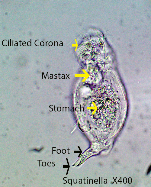
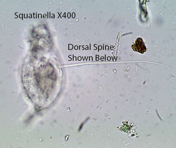
Squatinella,, about 120 microns long, has a small dorsal shield and lacks anterior dorsal spines typical of some species in this genus. In addition it has a long dorsal spine that is about the same length as the body. The long dorsal spine may make it more difficult for predators to handle and swallow the rotifer. It clearly extends the malleate trophi out of the mouth and appears to be scraping the substratum as it moves forward. . Movement is powered by the coronal cilia and aided by body contractions. When the toes are attached to the substratum they may act as a pivot point allowing the animal to turn and head off in a different direction.
http://vimeo.com/117688876 (Zottoli) X400.
http://vimeo.com/117688877 (Zottoli) X400.
6. Family Mytilinidae
Mytilina
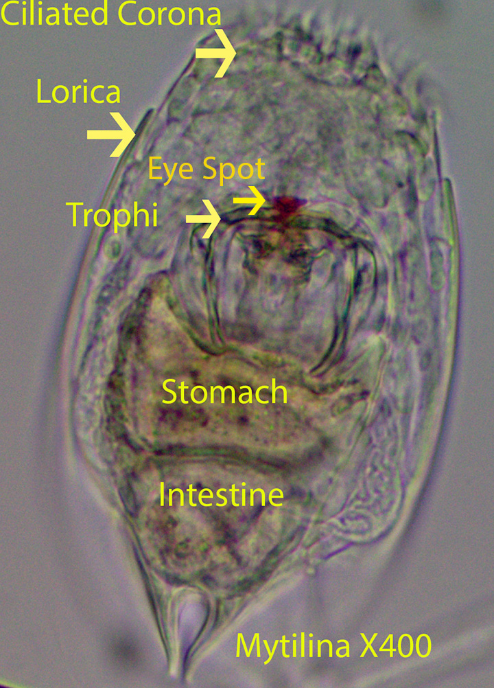

Mytilina , about 340 microns long, is characterized by a firm ,thick lorica ,made up of two lateral plates and one ventral plate. Spines extend from the anterior lateral corners of the lateral plates. The mastax with its malleate trophi are visible below the ciliated corona. One red eye-spot is located just above the mastax. The mastax pushes against the large brown stomach. The intestine lies just below the stomach. The posterior foot is short and connects to two large, blade-like toes.
http://vimeo.com/116348062 (Zottoli) X400
http://vimeo.com/116348064 (Zottoli) X400
http://vimeo.com/116350451 (Zottoli) X400
7. Family Notommatidae
a. Cephalodella Type 1
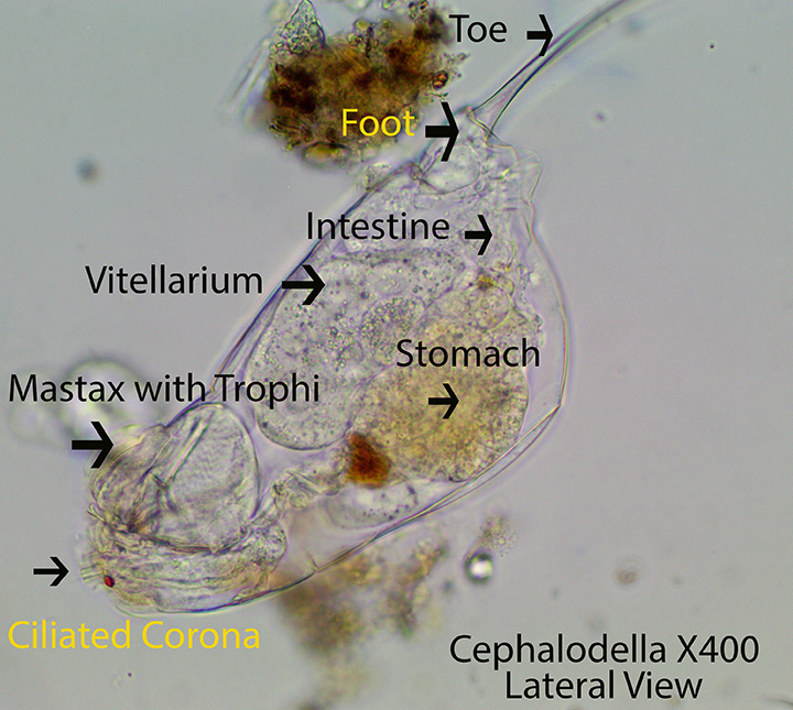
Cilia on the ventral part of the head seem to dislodge food particles (Detritus, Bacteria, small protozoans, etc.) from the substratum. The mastax with its virgate trophi, appears to draw food particles inward and passes them along into the short oesophagus and then into the stomach where digestion takes place. Digested material passes into the small intestine and then out through the anus. Cephalodella, about 200 microns in length, feeds on the surface of clumps of debris containing bacteria and small protozoans.
http://vimeo.com/116302454 Cephalodella Type 1 X400
http://vimeo.com/116304658 Cephalodella Type 1 X400
b. Cephalodella Type 2


Type 2, although smaller than Type 1 (60 microns long), has the same basic characteristics as Type 1.
http://vimeo.com/116302456 Cephalodella Type 2 X400
http://vimeo.com/116302457 Cephalodella Type 2 X400
http://vimeo.com/116302455 Cephalodella Type 2 X600
c. Eothinia

Eothinia, about 500 microns long, is situated within a protective, flexible Lorica (Illoricate). A pair of red eye-spots lies near the mastax that contains robust virgate trophi. The oblique anterior corona is populated with short cilia. The head is set off from the rest of the body. The dorsal surface of the body lacks deep folds. The short foot supports two short toes. A single sac-like salivary gland is positioned ventrally just behind the mastax. One, round digestive gland is attached to each side of the front edge of the stomach.
http://vimeo.com/118293677 (Zottoli) X400
d. Monommata

Monommata, about 350 microns long, is characterized by long toes (Longer than the body) and a short foot. A dark eye spot lies below the oblique corona and the mastax with its virgate trophi is located near the eye spot. The ventral mastax with its trophi can be protruded from the mouth and pull food (Detritus, bacteria, small protozoans, etc.) into the mouth and push it through the oesophagus into the stomach. The rotifer can move forward in a straight line powered by the coronal cilia. The animal can change direction by placing its toes on the substratum, bending its body and moving along a different path.
http://vimeo.com/116345099 (Zottoli X100 X400 Movement
http://vimeo.com/116345100 (Zottoli) X600. Feeding, Movement
e. Notommata

Notommata, about 350 microns long, has a ventral mastax with virgate trophi, that lies just behind the ventral coronal cilia. There is a distinct separation between the head and the body. A pair of small, lateral, ciliated lobes is located just behind the rounded anterior part of the head. The vitellarium is rounded. The body is flexible (Illoricate). The foot and toes are small and dorso-ventrally flattened. There are no frontal eye-spots or spines on the body. A relatively large dorsal papilla (Fleshy Extension) is present at the end of the body.
http://vimeo.com/116350452 (Zottoli) X400
http://vimeo.com/116350453 (Zottoli) X400
f. Taphrocampa

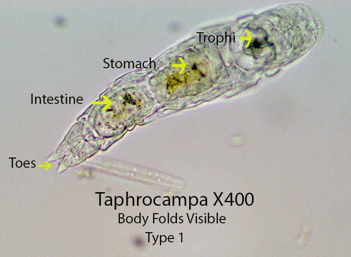
Taphrocampa, about 120 microns long, possesses a simple ciliated corona and a flexible lorica (Illoricate). The rotifer has a mastax containing virgate trophi. The dorsal side of the body has characteristic deep folds. The body ends with a short foot and two small toes. In the second video The virgate trophi are repeatedly thrust from the mouth, pulling in what seem to be green algae. Type 2 has deeper dorsal folds than Type 1, however they share the same anatomical features.
http://vimeo.com/117945645 (Zottoli) Taphrocampa Type 1 X400
http://vimeo.com/117945643 (Zottoli) Taphrocampa Type 1 Feeding X600
http://vimeo.com/117945640 (Zottoli) Taphrocampa Type 1 X400
http://vimeo.com/117945644 (Zottoli) Taphrocampa Type 2 X400
http://vimeo.com/117945642 (Zottoli) Taphrocampa Type 2 X600
8. Family Philodinidae
a. Philodina

A ciliated bi-lobed disc (Corona) is attached to the body as shown in the first photograph. A long sensory antenna visible in some of the videos, lies just below the corona. It can be retracted inside the mouth when the rotifer is disturbed. Disc cilia move the unattached animal smoothly through the water. When the posterior toes temporarily attach to the substratum the cilia create water currents , shown in most of the videos, that move food ( Bacteria, detritus, small protozoans, etc.) into the mouth. Food then passes down a short ciliated channel (Visible) into a constantly beating structure called the mastax (Visible). The mastax is made up of muscles and hard jaws (Ramate Trophi) that grasp and process the food before it is passed down a short oesophagus into a large brownish stomach (Visible). Here food is digested and indigestible remains pass into the short intestine (Visible) and then to the outside through the anus. The body is covered with a cuticle that is secreted by the hypodermis. The cuticle is ringed with thin annuli allowing the animal to extend or shorten the body in telescopic fashion. A short Foot (Visible ) extends from the posterior part of the body and gives rise to four terminal toes (Only two are visible). The toes secrete a mucus-like material that holds the rotifer in place while they feed. Two small red-eye spots are located just below the ciliated disc. A single, relatively large ,squarish ,bladder is located just below the short intestine. It contracts periodically, passing water taken into the body osmotically to the outside.
http://vimeo.com/116354419 (Zottoli) X400. Videos 418-420 show the ciliated mouth, the thin, ciliated channel that connects the mouth to the mastax, and the mastax that shreds food and passes it into the stomach. The posterior toes are also visible
http://vimeo.com/116354420 (Zottoli) X400. Videos 420 and 418 show the bladder contracting.
http://vimeo.com/116354418 (Zottoli) X400
http://vimeo.com/82147756 (Zottoli) (Philodina) (Bog) X400. This video, although it features a bog rotifer, shows the ciliated tube that leads into the stomach more clearly.
b. Rotaria
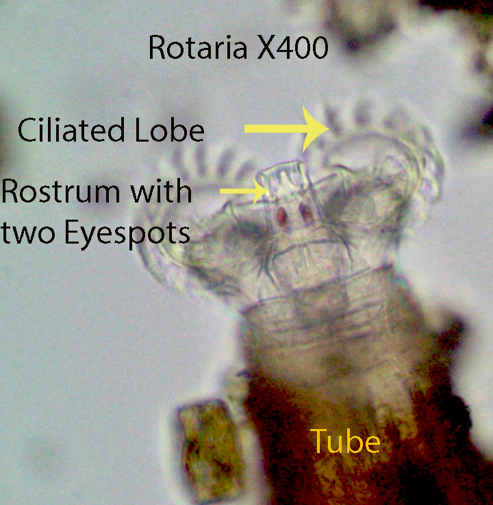
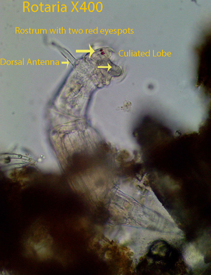
Rotaria, about 450 microns long, extends two ciliated coronal (Trochal) discs out into the water column while feeding. A rostrum that lies between the trochal discs has two distinctive red-eye spots . Circular water currents powered by the trochal cilia deliver food particles to the mouth where it is moved by cilia to the mastax. The mastax with its hard ramate trophi, macerate and push food through a short oesophagus into the stomach where digestion takes place. Food then passes into the intestine and undigested material is pushed to the outside through an anal opening. When trochal cilia are extended and the toes are not attached, the cilia draw the animal forward in a straight line. When the toes are attached to the substratum and the ciliated trochal discs are extended, the cilia circulate the surrounding water bringing food toward the mouth. A second type of movement characteristic of many rotifers involves first attaching their toes to the substratum and then extending their body in the direction of movement. During the next phase, they attach their head and pull the posterior end to just behind the head moving somewhat like an inch worm. The process is repeated again and again as the animal moves quickly forward. During this process coronal discs are retracted. There are three toes, one dorsal and two ventral. A sensory antenna, best seen in the last video, extends outward from the body just below the trochal discs.
http://vimeo.com/116206365 (Zottoli) X400
http://vimeo.com/116206368 (Zottoli) X400
http://vimeo.com/116269072 (Zottoli) X400
http://vimeo.com/116269073 (Zottoli) X400
http://vimeo.com/116269071 (Zottoli) X400
9. Family Synchaetidae
Synchaeta


Synchaeta, about 400 microns long, has a mastax with virgate trophi connected to large, visible, hypo-pharyngeal muscles. The lorica surrounding the rotifer is flexible (Illoricate). The ciliated corona is flanked on both sides by a group of lateral cilia. These most likely correspond to the “ear like lobes” listed as a characteristic of this genus. A pair of small, lateral, stiff setae are present at the point where the “head” ends and the “body” begins. The body tapers towards the posterior end joining a short foot and single toe. A large, brownish stomach lies behind the mastax and a small whitish vitellarium is situated next to the stomach in the anterior part of the body.
http://vimeo.com/116303740 (Zottoli) X400
http://vimeo.com/116303739 (Zottoli) X600. Note the large eggs inside the slightly compressed rotifer
10. Family Testudinellidae
Testudinella spp.
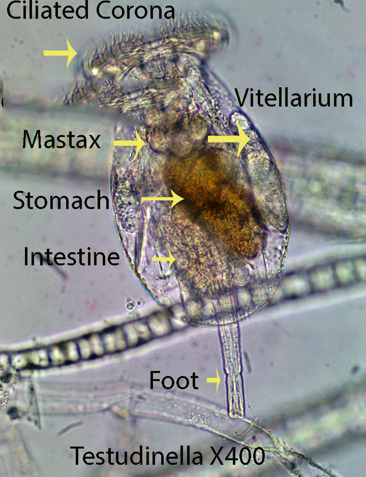
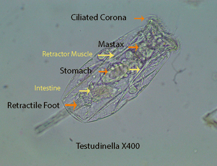

The disc-shaped rotifer , about 325 microns wide, is dorso-ventrally flattened. The ciliated corona can be extended from the protective lorica allowing the animal to move and feed. It also can be pulled into the lorica by retractor muscles during stressful times. The lorica is composed of a dorsal and a ventral plate, fused along the edges. An annulated, ventral , posterior foot, resembling an elephant’s trunk, can be extended from or withdrawn into the lorica. A ciliated cup, at the end of the foot can temporarily attach the rotifer to the substratum during feeding bouts.
http://vimeo.com/118069563 (Zottoli) Testudinella X100. Movement. The coronal cilia are responsible for movement. The foot attaches and detaches several times here.
http://vimeo.com/118069560 (Zottoli) Testudinella X400. Lateral View
http://vimeo.com/118069558 (Zottoli) Testudinella X400
http://vimeo.com/118072617 (Zottoli) Testudinella X00
11. Family Trichoceridae
Trichocera spp. Types 1 and 2

Trichocera Type 1 , about 170 microns long, has a ventral mastax with virgate trophi. They have a simple ciliated corona that is responsible for moving the bean-shaped rotifer from place to place (Refer to the first video). The rotifer often feeds by placing its head directly in front of clumps of debris. Cilia in concert with the mastax drive food (Detritus, bacteria and small protozoans, etc.) through a short oesophagus into the stomach. Two defining features are: Spine-like toes that are not the same length and a body that is slightly twisted. There are two red eye-spots, one of which is visible in the photograph above.
http://vimeo.com/118059076 (Zottoli) Trichocera X400 Type 1. Feeding. Extension of Trophi
http://vimeo.com/118059074 (Zottoli) Trichocera X400.X600 .Type 1. Anatomy
http://vimeo.com/118059073 (Zottoli) Trichocera X400.Type 1. Feeding
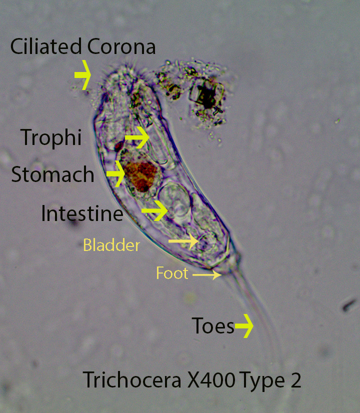
Trichocera Type 2, about 300 microns long, has the same basic characteristics as Type A, however it differs in size, shape and other characteristics that are not of concern here.
http://vimeo.com/118059075 (Zottoli) Trichocera X400 Type 2
12. Family Trichotriidae
a. Macrochaetus


Macrochaetus, about 120 microns long, lacks a dorsal head shield. The rotifer has a mastax with malleate trophi and is protected by a thick lorica that has small spines along the edges. The lorica covers the entire body including the head. Two dorsal spines arise from the foot while about 7 long pairs of dorsal spines arise from the body.
http://vimeo.com/116345098 Macrochaetus X400
http://vimeo.com/116348063 Macrochaetus X400
b. Trichotria
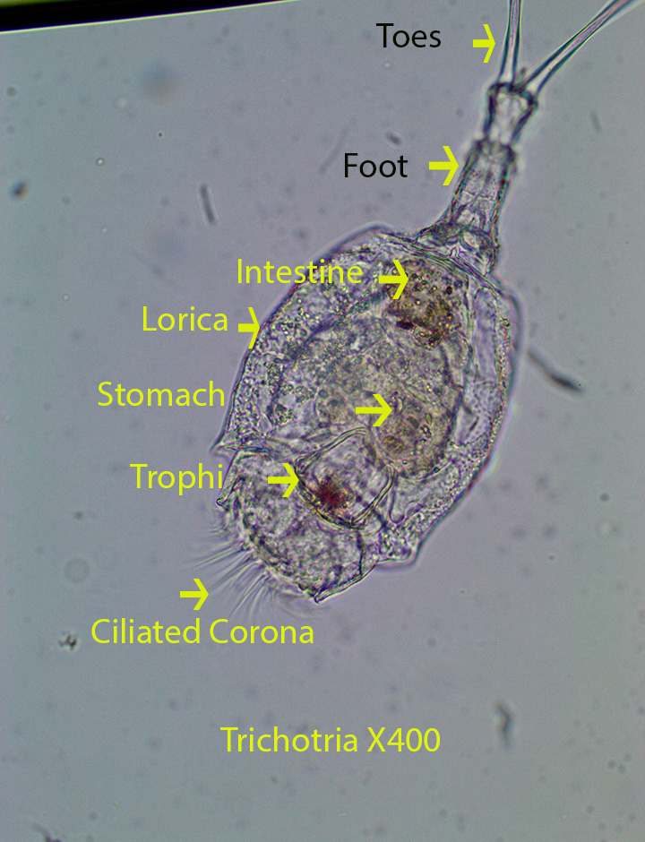

Trichotria, about 350 microns long, is covered completely by a smooth, one piece, rigid lorica. The rotifer which has a mastax with malleate trophi, lacks a circular anterior shield. There are two prominent thick spines on the foot.

Leave a comment