- Etiology:
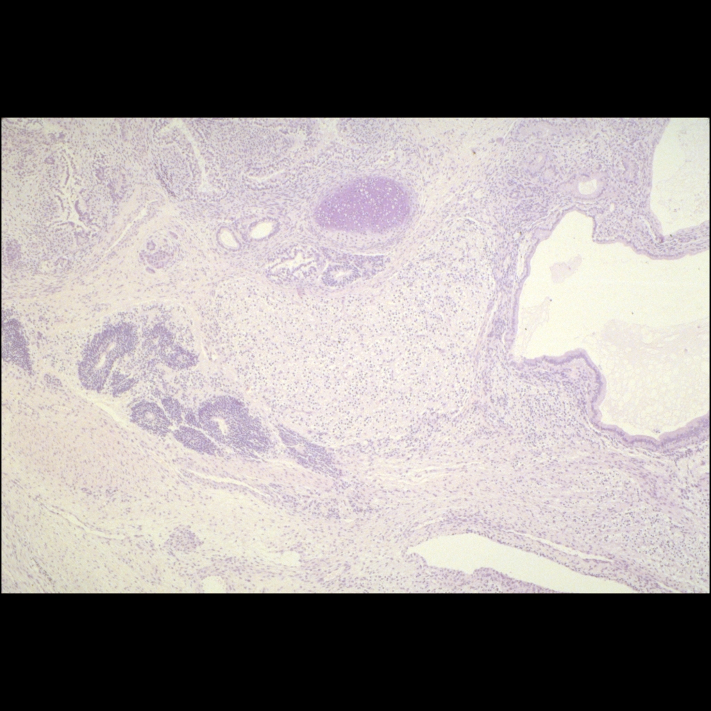
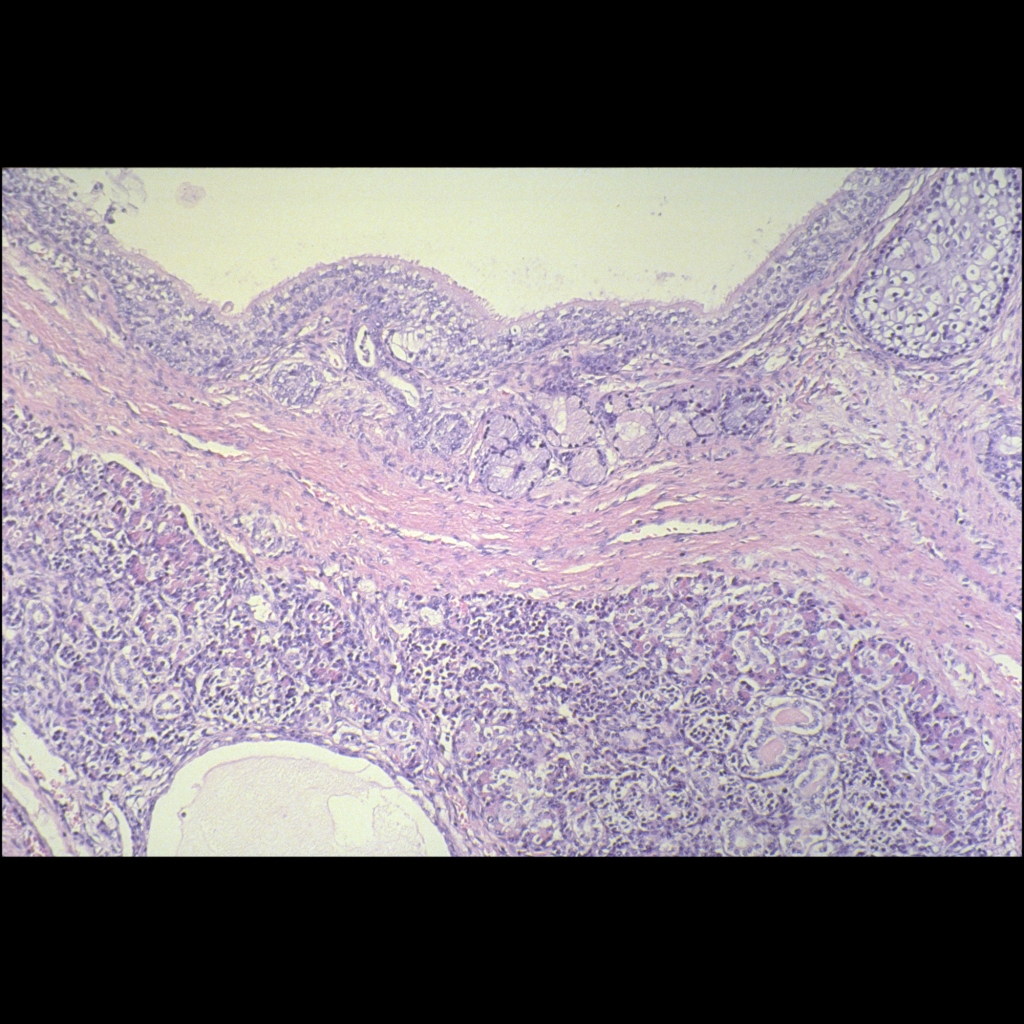
— Mature type – composed of fully differentiated adult-type tissues with absent or low mitoses
— Immature type – fetal-type incompletely differentiated tissues
— Malignant type – contains cancerous tissues such as sarcomas and carcinomas and other embryonal malignancies, malignant yolk sac endoderm can be aggressive component of teratomas - Imaging:
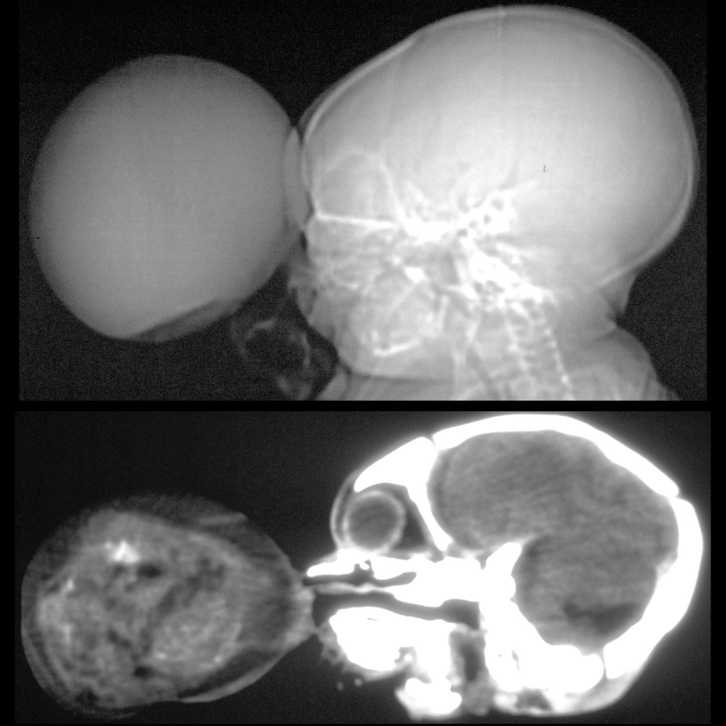
— Characteristically involve midline, nearly always heterogenous, often contain fat
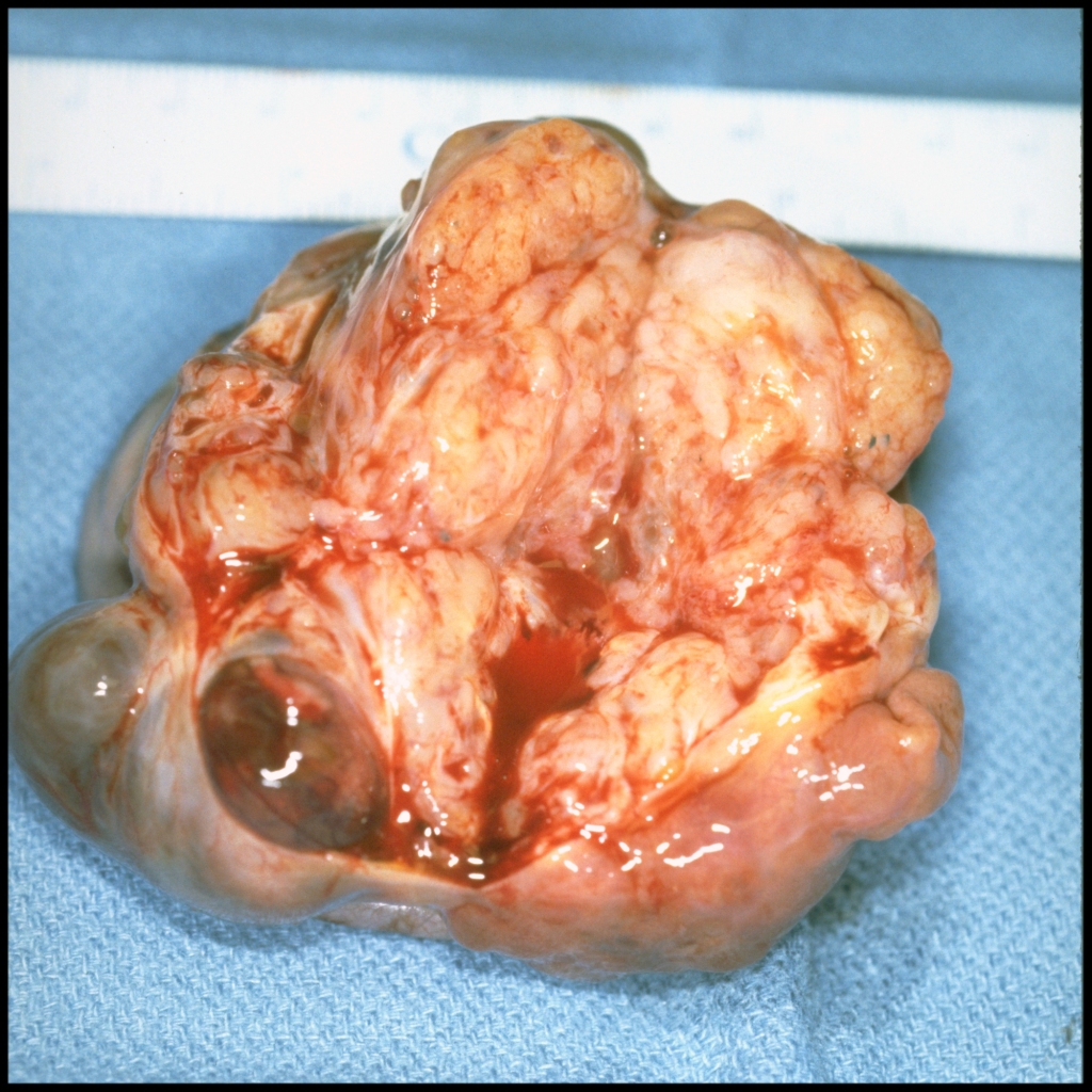
— Heterogenous appearance due to presence of fat, cysts (mucous-laden), calcium (bone and chondorid nodules), soft tissues
— Enhancing soft tissues present in all types of tumors with enhancement of capsule and heterogenous enhancement of soft tissue components - DDX:
- Complications:
- Treatment:
- Clinical: patients with malignant teratomas may have elevated levels of AFP or beta-HCG in serum and or CSF, 90% of teratomas are found in < 20 years with most 10-12 years old,
80% occur around third ventricle and thus most symptoms are due to hydrocephalus and increased intracranial pressure
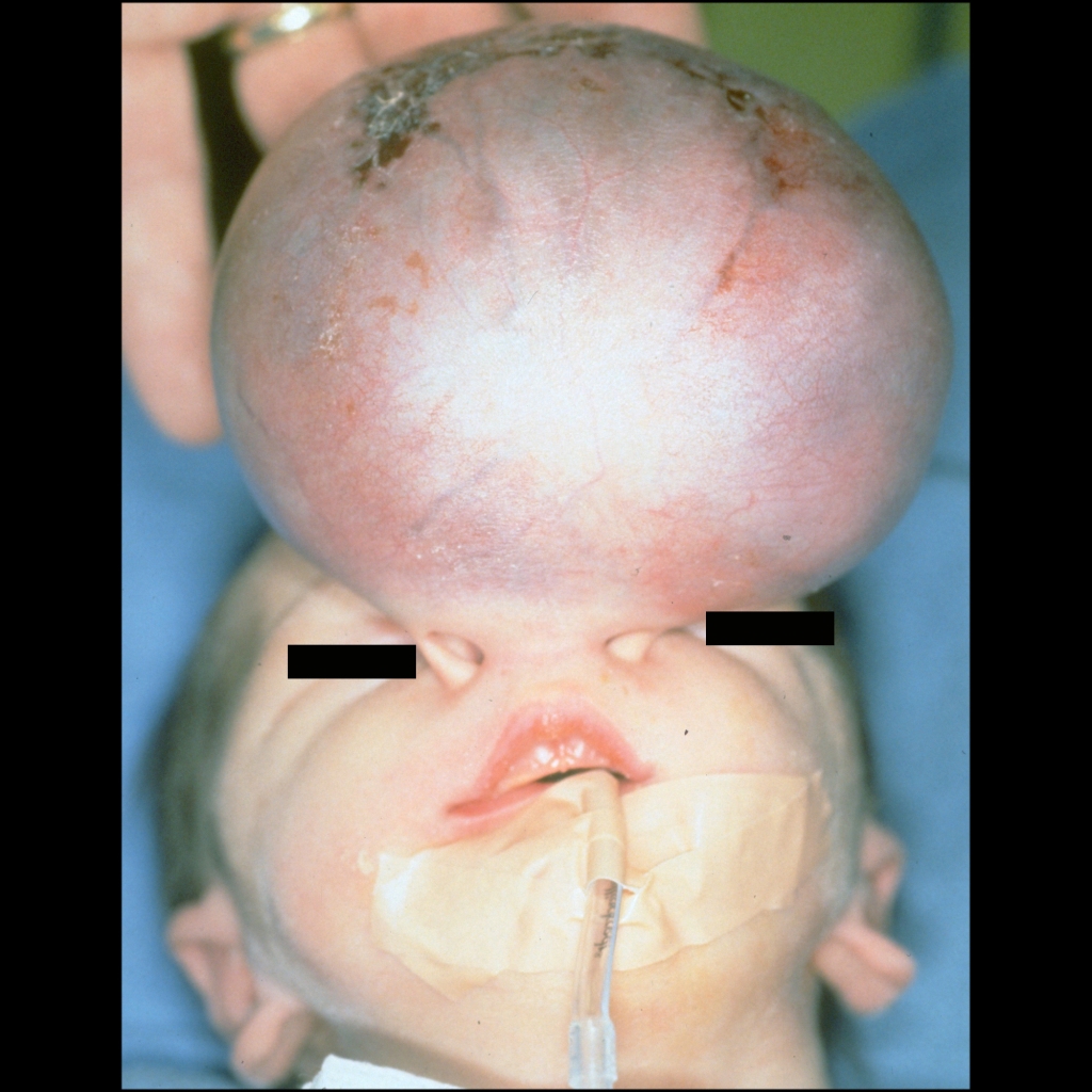
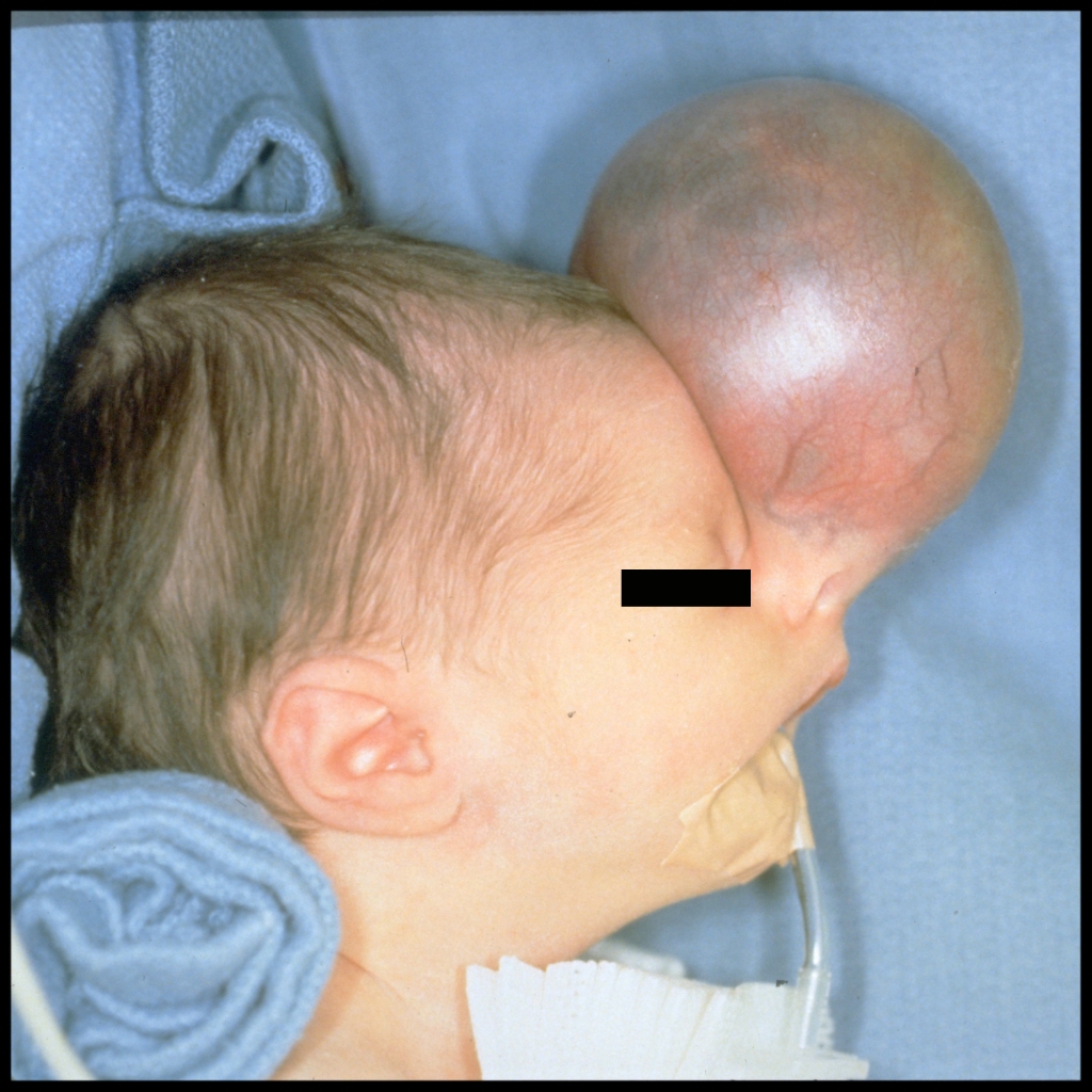
Congenital teratoma
- Intracranial teratomas are rare, accounting for 0.5-2.0% of intracranial tumors
- Comprise 50% of congenital brain tumors (those presenting in first 60 days of life)
- Typically benign tumors containing elements of all 3 germinal layers: ectoderm, mesoderm, endoderm
— Develop from embryonic cells which become misinvolved during formation of primitive streak in 3rd week of life
— Some of these cells become misenfolded as intracranial rests of tissue
Pineal teratoma
- Tissue rests typically found in midline, specifically pineal, suprasellar, 3rd ventricle regions
Suprasellar teratoma
- Differential diagnosis for T1 bright and T2 dark includes aneurysm, dermoid, lipoma, craniopharyngioma
Spinal teratoma
- Type I mainly external with small presacral component
- Type II external presentation with large intrapelvic component
- Type III external component with majority of tumor in pelvis and abdomen
- Type IV entirely presacral without externally visible component
- Sacrococcygeal region is most common site for teratomas in CNS
- Often associated with other abnormalities including anorectal and genital malformations, ventricular septal defect, hip dislocation, vertebral anomalies (spina bifida, sacral
Radiology Cases of Central Nervous System Teratoma
Radiology Cases of Nasal Teratoma
Clinical Cases of Central Nervous System Teratoma
Clinical Cases of Nasal Teratoma
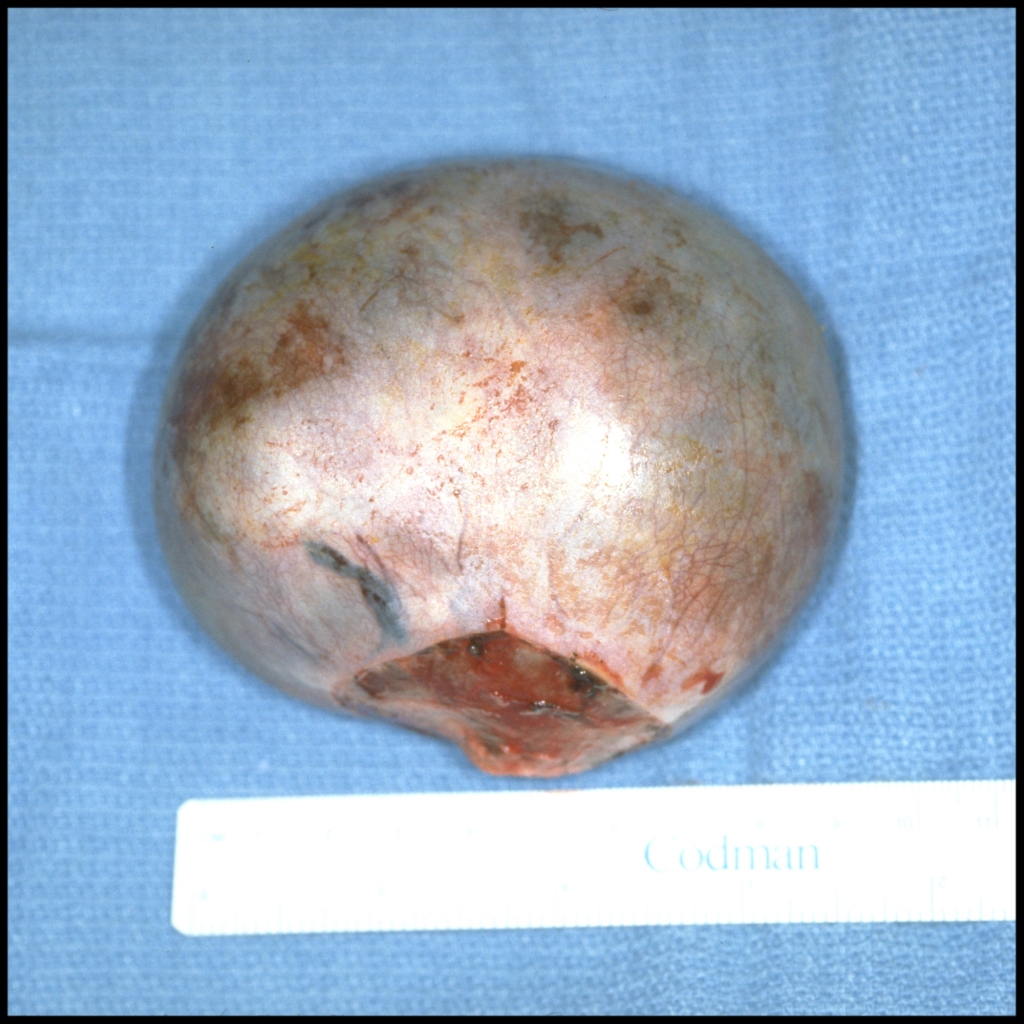
Gross Pathology Cases of Central Nervous System Teratoma
Gross Pathology Cases of Nasal Teratoma
Histopathology Cases of Central Nervous System Teratoma
Histopathology Cases of Nasal Teratoma