Teratoma
INTRODUCTION:
- All germ cell tumours show differentiation along embryonic rather than extra-embryonic pathways.
These are grouped together as teratomas, and divided into three categories:
- Mature (benign), e.g. dermoid cyst,
- Immature (essentially malignant), e.g. solid teratoma and
- Monodermal or highly specialized, e.g. struma ovarii
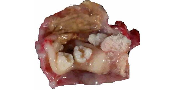
Dermoid Cysts:
- Unilocular with smooth surface
- Sebaceous material and hair, and the wall is lined by squamous epithelium which contains hair follicles and sebaceous glands
- Teeth, bone, cartilage, thyroid tissue and bronchial mucous membrane are often found in wall in inner surface is called a ‘focus’ or ‘embryonic node’.
- Origin:Ectodermal , mesoderm and endoderm
- Squamous epithelium usually lines the cyst, columnar and transitional types are also found.
- Arise in association with mucinous cystadenomas to form a combined tumour, part of which consists of a dermoid cyst while the rest has the characteristic structure of a mucinous cystadenoma
- Most common orbital cyst in children Dermoid cyst
- Most common ovarian tumour in pregnancy is Dermoid cyst
- Dermoid cyst is most prone to undergo torsion during pregnancy
- Extraovarian dermoid cysts arise occasionally in the lumbar region, uterovesical area, parasacral region and rectovaginal septum
- Epidermoid carcinoma (1.7%) and sarcomatous changes may occur


Solid Teratoma of the Ovary:
- Tumor containing cells of all three germ layers is called Teratoma
- Dermoid cyst of ovary is teratoma
- Teratoma arises from Totipotent cells
- Cut surface has a peculiar trabeculated appearance
- Large loculi are found beneath the capsule
- Solid part of the tumour contains :Plain muscle, brain tissue, glia, pia mater,cartilage , bone and intestinal mucous membrane
- Cystic spaces contains:Hair and sebaceous material
- In children differentiated mature teratoma may be benign
- Rokitansky’s protuberance where tissue elements such as tooth, bone, cartilage & various other odd tissues are present is seen in in one area of the cyst wall,as a solid prominence
- In post pubertal males all teratoma are regarded as malignant and capable of metastasis regardless the elements may be immature or mature.
- 10% are B/L & malignant
- Sacrococcygeal teratoma is associated with defect during gastrulation
- Maximum radio opaque shadow in ovary is seen in Teratoma
- Mostly Malignant tumours because of sarcomatous changes
Struma Ovarii:
- Struma ovarii consists of thyroid tissue similar to that of a thyroid adenoma.
- The tumour is solid,consisting almost entirely of thyroid tissue.
- The tumour resembles a mucinous cystadenoma but the material contained in the vesicles is colloid and gives reaction to iodine
STAGES:
The stage of a cancer tells you how far it has grown. In ovarian teratoma there are 4 stages, from 1 to 4:
- stage 1 means the cancer is only in the ovary (or both ovaries)
- stage 2 means the cancer has spread into the fallopian tube, womb, or elsewhere in the area circled by your hip bones (your pelvis)
- stage 3 means the cancer has spread to the lymph nodes or to the tissues lining the abdomen (called the peritoneum)
- stage 4 means the cancer has spread to another body organ some distance away, for example the lungs
DIAGNOSIS:
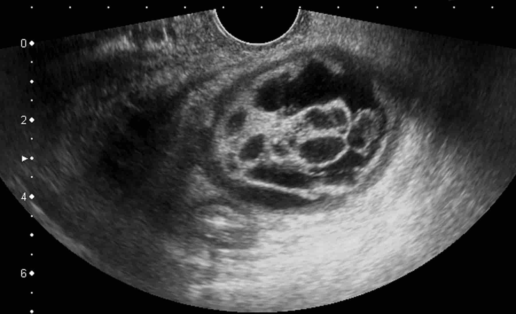
- As most cysts are asymptomatic (show no symptoms) they are more likely to be discovered during a routine pelvic examination or while undergoing an ultrasound scan for another reason such as pregnancy.
- An abdominal or transvaginal ultrasound will be carried out to determine the exact type, location, size and amount of cysts present.
- Ovarian mass with x-ray pelvis showing radio-opaque shadow suggest Dermoid cyst
Teratoma is a non seminomatous tumor of testis.
|
Marker |
Increased in |
|
Beta HCG |
Both seminoma and non-seminoma |
|
AFP |
Only in non-seminoma |
|
LDH |
Both seminoma and non-seminoma |
TREATMENT:
- If left untreated ovarian torsion can develop, this can restrict blood flow to the ovaries and eventually cause fertility problems.
- Unless a cyst ruptures causing an emergency, surgery is usually elective.
- The dermoid cyst in pregnancy should be treated At 14-16 weeks of pregnancy
Removed surgically:
- Cystectomy
- Total oophorectomy
- Partial oophorectomy
- Laparoscopy-assisted vaginal hysterectomy.
Exam Important
- Most common orbital cyst in children Dermoid cyst
- Sacro-coccygeal teratoma appear as swelling over sacral region
- Maximum radio opaque shadow in ovary is seen in Teratoma
- Ovarian mass with x-ray pelvis showing radio-opaque shadow suggest Dermoid cyst
- Rokitanski protruberences are seen in Teratoma
- Dermoid cyst of ovary is teratoma
- Dermoid cyst is most prone to undergo torsion during pregnancy
- Dermoid cyst of ovary contains derivatives from Endoderm, Mesoderm & Ectoder
- Most common ovarian tumour in pregnancy is Dermoid cyst
- Dermoid cyst of ovary Has sebaceous material
- Dermoid cyst of ovary Commonly more than 10 cm
- Teratoma arises from Totipotent cells
- In Benign cystic teratoma 10% are B/L & malignant
- Testicular teratoma markers are Beta HCG, AFP & LDH
- Testicular teratoma in adults is Malignant
- Lower abdominal mass which shows a well-formed tooth on plain x-ray is sugestive of A mature cystic teratoma
- Sacrococcygeal teratoma is associated with defect during gastrulation
- The dermoid cyst, diagnosed at 6 weeks of pregnancy should be treated At 14-16 weeks of pregnancy
- Tumor containing cells of all three germ layers is called Teratoma
Don’t Forget to Solve all the previous Year Question asked on Teratoma