US5908623A - Compositions and methods for the delivery of biologically active molecules using genetically altered cells contained in biocompatible immunoisolatory capsules - Google Patents
Compositions and methods for the delivery of biologically active molecules using genetically altered cells contained in biocompatible immunoisolatory capsules Download PDFInfo
- Publication number
- US5908623A US5908623A US08/450,862 US45086295A US5908623A US 5908623 A US5908623 A US 5908623A US 45086295 A US45086295 A US 45086295A US 5908623 A US5908623 A US 5908623A
- Authority
- US
- United States
- Prior art keywords
- cells
- ngf
- bhk
- rats
- capsules
- Prior art date
- Legal status (The legal status is an assumption and is not a legal conclusion. Google has not performed a legal analysis and makes no representation as to the accuracy of the status listed.)
- Expired - Lifetime
Links
Images


Classifications
-
- A—HUMAN NECESSITIES
- A61—MEDICAL OR VETERINARY SCIENCE; HYGIENE
- A61K—PREPARATIONS FOR MEDICAL, DENTAL OR TOILETRY PURPOSES
- A61K9/00—Medicinal preparations characterised by special physical form
- A61K9/0087—Galenical forms not covered by A61K9/02 - A61K9/7023
- A61K9/0092—Hollow drug-filled fibres, tubes of the core-shell type, coated fibres, coated rods, microtubules or nanotubes
-
- A—HUMAN NECESSITIES
- A61—MEDICAL OR VETERINARY SCIENCE; HYGIENE
- A61K—PREPARATIONS FOR MEDICAL, DENTAL OR TOILETRY PURPOSES
- A61K48/00—Medicinal preparations containing genetic material which is inserted into cells of the living body to treat genetic diseases; Gene therapy
-
- A—HUMAN NECESSITIES
- A61—MEDICAL OR VETERINARY SCIENCE; HYGIENE
- A61K—PREPARATIONS FOR MEDICAL, DENTAL OR TOILETRY PURPOSES
- A61K9/00—Medicinal preparations characterised by special physical form
- A61K9/0012—Galenical forms characterised by the site of application
- A61K9/0019—Injectable compositions; Intramuscular, intravenous, arterial, subcutaneous administration; Compositions to be administered through the skin in an invasive manner
- A61K9/0024—Solid, semi-solid or solidifying implants, which are implanted or injected in body tissue
-
- A—HUMAN NECESSITIES
- A61—MEDICAL OR VETERINARY SCIENCE; HYGIENE
- A61K—PREPARATIONS FOR MEDICAL, DENTAL OR TOILETRY PURPOSES
- A61K9/00—Medicinal preparations characterised by special physical form
- A61K9/0012—Galenical forms characterised by the site of application
- A61K9/0085—Brain, e.g. brain implants; Spinal cord
-
- C—CHEMISTRY; METALLURGY
- C07—ORGANIC CHEMISTRY
- C07K—PEPTIDES
- C07K14/00—Peptides having more than 20 amino acids; Gastrins; Somatostatins; Melanotropins; Derivatives thereof
- C07K14/435—Peptides having more than 20 amino acids; Gastrins; Somatostatins; Melanotropins; Derivatives thereof from animals; from humans
- C07K14/475—Growth factors; Growth regulators
- C07K14/48—Nerve growth factor [NGF]
-
- C—CHEMISTRY; METALLURGY
- C07—ORGANIC CHEMISTRY
- C07K—PEPTIDES
- C07K14/00—Peptides having more than 20 amino acids; Gastrins; Somatostatins; Melanotropins; Derivatives thereof
- C07K14/435—Peptides having more than 20 amino acids; Gastrins; Somatostatins; Melanotropins; Derivatives thereof from animals; from humans
- C07K14/665—Peptides having more than 20 amino acids; Gastrins; Somatostatins; Melanotropins; Derivatives thereof from animals; from humans derived from pro-opiomelanocortin, pro-enkephalin or pro-dynorphin
- C07K14/70—Enkephalins
-
- C—CHEMISTRY; METALLURGY
- C12—BIOCHEMISTRY; BEER; SPIRITS; WINE; VINEGAR; MICROBIOLOGY; ENZYMOLOGY; MUTATION OR GENETIC ENGINEERING
- C12N—MICROORGANISMS OR ENZYMES; COMPOSITIONS THEREOF; PROPAGATING, PRESERVING, OR MAINTAINING MICROORGANISMS; MUTATION OR GENETIC ENGINEERING; CULTURE MEDIA
- C12N15/00—Mutation or genetic engineering; DNA or RNA concerning genetic engineering, vectors, e.g. plasmids, or their isolation, preparation or purification; Use of hosts therefor
- C12N15/09—Recombinant DNA-technology
- C12N15/87—Introduction of foreign genetic material using processes not otherwise provided for, e.g. co-transformation
-
- A—HUMAN NECESSITIES
- A61—MEDICAL OR VETERINARY SCIENCE; HYGIENE
- A61K—PREPARATIONS FOR MEDICAL, DENTAL OR TOILETRY PURPOSES
- A61K38/00—Medicinal preparations containing peptides
Definitions
- the present invention relates to improved biocompatible capsules for delivery of biologically active molecules to a host.
- these improved capsules have an outer surface morphology characterized by a specific macropore distribution and macropore size range.
- the present invention relates to improved devices and methods for the long-term, stable expression of biologically active molecules and the delivery of those biologically active molecules and in particular the use of genetically altered cells contained in biocompatible immunoisolatory capsules to achieve such expression and delivery.
- Examples of disease or deficiency states whose etiologies include loss of secretory organ or tissue function include (a) diabetes, wherein the production of insulin by pancreatic islets of Langerhans is impaired or lost; (b) hypoparathyroidism, wherein the loss of production of parathyroid hormone causes serum calcium levels to drop, resulting in severe muscular tetany; (c) Parkinsonism, wherein dopamine production is diminished; and (d) anemia, which is characterized by the loss of production of red blood cells secondary to a deficiency in erythropoietin.
- the impairment or loss of organ or tissue function may result in the loss of additional metabolic functions.
- liver tissue is rendered incapable of removing toxins, excreting the products of cell metabolism, and secreting essential products, such as albumin and Factor VIII.
- essential products such as albumin and Factor VIII.
- these factors are biological response modifiers, such as lymphokines or cytokines, which enhance the patient's immune system or act as anti-inflammatory agents. These can be particularly useful in individuals with a chronic parasitic or infectious disease, and may also be useful for the treatment of certain cancers.
- NGF nerve growth factor
- BDNF brain-derived neurotrophic factor
- NT-3 neurotrophin-3
- NT-4/5 neurotrophin-4/5
- CNTF ciliary neurotrophic factor
- GDNF glial cell line-derived neurotrophic factor
- CDF/LIF cholinergic differentiation factor/Leukemia inhibitory factor
- EGF epidermal growth factor
- IGF insulin-like growth factor
- bFGF basic fibroblast growth factor
- PDGF platelet-derived growth factor
- the affected organ or tissue is one which normally functions in a manner responsive to fluctuations in the levels of specific metabolites, thereby maintaining homeostasis.
- the neurons of the hippocampus produce high levels of NGF which is directly supportive of the basal forebrain cholinergic neurons which innervate the hippocampus.
- a decrease in the level of NGF produced by these neurons may result in the loss of cholinergic input to this vital structure, resulting in age-associated memory impairment found in Alzheimer's disease.
- trophic factors In the nervous system, chronic, low-level delivery of trophic factors is sufficient to maintain the health of growth-factor dependent cell populations. In chronic disorders such as Alzheimer's disease and Huntington's disease, long-term delivery of one or more neurotrophic factors such as NGF, BDNF, NT-3, NT-4/5, CNTF, GDNF and CDF/LIF may be required to maintain neuronal viability. These growth factors cannot be delivered through systemic administration as they are unable to traverse the blood-brain barrier. Therefore, these neurotrophic factors must be delivered directly into the central nervous system (CNS).
- CNS central nervous system
- transplantation can provide dramatic benefits but is limited in its application by the relatively small number of organs suitable and available for grafting.
- the patient In general, the patient must be immunosuppressed in order to avert immunological rejection of the transplant, which generally results in loss of transplant function and eventual necrosis of the transplanted tissue or cells.
- it is desireable for the transplant to remain functional for a long period of time, even for the remainder of the patient's lifetime. It is both undesirable and expensive to maintain a patient in an immunosuppressed state for a substantial period of time.
- Another approach used in transplantation procedures is the implantation of cells or tissues within a semi-permeable physical barrier which will allow diffusion of nutrients, waste materials, and secreted products, but minimize the deleterious effects of the cellular and molecular effectors of immunological rejection.
- devices or capsules which protect tissues or cells producing a selected product from the immune system have been explored. These include extravascular diffusion chambers, intravascular diffusion chambers, intravascular ultrafiltration chambers, and implantation of microencapsulated cells (Scharp, World J. Surg., 8, pp. 221-9 (1984)).
- the use of encapsulated cells hinders elements of the immune system from entering the capsule, thereby protecting the encapsulated cells from immune destruction.
- This technology increases the diversity of cell types that can be employed in therapy.
- the semipermeable nature of the capsule membrane also permits the molecule of interest to easily diffuse from the capsule into the surrounding host tissue. This technique prevents the inherent risk of tumor formation and allows the use of unmatched human or even animal tissue, without immunosuppression of the recipient.
- the implant may be retrieved if necessary or desired. Such retrievability may be essential in many clinical situations.
- the outer surface morphology may affect a variety of parameters including the strength of the capsule, the retrievability of the capsule, as well as the ability of the capsule to support viable cells for extended periods of time.
- Capsules have been categorized as Type 1 (T1), Type 2 (T2) or Type 4 (T4) depending on their outer surface morphology.
- T1 Type 1
- T2 Type 2
- T4 Type 4
- Such membranes are described, e.g., in Lacy et al., "Maintenance Of Normoglycemia In Diabetic Mice By Subcutaneous Xenografts Of Encapsulated Islets", Science, 254, pp. 1782-84 (1991) and Dionne et al., PCT/US92/03327.
- the novel membranes of this invention have been designated T1/2, and are characterized by a hybrid outer surface morphology wherein the total area occupied by macropores, as well as the macropore diameter fall within a selected range.
- Retroviral vectors have generally been employed to genetically alter the cells used in such procedures (Gage et al., U.S. Pat. No. 5,082,670). However, it is known that retroviral expression vectors do not provide high-level long-term in vivo expression of heterologous proteins. A variety of factors contribute to the observed down-regulation of transgene expression under the control of retroviral promoters. These factors include quiescence of the genetically altered cells, methylation of CpG doublets within the promoters, and removal of selection pressure. Most expression vectors driven by mammalian promoters are also not best suited for traditional transplantation paradigms because of their inherent low-level promoter activity (See M. Schinstine and F. Gage, Molecular and Cellular Approaches for the Treatment of Neurological Disease, S. G. Waxman, ed., Raven Press pp. 311-323 (1993)).
- retroviruses for gene therapy.
- working with infectious virus particles poses safety risks for the laboratory workers and medical practitioners producing and administering the reagent.
- these concerns have led to a heightened perception of risk among researchers and medical practitioners as well as regulatory authorities.
- a method of delivering appropriate quantities of needed substances, such as growth factors, enzymes and hormones, from genetically altered cells, for an extended period of time is still unavailable and would be very advantageous to those in need of long-term treatment.
- needed substances such as growth factors, enzymes and hormones
- methods for maintaining the long term, stable in vivo expression of transgenes in transplanted cells are also unavailable and are needed (for example, see Schinstine and Gage (1993), supra, at p. 321).
- the present invention provides novel T1/2 hybrid capsules that may permit viability of encapsulated cells for an extended period of time upon implantation in a host mammal, and that are more easily retrievable.
- the present invention provides devices and methods for long-term, stable expression of a biologically active molecule using a biocompatible capsule containing genetically engineered cells for the effective delivery of biologically active molecules to effect or enhance a biological function within a mammalian host.
- This invention specifically provides improved methods and compositions which utilize cells transfected with recombinant DNA molecules comprising DNA sequences coding for biologically active molecules operatively linked to promoters that are not subject to down regulation in vivo upon implantation into a mammalian host.
- the methods of this invention allow for the long-term, stable and efficacious delivery of biologically active molecules from living cells to specific sites within a given mammal.
- This invention provides a general means for maintaining, for extended periods of time, the in vivo expression of transgenes.
- this invention provides devices and methods for treatment of age-related cognitive defects.
- a biocompatible capsule means that the capsule, upon implantation in a host mammal, does not elicit a detrimental host response sufficient to result in the rejection of the capsule or to render it inoperable, for example through degradation.
- an immunoisolatory capsule means that the capsule upon implantation into a mammalian host minimizes the deleterious effects of the host's immune system on the cells within its core.
- Biological activity refers to the biologically useful effects of a molecule on a specific cell.
- a biologically active substance is one which may exert its biological activity within the cell in which it is made (e.g., bcl-2 to prevent apoptosis) or it may be expressed on the cell surface and effect the cell's interactions with other cells or biologically active molecules (e.g., a neurotransmitter receptor or cell adhesion molecule) or it may be released or secreted from the cell in which it is made and exert its effect on a separate target cell (e.g., a neurotransmitter, hormone, growth factor, or cytokine).
- a separate target cell e.g., a neurotransmitter, hormone, growth factor, or cytokine
- Down regulation of a promoter means the reduction in the expression of the product of transgene to a level which leads to a lack of significant biological activity of the transgene product after in vivo implantation.
- a promoter not subject to down regulation means a promoter which, after in vivo implantation in a mammalian host, drives or continues to drive the expression of transgene at a level which is biologically active.
- long-term, stable expression of a biologically active molecule means the continued production of a biologically active molecule at a level sufficient to maintain its useful biological activity for periods greater than one month, preferably greater than three months and most preferably greater than six months.
- an "aged" individual is an individual in whom neural degeneration has occurred or is occurring, either as a result of the natural aging process, or as a result of a neurodegenerative disorder associated with the natural aging process.
- Neural degeneration as a result of the natural aging process means loss or decline of neural function compared to a previous state not attributable to a defined clinical abnormality or neurological/neurodegenerative disorder, such as Alzheimer's, Parkinson's or Huntington's.
- FIG. 1 depicts the restriction map of the pNUT- ⁇ -NGF expression vector.
- FIG. 2 depicts the percentage of septal cholinergic neurons surviving after fimbria-fornix transection lesion with and without NGF secreting BHK cell implants.
- FIG. 3 depicts the effects of encapsulated NGF-secreting BHK cells on apomorphine-induced rotations in unilaterally lesioned QA rats. Animals were tested for rotation behavior on three separate occasions. Data are presented as the mean ( ⁇ ) SEM number of complete ipsilateral rotations during a 30 minute test session or each treatment group.
- the solid circles/solid lines represent data for animals receiving QA alone, open circles/solid lines represent data for animals receiving QA and NGF secreting BHK cell implants, and solid circles/dashed lines represent data for animals receiving QA and Non-NGF secreting BHK cell implants.
- FIG. 4(A-C) illustrates the effects of encapsulated NGF-secreting BHK cells on spontaneous locomotor activity in bilaterally lesioned QA rats. Data are presented as the mean ( ⁇ ) SEM percent of presurgery activity levels for each activity measure.
- HA horizontal activity
- TD total distance
- NM number of movements
- AD average distance
- MT movement time
- AS average speed
- VA vertical activity
- VM number of vertical movements
- VT vertical time
- SC stereotypy time
- NS number of stereotypic movements.
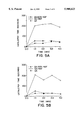
- FIG. 5(A-B) shows the effects of encapsulated NGF-secreting BHK cells on haloperidol (Panel A) and SCH23390-induced (Panel B) catalepsy in bilaterally lesioned QA rats. Data are presented as the mean ( ⁇ ) amount of time spent in catalepsy for each of the treatment groups.
- the solid circles/solid lines represent data for animals receiving QA and Non-NGF secreting BHK cell implants, open circles/solid lines represent data for animals receiving QA and NGF secreting BHK cell implants, and solid circles/dashed lines represent data for animals receiving QA alone.
- FIG. 6(A-C) depicts von Frey Somatosensory Thresholds, measured by touches to response at each of three tensile strengths.
- Old rats (26.7 mos.) implanted with encapsulated BHK-NGF cells were more responsive to the light stimulus than old rats in the control groups, but they were no more responsive than the young rats. Closed circles represent controls; squares represent NGF-BHK cell data.
- FIG. 7(A-B) shows the results of a working memory version of the Morris water maze test, with a two hour interval between trials.
- Panel A-Better 50% Pre-implant
- Panel B-Worse 50% Pre-implant. Closed circles represent controls
- squares represent NGF-BHK cell data.
- FIG. 8 shows that in aged monkeys receiving encapsulated BHK-NGF cells, lesion-induced degeneration of septal neurons was significantly attenuated compared to controls.
- This invention is directed to improved capsules that permit viability of encapsulated cells for an extended period of time and that are easily retrievable.
- This invention is also directed to improved devices and methods, which use genetically altered cells contained in biocompatible capsules, for the expression of biologically active molecules and long-term, stable delivery of biologically active molecules to host mammals.
- the devices and methods of the instant invention are useful for long-term, stable expression of a wide range of biologically active molecules, including high molecular weight products up to 200 kD, to an individual in need of them, and/or to provide needed metabolic functions to an individual, such as the removal of harmful substances.
- Biologically active molecules used in the devices and methods of the instant invention include a wide variety of factors normally secreted by various organs or tissues. For example, insulin can be delivered to a diabetic patient, dopamine to a patient suffering from Parkinson's disease, Factor VIII to a Type A hemophiliac, or an analgesic to a patient in pain.
- trophic factors such as erythropoietin, growth hormone, Substance-P, neurotensin, NGF, BDNF, NT-3, NT-4/5, CNTF, GDNF, CDF/LIF, EGF, IGF, PDGF, bFGF, and aFGF.
- the devices and methods of the instant invention are also useful for long-term, stable expression of biologically active molecules including hemoglobin, tyrosine hydroxylase, prohormone convertase, bcl-2, dopa decarboxylase, and dopamine beta-hydroxylase.
- Another family of products suited to delivery by the instant invention comprises biological response modifiers, including lymphokines and cytokines.
- the encapsulated cells described herein can also be used to restore or augment vital metabolic functions, such as the removal of toxins or harmful metabolites (e.g., cholesterol) from the bloodstream by cells such as hepatocytes.
- vital metabolic functions such as the removal of toxins or harmful metabolites (e.g., cholesterol) from the bloodstream by cells such as hepatocytes.
- the methods of the instant invention make possible the implantation of cells without the concomitant need to immunosuppress the recipient for the duration of treatment. Through use of the methods of this invention, homeostasis of particular substances can be restored and maintained for extended periods of time.
- the biologically active molecules contemplated within the scope of this invention include molecules that are secreted from the capsule, or from an otherwise transplanted cell, and either directly or indirectly result in a biological effect in the mammalian host, as well as those biologically active molecules that directly or indirectly result in a biological effect on cells contained within the capsule.
- a preferred embodiment of this invention is an improved method for delivering neurotrophic factors to the central nervous system (CNS) of host mammals.
- CNS central nervous system
- an improved method for long-term, stable expression and delivery of nerve growth factor (NGF) to a specific region of the CNS of a mammalian host is provided.
- NGF nerve growth factor
- genes encoding numerous biologically active molecules have been cloned and their nucleotide sequences published. Many of those genes are publicly available from depositories such as the American Type Culture Collection (ATCC) or various commercial sources. Genes encoding the biologically active molecules useful in this invention that are not publicly available may be obtained using standard recombinant DNA methods such as PCR amplification, genomic and CDNA library screening with oligonucleotide probes. Any of the known genes coding for biologically active molecules may be employed in the methods of this invention. See, e.g., U.S. Pat. No. 5,049,493; Gage et al., U.S. Pat. No. 5,082,670; and Genentech U.S. Pat. No. 5,167,762.
- genes particularly useful in this invention are the genes encoding human proenkephalin A, human prohormone convertase 2, human prohormone convertase 3, human BDNF, POMC (pro-opiomelanocortin), ⁇ -endorphin, prodynorphin, mature human BDNF with the human NGF signal sequence, human CNTF, human NT3, human NGF, rat GDNF, mature human NT5 with the human NGF signal sequence, bovine dopamine ⁇ hydroxylase, bovine dopamine decarboxylase, and thymidine kinase.
- a gene of interest i.e., a gene that encodes a suitable biologically active molecule
- a suitable expression vector can be inserted into a cloning site of a suitable expression vector by using standard techniques. It will be appreciated that more than one gene may be inserted into a suitable expression vector. These techniques are well known to those skilled in the art.
- the expression vector containing the gene of interest may then be used to transfect the cell line to be used in the methods of this invention.
- Standard transfection techniques such as calcium phosphate co-precipitation, DEAE-dextran transfection or electroporation may be utilized.
- Commercially available mammalian transfection kits may be purchased from e.g., Stratagene.
- a wide variety of host/expression vector combinations may be used to express the gene encoding the biologically active molecule of interest.
- Long-term, stable in vivo expression is achieved using expression vectors (i.e., recombinant DNA molecules) in which the gene encoding the biologically active molecule is operatively linked to a promoter that is not subject to down regulation upon implantation in vivo in a mammalian host. Accordingly, such expression vectors would typically not contain a retroviral promoter.
- Suitable promoters include, for example, the early and late promoters of SV40 or adenovirus and other known non-retroviral promoters capable of controlling gene expression.
- Useful expression vectors may consist of segments of chromosomal, non-chromosomal and synthetic DNA sequences, such as various known derivatives of SV40 and known bacterial plasmids, e.g., pUC, pBlue ScriptTM plasmids from E. coli including pBR322, pCR1, pMB9, pUC, pBlue ScriptTM and their derivatives.
- segments of chromosomal, non-chromosomal and synthetic DNA sequences such as various known derivatives of SV40 and known bacterial plasmids, e.g., pUC, pBlue ScriptTM plasmids from E. coli including pBR322, pCR1, pMB9, pUC, pBlue ScriptTM and their derivatives.
- Expression vectors containing the geneticin (G418) or hygromycin drug selection genes are also useful in practicing this invention. These vectors can employ a variety of different enhancer/promoter regions to drive the expression of both a biologic gene of interest (e.g., NGF) and/or a gene conferring resistance to selection with toxin such as G418 or hygromycin B.
- a biologic gene of interest e.g., NGF
- a gene conferring resistance to selection with toxin such as G418 or hygromycin B.
- the G418 resistance gene codes for aminoglycoside phosphotransferase (APH) which enzymatically inactivates G418 (100-500 ⁇ g/ ⁇ l) added to the culture medium. Only those cells expressing the APH gene will survive drug selection usually resulting in the expression of the second biologic gene as well.
- the hygromycin B phosphotransferase (HPH) gene codes for an enzyme which specifically modifies hygromycin toxin and inactivates it. Genes cotransfected with or contained on the same plasmid as the hygromycin B phosphotransferase gene will be preferentially expressed in the presence of hygromycin B at 50-200 ⁇ g/ml concentrations.
- promoters can be employed to direct the expression of the genes for G418 and hygromycin B and/or the biologic gene of interest.
- These promoters include, but are not limited to, the promoters of hDBH (human dopamine beta hydoxylase) (Mercer et al., Neuron, 7, pp. 703-716, (1991)), hTH (human tyrosine hydroxylase) (Kaneda, et al., Neuron, 6, pp. 583-594, (1991)), hPNMT (human phenylethanaolamine N-methyltransferase) (Baetge et al., PNAS, 85, pp.
- hDBH human dopamine beta hydoxylase
- hTH human tyrosine hydroxylase
- hPNMT human phenylethanaolamine N-methyltransferase
- mGFAP mouse glial fibrillary acidic protein
- MBP myelin basic protein
- mNF-L mouse neurofilament-light subunit
- hPo human P 0' , the promoter for the gene encoding the major myelin glycoprotein in the peripheral nervous system
- mMT, rNSE rat neuron-specific enolase
- expression vectors examples include the commercially available pRC/CMV, pRC/RSV, and pCDNA1NEO (InVitrogen).
- the viral promoter regions directing the transcription of the drug selection and biologic genes of interest are replaced with one of the above promoter sequences that are not subject to the down regulation experienced by viral promoters within the CNS.
- the GFAP promoter would be employed for the transfection of astrocytes and astrocyte cell lines
- the TH promoter would be used in PC12 cells
- MBP promoter would be used in oligodendrocytes.
- the pNUT expression vector is used (see FIG. 1).
- the pNUT expression vector can be modified such that the DHFR coding sequence is replaced by the coding sequence for G418 or hygromycin drug resistance.
- the SV40 promoter within the pNUT expression vector can also be replaced with any suitable constitutively expressed mammalian promoter, such as those discussed above.
- a wide variety of cells may be used. These include well known, publicly available immortalized cell lines as well as dividing primary cell cultures. Examples of suitable publicly available cell lines include, baby hamster kidney (BHK), chinese hamster ovary (CHO), mouse fibroblast (L-M), NIH Swiss mouse embryo (NIH/3T3), African green monkey cell lines (including COS-1, COS-7, BSC-1, BSC-40, BMT-10 and Vero), rat adrenal pheochromocytoma (PC12 and PC12A), AT3, rat glial tumor (C6), astrocytes and other fibroblast cell lines.
- BHK baby hamster kidney
- CHO chinese hamster ovary
- L-M mouse fibroblast
- NIH/3T3 NIH Swiss mouse embryo
- African green monkey cell lines including COS-1, COS-7, BSC-1, BSC-40, BMT-10 and Vero
- PC12 and PC12A rat adrenal pheochromocytoma
- Primary cells that may be used include, bFGF-responsive neural progenitor stem cells derived from the CNS of mammals (Richards et al., PNAS 89, pp. 8591-8595 (1992); Ray et al., PNAS 90, pp. 3602-3606 (1993)), primary fibroblasts, Schwann cells, astrocytes, ⁇ -TC cells, Hep-G2 cells, oligodendrocytes and their precursors, myoblasts and the like.
- the cell types that can be employed for encapsulated cell therapy within the scope of this invention include cells from allogeneic and xenogeneic sources.
- One of the principal advantages of our encapsulated approach rests with the immunoisolatory properties of the membranes of this invention, and their ability to support cells that otherwise would not be appropriate for transplantation (i.e., non-human sources, immortalized and/or tumor cell lines).
- a particular advantage to using xenogeneic over allogeneic cells is that in the unlikely event of membrane failure, the xenogeneic cells are more likely to be targeted for destruction by the immune system when compared to allogeneic cells.
- xenogeneic sources are easy to obtain and their use precludes the necessity for the use of human tissue which is difficult to obtain and fraught with societal and ethical considerations.
- human tissue may contain adventitious agents that are more readily transmitted to the transplantation recipient.
- use of xenogeneic tissue and cell lines for transplantation in humans removes the risks associated with the handling and processing of human tissue.
- the pNUT amplification expression system is used to transfect BHK cells.
- the pNUT vector containing the gene of interest can also be employed to transfect a large number of other standard immortalized or transformed tissue culture cell lines such as COS, L-cells, CHO, and the like, as discussed above.
- the pNUT expression vector can be employed to transfect primary astrocytic, oligodendrocytic or neuronal cell lines (e.g., bFGF-responsive neural progenitor-stem cells, as discussed above).
- the cell lines transformed according to this invention are capable of providing long-term, stable expression of a biologically active molecule(s).
- Such long-term, stable expression can be achieved by increasing or amplifying the copy number of the transgene encoding the biologically active molecule(s), using amplification methods well known in the art.
- amplification methods include, e.g., DHFR amplification (see, e.g., Kaufman et al., U.S. Pat. No. 4,470,461) or glutamine synthetase ("GS") amplification (see, e.g., U.S. Pat. No. 5,122,464, and European published application EP 338,841).
- Another method for obtaining long-term, stable expression of genes in mammalian cells is by double transfection using two separate drug-selection markers, hygromycin or G418 resistant expression vectors can be employed to sequentially or simultaneously transfect a number of the cell lines above to achieve a variety of gene copy inserts and hence expression levels of the gene of interest.
- cells are transfected with plasmid pRC/CMV containing the coding sequence for the ⁇ -NGF or other gene of interest operatively linked to the GFAP promoter/enhancer element replacing the CMV promoter sequences normally found in this expression vector.
- plasmid pRC/CMV containing the coding sequence for the ⁇ -NGF or other gene of interest operatively linked to the GFAP promoter/enhancer element replacing the CMV promoter sequences normally found in this expression vector.
- the clones Upon selection of stable clones expressing the transfected transgene the clones would be retransfected with the same pRc/GFAP NGF expression vector containing the bacterial hygromycin B phosphotransferase gene (Gritz and Davies, Gene, pp. 179-188 (1983)) in place of the G418 resistance gene.
- the cells could be assayed for the clones with the highest number of integrated copies and/or expression of the transfected transgene in both RNA and protein products.
- other pairs of drug selection expression vectors may be employed to produce cell lines transfected with multiple vectors which have stably integrated high copy numbers of the genetic material and express various levels of biologically active molecules of interest.
- a multiplicity of cells may be used in the methods of this invention, such that implantation of a polymer-capsule can be sufficient to provide an effective amount of the needed substance or function to an individual.
- more than one biologically active molecule may be stably expressed and/or delivered over long periods from a single capsule.
- One way to accomplish this result is to encapsulate a single cell line which has been genetically altered to express more than one heterologous gene.
- Another way to accomplish this result is to encapsulate in a single capsule a mixture of cells, wherein some cells have been genetically modified to express one biologically active molecule and other cells have been genetically modified to express a second biologically active molecule. It will be appreciated that a non-genetically engineered cell line can be utilized to provide the second biologically active molecule.
- a cell line may also be genetically engineered to express different biologically active molecules.
- the sub clones of the parental cell lines, each expressing a different transgene, may then be pooled and encapsulated to achieve the desired effect on a long-term basis.
- This invention also contemplates using capsules containing cells that are genetically modified with a heterologous gene, which gene enables the cells to remain viable within the capsule upon implantation within a host mammal.
- the methods of this invention are also directed to methods of delivery of molecules within the implanted capsules.
- pNUT expression vector containing the human ⁇ -NGF gene operatively linked to the mouse metallothionein promoter to transfect BHK cells via the calcium phosphate co-precipitation method.
- a variety of biocompatible immunoisolatory capsules are suitable for delivery of molecules according to this invention. Such capsules will allow for the passage of metabolites, nutrients and therapeutic substances while minimizing the detrimental effects of the host immune system.
- the capsule of this invention will be similar to those described in Aebischer et al., PCT publication WO 92/19195, incorporated herein by reference.
- the T1/2 membranes described herein are used to encapsulate the cells that are modified according to the methods of this invention. It will be appreciated that the T1/2 membranes described herein can also be used for encapsulation of any other suitable cell or cell lines. Thus, the T1/2 membranes of this invention can be used to encapsulate primary (non-dividing) cells, as well as dividing cells.
- Useful biocompatible polymer capsules comprise (a) a core which contains a cell or cells, either suspended in a liquid medium or immobilized within a hydrogel or extracellular matrix, and (b) a surrounding or peripheral region of permselective matrix or membrane (jacket) which does not contain isolated cells, which is biocompatible, and which is sufficient to protect isolated cells if present in the core from detrimental immunological attack.
- the core of the polymer capsule is constructed to provide a suitable local environment for the continued viability and function of the cells isolated therein.
- cells or cell lines are most advantageously isolated within a capsule having a liquid core.
- cells can be isolated within a capsule whose core comprises a nutrient medium, optionally containing a liquid source of additional factors to sustain cell viability and function, such as fetal bovine or equine serum.
- Microcapsules may sometimes be suitable for use in the methods and compositions of this invention.
- the fabrication of microcapsules have been described in Espevik et al., PCT publication WO 9107951, and Sefton, U.S. Pat. No. 4,353,888 incorporated herein by reference.
- the core may be composed of a matrix formed by a hydrogel which stabilizes the position of the cells in cell clumps.
- hydrogel herein refers to a three dimensional network of cross-linked hydrophilic polymers.
- the network is in the form of a gel, substantially composed of water, preferably but not limited to gels being greater than 90% water.
- compositions which form hydrogels fall into three classes.
- the first class carries a net negative charge (e.g., alginate).
- the second class carries a net positive charge (e.g., collagen and laminin).
- Examples of commercially available extracellular matrix components include MatrigelTM and VitrogenTM. Fibroblasts generally survive well in a positively charged matrix and are thus suitably enclosed in extracellular-matrix type hydrogels.
- the third class is net neutral in charge (e.g., highly crosslinked polyethylene oxide, or polyvinylalcohol). Any suitable matrix or spacer may be employed within the core, including precipitated chitosan, synthetic polymers and polymer blends, microcarriers and the like, depending upon the growth characteristics of the cells to be encapsulated.
- the capsules are immunoisolatory.
- the surrounding or peripheral region of the capsule should confer protection of the cells from the immune system of the host in whom the capsule is implanted, by preventing harmful substances of the host's body from entering the core of the vehicle, and by providing a physical barrier sufficient to prevent detrimental immunological contact between the isolated cells and the host's immune system.
- the thickness of this physical barrier can vary, but it will always be sufficiently thick to prevent direct contact between the cells and/or substances on either side of the barrier.
- the thickness of this region generally ranges between 5 and 200 microns; thicknesses of 10 to 100 microns are preferred, and thickness of 20 to 75 microns are particularly preferred.
- Types of immunological attack which can be prevented or minimized by the use of the instant vehicle include attack by macrophages, neutrophils, cellular immune responses (e.g. natural killer cells and antibody-dependent T cell-mediated cytoloysis (ADCC), and humoral response (e.g., antibody-dependent complement mediated cytolysis).
- cellular immune responses e.g. natural killer cells and antibody-dependent T cell-mediated cytoloysis (ADCC)
- humoral response e.g., antibody-dependent complement mediated cytolysis
- immunoisolatory capsules allows the implantation of xenogeneic cells or tissue, without a concomitant need to immunosuppress the recipient.
- Use of immunoisolatory capsules also allows use of unmatched cells (allografts).
- the type and vigor of an immune response to xenogeneic cells is expected to differ from the response encountered when syngeneic or allogeneic tissue is implanted into a recipient. This response may proceed primarily by cell-mediated, or by complement-mediated attack; the determining parameters in a particular case may be poorly understood.
- the exclusion of IgG from the core of the vehicle is not the touchstone of immunoprotection, because in most cases IgG alone is insufficient to produce cytolysis of the target cells or tissues.
- immunoisolatory macrocapsules it is possible to deliver needed high molecular weight products or to provide metabolic functions pertaining to high molecular weight substances, provided that critical substances necessary to the mediation of immunological attack are excluded from the immunoisolatory capsule.
- These substances may comprise the complement attack complex component Clq, or they may comprise phagocytic or cytotoxic cells; the instant immunoisolatory capsule provides a protective barrier between these harmful substances and the isolated cells.
- an immunoisolatory capsule can be used for the delivery even from xenogeneic cells, products having a wide range of molecular sizes, such as insulin, parathyroid hormone, interleukin 3, erythropoietin, albumin, transferrin, enkephalins, endorphins, catecholamines, Factor VIII, NGF, BDNF, NT-3, NT-4/5, CNTF, GDNF, CDF/LIF, EGF, IGF, bFGF, aFGF, PDGF, TGF and the like.
- xenogeneic cells products having a wide range of molecular sizes, such as insulin, parathyroid hormone, interleukin 3, erythropoietin, albumin, transferrin, enkephalins, endorphins, catecholamines, Factor VIII, NGF, BDNF, NT-3, NT-4/5, CNTF, GDNF, CDF/LIF, EGF, IGF, bF
- polymers and polymer blends can be used to manufacture the capsule jacket, including polyacrylates (including acrylic copolymers), polyvinylidenes, polyvinyl chloride copolymers, polyurethanes, polystyrenes, polyamides, cellulose acetates, cellulose nitrates, polysulfones, polyphosphazenes, polyacrylonitriles, poly(acrylonitrile/covinyl chloride), as well as derivatives, copolymers and mixtures thereof.
- polyacrylates including acrylic copolymers
- polyvinylidenes including acrylic copolymers
- polyvinyl chloride copolymers polyurethanes
- polystyrenes polyamides
- cellulose acetates cellulose nitrates
- polysulfones polyphosphazenes
- polyacrylonitriles poly(acrylonitrile/covinyl chloride)
- the capsule can be any configuration appropriate for maintaining biological activity and providing access for delivery of the product or function, including for example, cylindrical, rectangular, disk-shaped, patch-shaped, ovoid, stellate, or spherical. Moreover, the capsule can be coiled or wrapped into a mesh-like or nested structure. If the capsule is to be retrieved after it is implanted, configurations which tend to lead to migration of the capsules from the site of implantation, such as spherical capsules small enough to travel in the recipient's blood vessels, are not preferred. Certain shapes, such as rectangles, patches, disks, cylinders, and flat sheets offer greater structural integrity and are preferable where retrieval is desired.
- the instant capsule must provide, in at least one dimension, sufficiently close proximity of any isolated cells in the core to the surrounding tissues of the recipient, including the recipient's bloodstream, in order to maintain the viability and function of the isolated cells.
- the diffusional limitations of the materials used to form the capsule do not in all cases solely prescribe its configurational limits.
- Certain additives can be used which alter or enhance the diffusional properties, or nutrient or oxygen transport properties, of the basic vehicle.
- the internal medium can be supplemented with oxygen-saturated perfluorocarbons, thus reducing the needs for immediate contact with blood-borne oxygen. This will allow isolated cells to remain viable while, for instance, a gradient of angiotensin is released from the capsule into the surrounding tissues, stimulating ingrowth of capillaries.
- the implantable capsule is of a sufficient size and durability for complete retrieval after implantation.
- Such macrocapsules have a core of a preferable minimum volume of about 1 to 10 ⁇ l and depending upon use are easily fabricated to have a volume in excess of 100 ⁇ l.
- the preferred capsule will have an inner single ultrafiltration membrane with a permselective pore-size permeability range of 60-98% BSA rejection coefficient and 50-90% ovalbumin rejection coefficient.
- the capsule may be in the form of a flat sheet sealed at the periphery or of a hollow fiber sealed at the ends as described in PCT application WO 92/19195. In a flat sheet format the two walls will be separated by a gap thickness of less than 1000 microns, preferably less than 300 microns. Wall thickness should be between about 25-200 microns, preferably between about 30-75 microns.
- the fiber In a hollow fiber configuration, the fiber will have an inside diameter of less than 1500 microns, preferably less than 300-600 microns. In either geometry, the hydraulic permeability will be in the range of 1-100 mls/min/M 2 /mm Hg, preferably in the range of 25 to 70 mls/min/M 2 /mmHg.
- the glucose mass transfer coefficient of the capsule defined, measured and calculated as described by Dionne et al., ASAIO Abstracts, p. 99 (1993), and Colton et al., The Kidney, eds., Brenner BM and Rector FC, pp. 2425-89 (1981) will be greater than 10 -6 cm/sec, preferably greater than 10 -4 cm/sec.
- the morphology of the outer wall surface of the capsule is variable.
- Previously described T1, T2, T4 membranes and the novel T1/2 membranes of this invention differ in their outer wall surface morphology. All these membranes are characterized by an inner permselective skin.
- T1 membranes are characterized by an "open” or fenestrated non-permselective outer surface wall, and a trabecular wall structure between the outer and inner wall surfaces. See, e.g. Lacy et al., Science, 254, pp. 1782-284 (1991).
- the fenestrations of "macropores" in the outer wall surface of a T1 membrane typically occupy about 20%-40% of the total outer surface wall area.
- the macropores are 10 ⁇ mm-15 ⁇ m in diameter or greater.
- T2 membranes have a similar trabecular structure between the inner and outer walls but are characterized by a more "closed” or smoother outer surface wall. T2 membranes, typically are characterized by fewer than 10% macropores on the outer surface wall and virtually no macropores in the 5-15 ⁇ m diameter size range.
- T4 membranes are further distinct in that the outer surface is also permselective, unlike the T1 or T2 membranes.
- T4 membranes are useful for CSF implantation sites, such as the ventricles or sub-arachnoid space, as well as other fluid bathed implantation sites.
- T1 and T1/2 capsules are especially suited for long term implants. We prefer T1/2 capsules.
- T1/2 capsules are characterized by a total macropore distribution of between approximately 2-20%, preferably 2-15% of the total outer surface wall area.
- the macropores should fall within the size range of approximately 5 ⁇ m to about 15 ⁇ m in diameter. The relative distribution of pore sizes within this range can vary.
- T1/2 hollow fiber membrane capsules made from PAN/PVC were utilized.
- the total macropore area was about 12% of the total outer wall surface area.
- Approximately 20% of the macropores ranged between 5-10 ⁇ m in diameter and about 80% of the macropores were about 10 ⁇ m in diameter.
- T1/2 hollow fiber membrane capsules were fabricated having a total macropore area of about 2.4% of the total outer wall surface area. Approximately 17% of the macropores were about 5 ⁇ m in diameter, about 33% were about 10 ⁇ m in diameter, and about 50% were about 15 ⁇ m in diameter.
- T1/2 hollow fiber membrane capsules were fabricated having a total macropore area of approximately 10% of the total outer wall surface area. Greater than 99% of these macropores ranged between 10-15 ⁇ m in diameter.
- the quantification of outer surface macropore morphology can be accomplished using any standard method. We used two methods.
- sections of the hollow fiber membrane are mounted for scanning electron microscopy.
- the outer surface of the fibers is then sputter coated with gold.
- Images of 1000 ⁇ magnification are generated.
- One scanning electron micrograph at 1000 ⁇ comprises an area of approximately 9.2 k ⁇ m 2 (115 ⁇ m ⁇ 80 ⁇ m).
- a 10 ⁇ m diameter pore is 1 cm in diameter.
- all pores can be classified as either 15 ⁇ m, 10 ⁇ m, 5 ⁇ m or 2.5 ⁇ m in diameter.
- the total percent of the outer surface wall open i.e., as macropores
- the T1/2 fibers of this invention may be prepared by any suitable method known in the art.
- One method involves coextrusion of a polymeric casting solution and a coagulant through a coaxial spinneret by a suitable adjustment of luminal and casting solution flow rates using well known techniques described by Cabasso, I., Encyclopedia of Chemical Technology, 12, pp. 492-517 (1980).
- the coagulant which can include biological tissue fragments, organelles, or suspensions of cells and/or other therapeutic agents, as described in Dionne, WO 92/19195 and U.S. Pat. Nos. 5,158,881, 5,283,187 and 5,284,761, incorporated herein by reference.
- T1 membranes may be formed by coextrusion of a polymer solution and coagulant solution through air before entering a quench bath.
- T2 membranes may be formed by coextruding the polymer, and coagulation solutions into humidified air or a mist and then into a bath.
- T4 membranes may be formed by coextrusion of the polymer and coagulant solutions directly into a coagulant bath, so that formation of the permselective membrane occurs on both outer and inner wall surfaces simultaneously.
- T1/2 membranes may be formed using similar methods used to form T2 membranes.
- the mist or humidity at the coextrusion port may be controlled according to known methods to produced the desired outer surface morphology.
- the nozzle distance from a quench bath may be varied, according to routine methods.
- the absolute and/or relative flow rates of polymer and coagulant may be adjusted to achieve the desired outer wall surface morphology.
- the polymer and coagulant solution compositions and temperatures can be varied to achieve the desired outer surface wall morphology.
- the casting solution may be 10-15% PAN/PVC in DMSO (w/w) and the coagulant may be water, or other aqueous medium.
- the casting solution may be, e.g., 16% PAN/PVC, and the coagulant may be, e.g, 40% NMP, 60% H 2 O at 23° C.
- any suitable method of sealing the capsules may be used, including the employment of polymer adhesives and/or crimping, knotting and heat sealing. These sealing techniques are known in the art.
- any suitable "dry” sealing method can also be used. In such methods, a substantially non-porous fitting is provided through which the cell-containing solution is introduced. Subsequent to filling, the capsule is sealed. Such a method is described in copending U.S. application Ser. No. 08/082,407, herein incorporated by reference.
- the methods and devices of this invention are intended for use in a mammalian host, recipient, subject or individual, preferably a primate, most preferably a human.
- implantation sites include the central nervous system, including the brain, spinal cord, and aqueous and vitreous humors of the eye.
- Preferred sites in the brain include the striatum, the cerebral cortex, subthalamic nuclei and nucleus Basalis of Maynert.
- Other preferred sites are the cerebrospinal fluid, most preferably the subarachnoid space and the lateral ventricles.
- This invention also contemplates implantation into the kidney subcapsular site, and intraperitoneal and subcutaneous sites, or any other therapeutically beneficial site.
- methods are provided for the treatment of diseases caused by neural degeneration.
- diseases caused by neural degeneration include Alzheimer's disease, Huntington's disease, AIDS-related dementia, Amyotrophic Lateral Sclerosis (ALS) and Parkinson's disease.
- ALS Amyotrophic Lateral Sclerosis
- Some animal models for neurodegenerative conditions are based on the premise that a specific insult may damage or kill neurons. In some cases this may even lead to a cascade of neuronal death which affects trophically interdependent neurons along pathways responsible for specific brain functions.
- a strategy for treatment of neural degenerative conditions involves the localized administration of growth or trophic factors in order to (1) inhibit further damage to postsynaptic neurons, and (2) improve viability of cells subjected to the insult.
- Factors known to improve neuronal viability include NGF, BDNF, NT-3, NT-4/5, CNTF, GDNF, CDF/LIF, bFGF, aFGF, IGF, neurotensin, and Substance-P.
- HD Huntington's disease
- the manifestation of the disorder typically occurs in middle life, about 35-45 years of age, followed by an intractable course of mental deterioration and progressive motor abnormalities with death usually occurring within 15 years.
- Research into the neural pathology in HD has revealed a complex mosaic of related and interdependent neurochemical and histopathological alterations.
- Glutamate is one of the major excitatory neurotransmitters found in the CNS. It can act, however, as a potent neurotoxin and a number of attempts have been made to develop animal models of HD based on the relatively specific cytotoxic effects of glutamate and other excitotoxic compounds. These compounds include structural analogs of glutamate, such as kainic acid (KA), ibotenic acid (IA), and the endogenous tryptophan metabolite quinolinic acid (QA).
- KA kainic acid
- IA ibotenic acid
- QA endogenous tryptophan metabolite quinolinic acid
- Nerve growth factor-secreting cells such as BHK cells engineered to express human NGF represent a therapy for quinolinic acid induced neurodegeneration.
- Another animal model involves lesion of the fimbria-fornix (rodents) or fornix (primates).
- rodents the fimbria-fornix
- fornix primates
- neurons of the septohippocampal system are axotomized which leads to NGF-dependent degeneration and cell death in the septal cholinergic neurons.
- These lesions cause degenerative changes in brain areas similarly affected in Alzheimer's disease in humans.
- NGF may be delivered to the affected area by the implantation of a capsule containing genetically altered cells which secrete NGF.
- Other neurotrophic factors such as CNTF, BDNF, bFGF, CDF/LIF may also protect similar or non-overlapping populations of septal cholinergic neurons from atrophy and/or death.
- the cells are fibroblasts which have been genetically engineered to produce recombinant human NGF.
- the methods and compositions of this invention may be used for the treatment of age-related cognitive defects resulting from neural degeneration. Such treatment may augment cognitive performance, thus providing a symptomatic benefit. Alternatively, treatment may provide a neuroprotective effect, although not a symptomatic benefit.
- Age-related cognitive dysfunction and dementia in humans has been related to neuronal degeneration, especially of cholinergic basal forebrain neurons, and the decline of cortical and hippocampal levels of ChAT (Coyle et al., Science, 219, pp. 1184-90 (1983); Whitehouse et al., Science, 215, pp. 1237-39 (1982); Phelps et al., Neurobiol. Aging, 10, pp.
- NGF does not readily cross the blood brain barrier
- its administration into the CNS requires the use of invasive procedures which compromise the integrity of the blood brain barrier.
- the NGF was administered with osmotic minipumps or through chronic intraventricular cannulae.
- Those techniques require repeated infusions into the brain, either through injections via the cannulae, or from pumps which must be replaced every time the reservoir is depleted. Every occasion in which the pump reservoir must be replaced or the injection syringe reinserted through the annulae represents another opportunity that contaminants might be introduced into the brain, which is especially susceptible to infection.
- fetal tissue is clouded by ethical concerns and unencapsulated non-fetal cells may be rejected or produce tumors.
- tissue taken from fetal sources may be highly variable.
- exogenous NGF can be supplied with a relatively low risk of infection, without the use of fetal tissue, and without the risk of tissue rejection or tumor development.
- the beneficial effects of exogenous NGF for the treatment of age related cognitive defects may be obtained with doses much lower than previously reported to be effective for exogenous hNGF delivery.
- NGF neurotrophic factor
- capsular delivery of NGF, synthesized in vivo, to the brain ventricles, brain parenchyma, or other suitable CNS location, ranging from 1-1500 ng/day is desirable.
- the actual dosage of NGF, or other suitable factor can be varied by implanting a fewer or greater number of capsules.
- 1-1500 preferably 10-600, most preferably 50-500, ng NGF/human/day, for ventricular delivery and 1-1500, preferably 10-150, ng NGF/human/day for parenchymal delivery.
- NGF delovered was 75 ⁇ g/day.
- hNGF human NGF
- encapsulated cells that secrete a growth factor may be used enhance the survival and growth of unencapsulated cell grafts.
- a growth factor e.g., including NGF, BDNF, platelet derived factor "PDGF”, fibroblast growth factor “IFGF”, epidermal growth factor “EGF”, NT-3, NT-4/5, CNTF, gDNF
- NGF neurotrophic factor
- BDNF platelet derived factor
- IFGF fibroblast growth factor
- EGF epidermal growth factor
- NT-3, NT-4/5, CNTF, gDNF may be used enhance the survival and growth of unencapsulated cell grafts.
- the growth factor may be specific for the unencapsulated cell type, or may have a general effect.
- a particular embodiment contemplates co-implantation of encapsulated growth factor secreting cells with unencapsulated neurotransmitter or analgesic (e.g., enkephalins, endorphins or catecholamines) secreting cells into the central nervous system, in particular the lateral ventricles or striatum.
- analgesic e.g., enkephalins, endorphins or catecholamines
- effect of the encapsulated growth factor secreting cells on the unencapsulated cells may vary according to the implant location. We prefer intrastriatal implants.
- the human gene for ⁇ -NGF coding for the complete amino acid sequence of the pre-pro form of NGF was subcloned behind the mouse metallothionein promoter in an expression construct that contains the mutant form of dihydrofolate reductase (see, e.g., Kaufman U.S. Pat. No. 4,470,461) driven by the SV40 promoter (FIG. 1).
- Two human genomic clones coding for the 5' and 3' ends of the ⁇ -NGF gene were purchased from ATCC (phbeta N8D8, phbeta N8B9).
- a 440 bp 5' Sca1-EcoR1 fragment from phbeta N8D8 was ligated to a 3' 2.0 kb EcoR1 fragment isolated from phbeta N8B9.
- the spliced NGF genomic sequence contained ⁇ 37 bp of the 3' end of the first intron, the double ATG sequence believed to be the protein translation start for pre-pro-NGF and the complete coding sequence and entire 3' untranslated region of the human gene (Hoyle et al., Neuron 10, pp.
- the combined 2.51 kb ⁇ -NGF construct was subcloned into the DHFR based pNUT expression vector (Baetge et al., Proc. Natl. Acad. Sci. USA, 83, pp. 5454-5458 (1986)) immediately downstream from the mouse metallothionein-I promoter (-650 to +7) and the first intron of the rat insulin II gene (Palmiter R. D. et al., Proc. Natl. Acad. Sci. USA, 88, pp. 478-482 (1991)).
- the pNUT- ⁇ -NGF construct see FIG.
- BHK cells were introduced into BHK cells using a standard calcium phosphate-mediated transfection method.
- Transfected BHK cells were selected in medium containing 200 ⁇ M methotrexate (Sigma) for 3-4 weeks and resistant cells were maintained as a polyclonal population either with or without 200 ⁇ M methotrexate.
- Nerve growth factor causes a marked outgrowth of neurite processes in PC12 cells and as such provides a rapid and sensitive assay for NGF bioactivity.
- CM conditioned medium
- BHK-control parental BHK cells
- BHK-NGF cells were added to the PC12A (Schweitzer and Kelly, J. Cell Biol., 101, pp. 667-676 (1985)) cells grown on 6 well standard tissue culture plates.
- 25 S mouse NGF was added to some of the wells to induce neurites (50 ng/ml).
- the PC12A cells were scored for neurites that were ⁇ 3 times the length of the cell body diameter over a period of 1-4 days.
- NGF bioassays were also performed upon retrieval of implanted control and NGF-secreting, BHK cell-loaded, capsules by adding CM from the capsules to naive PC12A cells taken from capsules incubated with fresh medium for 24 hours.
- CM from BHK-NGF cells produced a robust neurite outgrowth in PC12A cells within 24 hours indicating that the NGF produced from the BHK cells was bioactive.
- BHK-controls showed no such capacity to elicit neurite outgrowth in the PC12A cells in parallel experiments.
- NGF blocking antibody mouse anti- ⁇ -NGF; Boehringer-Mannheim Cat. #1008-220
- CM recombinant hNGF
- bFGF basic fibroblast growth factor
- NGF expression from the encapsulated and the unencapsulated BHK-NGF cells was performed as follows: All of the reagents were obtained from Boehringer Mannheim Biochemicals unless otherwise noted. Nunc-Immuno MaxiSorp ELISA plates were coated with 150 ⁇ l per well of anti-mouse- ⁇ (2.5S) nerve growth factor at 1 ng/ml in coating buffer (1 ⁇ PBS without CACl 2 and without MgCl 2 /0.1% sodium azide; pH 9.6). The coated plates were incubated at 37° C. for at least 2 hours or alternatively at 4° C. overnight.
- the coating solution was withdrawn from the wells and the wells were washed three times with 300 ⁇ l wash buffer (50 mM Tris-HCl/200 mM NaCl/10 mM CaCl 2 /1% Triton X-100/0.1% sodium azide; pH 7.0).
- the wells were then blocked with 300 ⁇ l of coating solution with 10 mg/ml of bovine serum albumin (BSA) at room temperature for at least 30 minutes.
- BSA bovine serum albumin
- Conditioned medium samples were diluted 1:1 in 2 ⁇ sample buffer (the sample buffer is the same as wash buffer, only with 2% BSA). 100 ⁇ l of the prepared samples were loaded into the wells.
- the plates were covered and then incubated for at least 2 hours at 37° C. or overnight at 4° C.
- the solutions were removed from the wells by suction and washed three times with 300 ⁇ l of wash buffer.
- 100 ⁇ l of 4 U/ml of anti-mouse- ⁇ (2.5S) nerve growth factor- ⁇ -gal conjugate was added.
- the plates were incubated at 37° C. for at least 1 hour.
- the solutions were removed from the wells by suction and washed three times with 300 ⁇ l of wash buffer.
- chlorophenol red- ⁇ -D-galactopyranoside substrate solution 40 mg CPRG in 100 mM Hepes/150 mM NaCl/2 mM MgCl 2 / 0.1% sodium azide/1% BSA; pH 7.0
- CPRG chlorophenol red- ⁇ -D-galactopyranoside substrate solution
- PC12A cells were plated at a density of 100,000 cells per ml on poly ornithine-treated glass coverslips (12 mm) and allowed to equilibrate for at least 24 hours in a 24-well plate. Cells were grown in the same medium as previously described in the section on neurite outgrowth bioassay.
- conditioned medium from capsules containing BHK and BHK-NGF cells or recombinant human NGF (50 ng/ml) was added to the PC12A cells for 2 hours and allowed to incubate at 37° C. and 5% CO 2 . Following this incubation, the coverslips were fixed with 4% paraformaldehyde (in 0.1M PBS, pH 7.4), washed 2 ⁇ with 10 mM glycine in PBS, permeabilized with 1% triton X100 (in PBS for 10 minutes) and 1% nonidet P40 (in PBS for 10 minutes).
- the cells on coverslips were washed 3 ⁇ 5 minutes with PBS, blocked with 5% normal goat serum (NGS) in PBS for 1 hour and incubated in a rabbit polyclonal antiserum (Oncogene Science) raised against c-fos diluted 1:10 in 1% NGS in PBS for 3 hours.
- the coverslips were then washed 2 ⁇ 5 minutes with PBS and incubated with a fluorescein-conjugated goat anti-rabbit IgG antibody.
- the coverslips were washed 2 ⁇ with PBS and mounted with Citifluor® antifadent and viewed by fluorescence microscopy. Fluorescence was monitored by microscopy and c-fos induction measured by the presence of fluorescently labeled nuclei.
- the fibers produced (XP 11) were a T1/2 membrane type, having an inner diameter of 450 ⁇ 25 ⁇ m, a hydraulic permeability of 53 ml/(m 2 min mmHg), a BSA rejection coefficient of 88.7 ⁇ 2.1%, an ovalbumin rejection coefficient of 82.0 ⁇ 1.7%, and a glucose mass transfer coefficient of about 8 ⁇ 10 -4 cm/s.
- devices were fabricated by mounting a length of 6-7 mm dry hollow fiber, with a distal seal, onto a light-cured acrylate hub with a septal fixture at the proximal end which has loading access for cells to be injected into the lumen of the device. Glycerol was removed from the devices with 70% filter sterilized ethanol and placed in HBSS prior to the encapsulation procedure.
- BHK-control cells were loaded into prefabricated devices at a density of approximately 10 7 /ml.
- the BHK cell suspensions at a density of 2 ⁇ 10 7 /ml were mixed 1:1 with physiologic Vitrogens® (Celtrix, Palo Alto, Calif.), and infused into the pre-fabricated devices through the septal access port. After infusing 2-2.5 ⁇ l of the cellular suspension, the septum was cracked off and the access port was sealed using a light-cured acrylate (Zeneca).
- BHK cell-loaded devices were maintained in a serum-free defined medium, PC1 (Hycor), for 4-5 days prior to implantation. After 3 or 4 days in vitro, the cell-loaded capsules were washed twice in HBSS, and placed in 1 ml of fresh medium to be analyzed for NGF by ELISA.
- CM conditioned medium
- the capsules were implanted into the striatum of adult Lewis rats for one, three and six month periods. Upon explanation, the capsules were tested for NGF production by the neurite outgrowth assay.
- the BHK-NGF loaded capsules were able to produce neurite outgrowth in PC12A cells equivalent to or greater than 50 ng/ml of NGF. No NGF activity was detectable in the CM from the BHK-control capsules.
- ELISA quantitation of the samples from the encapsulated BHK-NGF cells release up to about 20 ng/24 hr/capsule after 3 and 6 months in vivo.
- rats were anesthetized with an intramuscular injection of a ketamine, xylazine and acepromazine mixture and positioned in a Kopf stereotoxic instrument (see Emerich et al. 1992). A sagittal incision was made in the scalp and a craniotomy performed extending 2.0 mm posterior and 3.0 mm lateral from Bregma.
- An aspirative device with a 20 gauge tip was mounted to a stereotaxic frame (Kopf Instruments) and the medial parietal cortex, cingulate cortex, corpus callosum, dorsal hippocampus, dorsal thalamus and fimbria-fornix were aspirated by placing the suction tip 1.40 mm posterior to Bregma and lowering it immediately lateral to the sagittal sinus to a depth of 5.0 mm. The tip was then moved laterally in 0.5 mm increments until a position of 3.0 mm lateral to Bregma was attained.
- a stainless steel obdurator was placed within the cannula, and the obdurator held in place while the outer cannula was raised to passively place the capsule within the previously prepared cavity.
- the stereotaxic coordinates for implantation were: 0.5 mm posterior to Bregma, 1.0 mm lateral to the sagittal suture and 7.5 mm below the cortical surface.
- ChAT choline acetyltransferase
- Sections were mounted, dehydrated and coverslipped. Adjacent sections were stained for hematoxylin and eosin (H+E). To verify the extent of lesion produced following aspirations of the fimbria-fornix, every 10th section was taken throughout the hippocampus and stained for acetylcholinesterase according to the method of Van Ootegan et al. (Brain Res. Bull., 12, pp. 543-553 (1984)). For quantification of cholinergic cell loss, ChAT-positive neurons were counted in the medial septum and vertical limb of the diagonal band at a magnification of 10 ⁇ . Representative sections (3 per brain) located approximately 0.7, 0.5 and 0.2 mm anterior to Bregma from each animal were used for this analysis.
- AchE immunoreactivity in the hippocampus was used as a second indicator of lesion completeness and ChAT-immunoreactivity in the septum provided evidence for cholinergic cell body sparing or atrophy as a result of transplant and lesions.
- a representative comparison of AchE immunostaining in the hippocampus of the lesioned-vs-control side demonstrated nearly complete loss of AchE afferents to the lesioned side.
- Example 2 demonstrated that the chronic delivery of hNGF from the BHK cells of this invention into the lateral ventricle of adult rats with fimbria-fornix lesions protects the medial septal cholinergic neurons that otherwise would have died as a result of the lesion. Similar experiments are described here performed in non-human primates. Lesioning of the fornix in monkeys (Cebus apella) is described in (Kordower and Fiandaca, J. Comp. Neurol., 298, pp. 443, (1990)).
- the data shown in Table III represents analysis of two BHK-NGF implanted monkeys and one control monkey. Subsequent analysis of NGF release data from an additional 6 animals in the same study revealed that the average level of hNGF produced by the capsules within prior to implantation was 21.4 ⁇ 2.0 ng/capsule/24 hours and 8.5 ⁇ 1.2 ng/capsule/24 hours in the retrieved capsules as measured by the NGF ELISA. Because each monkey was implanted with five capsules, the total amount of NGF produced per animal was 107 ng/24 hours and 41.5 ng/24 hours prior to implant and 1 month following implant, respectively. The BHK-control capsules produced no detectable hNGF (assay sensitive to 25 pg NGF/ml).
- the polymer capsules were left in situ from one animal receiving BHK-control implants. In all other monkeys, the BHK cell-loaded devices were retrieved from the lateral ventricles 23-28 days following implantation with little to no host tissue adhering to the capsules.
- monkeys receiving implants of BHK-hNGF cells exhibited a only a modest loss of cholinergic neurons within the septum (19 and 20%, respectively) and VLDB (7%). Furthermore, only implants of hNGF-secreting cells induced a dense sprouting of cholinergic fibers within the septum, which ramified against the ependymal lining of the ventricle adjacent to the transplant site. Examination of the retrieved capsules revealed an abundance of cells that produced detectable levels of hNGF in a sufficient concentration to differentiate PC12A cells in culture.
- Lewis rats were anesthetized with sodium pentobarbital (45 mg/kg, i.p.), and positioned in a Kopf stereotaxic instrument. A sagittal incision was made in the scalp and two holes drilled for the placement of XP-11 polymer capsules containing NGF-secreting BHK cells (as described in Example 1) into the lateral ventricle. Rats were either uni- or bilaterally implanted by placing the capsule within an 18 gauge Teflon catheter mounted to the stereotaxis frame and lowering it to the appropriate site. The stereotaxic coordinates for implantation were: 0.5 mm anterior to Bregma, 1.5 mm lateral to the sagittal suture and 8.0 mm below the cortical surface.
- QA was dissolved in 2N sodium hydroxide and diluted with phosphate buffer at pH 7.2 to a final pH of 7.4 and concentration of 225 nmol/ul. QA was infused into each striatum, using a 28-gauge Hamilton syringe, over five minutes in a volume of 1 ⁇ l.
- the injection cannula was left in place for an additional two minutes to allow for diffusion of the perfusate.
- This procedure resulted in the formation of three experimental groups: 1) quinolinic acid only (QA), quinolinic acid+NGF-secreting BHK cells (QA/NGF), and quinolinic acid+non-NGF-secreting BHK cells (QA/NON-NGF).
- QA quinolinic acid only
- QA/NGF quinolinic acid+NGF-secreting BHK cells
- QA/NON-NGF quinolinic acid+non-NGF-secreting BHK cells
- locomotor variables were collected: (1) horizontal activity (HA): the total number of interruptions of the horizontal sensors; (2) total distance (TD) ; the distance travelled by the animal in inches; (3) number of movements (NM): this parameter increased by one each time a movement was registered by the breaking of a beam separated by at least one second from the last interruption; (4) movement time (MT): the time in seconds that the animal spent in motion; (5) average speed (AS): the average speed of the animal's movement in cm/second; (6) average distance (AD): the average distance the animal moved in inches during a movement bout; (7) vertical activity (VA): the total number of beam interruptions in the vertical sensors; (8) vertical time (VT): this parameter increased while an animal was breaking the beam of a vertical sensor; (9) number of vertical movements (VM): this increased with vertical sensor-beam interruptions that were separated by at least one (1) second; (10) number of stereotypic movements (NS): this parameter increased when the same beams were broken repeatedly with one second in between; (11) stereotypy time
- mice were tested for hypokinesia (catalepsy) following administration of the D1 and D2 dopamine receptor antagonists haloperidol and SCH2388.
- Catalepsy was measured using the bar test in which the rear feet of the animals were placed on a platform and their front feet were placed on a horizontal bar (0.6 cm in diameter) suspended 9.0 cm above the platform. The degree of catalepsy produced in the animals was measured by how long it took for each animal to remove itself from the bar. A maximum of 300 seconds was allowed. Animals were randomly assigned to one of three treatment groups and injected with either haloperidol (1.0 mg/kg), SCH23388 (1.0 mg/kg) or control vehicle (0.9% saline). Bar tests were conducted again at 1, 2, 3 and 4 hours after administration of drug and/or control vehicle. All animals were tested under each treatment condition with tests separated by a 3-4 day interval.
- mice Consistent with the behavior protection observed following unilateral QA, animals exhibited an attenuation of hyperactivity produced by bilateral injections of QA. Animals which received QA alone or QA and non-NGF BHK cell implants (i.e., BHK-controls) exhibited comparable increases in activity which ranged from approximately 100-350% of pre-implant levels (FIG. 4). In contrast, those animals which received QA together with NGF-secreting BHK cells (Table IV, QA BILAT) showed activity levels which were markedly attenuated relative to NON-NGF treated animals and ranged from approximately 25-150% of preimplant values.
- monkeys received quinolinic acid injections into the caudate nucleus and into the putamen in the immediate vicinity of the capsule implants.
- Monkeys were videotaped under normal conditions and following apomorphine treatment once per week beginning one week prior to the implant and starting 3 weeks after lesions for two weeks. Just prior to sacrifice, the capsules were removed.
- the levels of hNGF release from each capsule will be quantified via ELISA.
- the biological activity relevance of this hNGF release from the capsule upon retrieval will be assessed via PC12 cell bioassay, and the variability of the implanted BHK cells will be assessed via Nissl staining.
- the monkey brains will be immunohistologically processed for ChAT, GAD, NADPH, NPY, somatostatin, and Nissl.
- the area of the lesion will be quantified on Nissl stained sections and the number of ChAT-ir, GAD-ir, NPY-ir, somatostatin-ir, NADPH stained cells will be quantified bilaterally.
- mice NGF intra ventricular infusion using a mini pump system (Medtronic).
- the infusion system was implanted subcutaneously into the abdominal wall and connected by a subcutaneous catheter to a intraventricular catheter which was inserted through a 2 mm burr hole in the cranium into the right lateral ventricle.
- a total of 32.4 ml of mouse NGF was infused over a 3 month period.
- capsules are stereotaxically placed in the ventricular space located between the septum and caudate or in selected neocortical/hippocampal regions.
- the capsules release between 50-500 ng of human NGF/24 hours.
- the capsules will remain implanted for 3-12 months over which time the patients can be monitored for behavioral and biochemical improvements.
- Patients can be assessed using a series of cognitive function tests including but not limited to: (1) mini-mental state examination, used to assess the level of cognitive functioning (Folstein et al., J. Psychiatr. Res., 12, pp. 189-198 (1975); (2) Face recognition exam (Backman et al., Psychol.
- Neural stem cells are generated from a 1 mg biopsy of brain tissue obtained from the subventricular zone of the do the lateral ventect of the lateral ventricles from the prospective recipient.
- a proliferating population of embryonic hippocampal precursor cells is generated from a combination of cerebral cortex, hippocampus, diencephalon, striatum, and/or septum in the presence of bFGF according to the methods of Richards (Richards et al., Proc. Natl. Acad. Sci. USA, 89, pp. 8591-8595, (1992)).
- the proliferating precursor cells are transfected with the pNUT vector containing the enkephalin gene driven from a constitutive promoter such as metallothionein, and selected with methotrexate as described above (see also, e.g., Kaufman et al., U.S. Pat. No. 4,740,461).
- Successful transformants are identified by their resistance to the selective agent, and expression levels of the heterologous gene are confirmed by radioimmunoassay (RIA).
- bovine adrenal chromaffin cells may be encapsulated, for example, in the T1/2 membrane capsules of this invention and implanted in the brain ventricles or sub-arachnoid space of a human.
- T1, T2 and T1/2 hollow fiber membranes were fabricated using a coextrusion spinning technique, similar to that described in U.S. Pat. Nos. 5,158,881, 5,284,761 and 5,283,187. Table V shows the characteristics of these T1, T2 and T1/2 membranes.
- the casting solution for these membranes was 12.5% PAN/PVC (Dynel) in DMSO (w/w). The solution was filtered through a 0.22 ⁇ m filter before use. The fibers were spun into a deionized (MilliQ) water bath at 23° C. with deionized (MilliQ) water as the coagulant solution.
- the nozzle dimensions were: annulus (faceplate) 0.650 mm, capillary 0.475 mm.
- a third T1/2 membrane was fabricated by coextrusion.
- the polymeric casting solution was 16.0% PAN/PVC (Mw approximately 40K) in NMP (w/w), with 10% H 2 O.
- the bore coagulant solution and bath solution was 40% NMP, 60% H 2 O at 23° C.
- the polymer flowrate was 40 # 28 psi as measured on a Gilmont #0 flowmeter.
- the coagulant flowrate was 97 @ 15 psi as measured on a Gilmont #4 flowmeter.
- the nozzle dimensions were: capillary O.D. 400 ⁇ m and annulus I.D. 650 ⁇ m.
- the air gap between the nozzle and the quench bath was 10.3 inches.
- the atmosphere in the air gap was humidified in a mist chamber, using a room humidifier.
- T1/2 hollow fibers are asymmetric; they have a dense inner permselective barrier and a large macrovoid trabecular wall structure.
- the outer surface is only slightly open to host tissue infiltration with the remainder being porous (i.e., not selective) but closed to host tissue ingrowth.
- the cells were produced as described in Example 1.
- T1/2 (XP1193-001) capsules 7.0 ⁇ 0.5 mm in length were fabricated as described in Example 7. Inner diameters of the capsules ranged in size from 425-500 ⁇ m, and the walls were 50-65 ⁇ m thick. These membranes had a nominal molecular weight cut-off of approximately 100 kD as measured by dextran convective seiving experiments.
- the fiber devices had a septal fixture at the proximal end for cellular loading access and were sealed at the distal end.
- BHK cells were prepared as a single cell suspension and infused into the septal port at a density of 10 4 cells per ⁇ l after mixing 1:1 with physiologic collagen. After infusing 202.5 ⁇ l of the cellular suspension, the septum was removed, and the access port was sealed.
- BHK cell loaded devices were maintained in PC-1 medium 4-5 days before implantation. After 3 or 4 days, the capsules were rinsed in Hanks' balanced salt solution and placed in 1 ml of fresh PC-1 medium overnight to be analyzed for hNGF by ELISA.
- hNGF released from BHK-NGF loaded capsules was performed by a two-site enzyme immunoassay.
- the protocol was a modification of that described by Boehringer Mannheim using Nunc-Immuno Maxisorp ELISA plates. After color development (30 min), the samples were analyzed on a plate reader and measured against recombinant mouse NGF protein standards. Capsules exhibited a range of hNGF production. In order to ensure that all rats received equivalent amounts of exogenous hNGF, each rat was implanted with one fairly high and one fairly low hNGF-producing capsule.
- Rats were anesthetized with a 1.0 ml/kg im injection of a mixture of ketamine (33 mg/ml), xylazine (1.7 mg/ml), and acepromazine (10 mg/ml). They were positioned in a Kopf stereotaxic instrument, a sagittal incision was made in the scalp and a burr hole drilled at the appropriate coordinates for placement of the polymer capsules into the ventricles. Rats were implanted by placing the capsule within an 18-gauge Teflon catheter mounted to the stereotaxic frame.
- a stainless steel obdurator was placed within the cannula, the device lowered into the brain, and the obdurator held in place while the outer cannula was raised to passively place the capsule within the lateral ventricles.
- the stereotaxic coordinates for implantation were: 0.5 mm anterior to bregma, 1.5 mm lateral to the sagittal suture, and 7.5 mm below the cortical surface. Rats not receiving implants were given sham surgeries (anesthetized, scalp lacerated, skull drilled, and dura punctured).
- Rats were weighed once each week from the time they arrived in the animal care facility.
- the tank was placed against a wall in a room illuminated by two 18 watt fluorescent bulbs and four 50 watt incandescent bulbs positioned around the room below the tank rim. Large 122 cm ⁇ 183 cm black and white visual cues (checkerboard and bullseye) were placed on opposite walls near the tank.
- the stationary platform was placed halfway between the center and the outside wall of the tank for all trials. Random starting positions throughout the four quadrants were used. Trials were terminated when the rat found the platform or after 2 minutes. At the end of each trial, the rat was placed on the platform for 10 seconds. Prior to surgery, rats underwent one session per day for the first 7 sessions and two sessions per day for the next 8 sessions. Post-surgery, rats underwent one session per day for 5 days, on days 15-19 post-implant.
- the collapsible platform was placed in the center of the same quadrant used in reference memory trials.
- the platform was in the down position at the beginning of the trial and raised to the up position after 30 seconds. Random starting positions were used. Trials were terminated when the rat found the platform or after 2 minutes. At the end of each trial, rats were placed on the platform for 10 seconds. Data was collected for the first 30 seconds only. The number of times the rat swam over the platform location during the initial 30 seconds (when the platform was collapsed to the bottom of the tank) were recorded. Rats underwent one trial per day for 5 days on the same days that post-implant reference memory trials were conducted. The order of the probe trials and reference memory trials alternated from one day to the next.
- the stationary platform with a black cover secured to the top with rubber bands was placed in the center of the same quadrant used in reference memory testing.
- Three wooden dowels were taped one on top of the other and attached to the black cover. Random starting positions were used. Rats underwent 3 trials with no inter-trial interval after the completion of probe trial testing.
- the stationary platform was placed in a new position randomly selected from all 4 quadrants in each session. Random starting positions were used.
- One session consisted of three trials with no intertrial interval, on four days from day 26 to day 29 post-implant.
- Another series of working memory Morris water maze tests were conducted with a 2 hr intertrial interval for 6 days from day 30 through day 35 post-implant.
- Trials were terminated when the rat found the platform or after 2 minutes. At the end of the first two trials, rats were placed on the platform for 10 seconds.
- the Hotplate was heated to 50° C. and covered by a Plexiglas box. Rats were placed on the plate and removed immediately when signs of discomfort were displayed (e.g. licking its paws, quick paw flick). The trial was discontinued after 60 seconds, even if no signs of discomfort were displayed. The latency to respond was recorded as the dependent measure. Forty-five days before being implanted, rats were given one trial each day for 5 days, and the means of these 5 trials were computed as a measure of baseline performance. Fifteen days after surgery, another 5 sessions were conducted, one per day. Finally, 35 days post-operatively another session was conducted in the hot plate.
- signs of discomfort e.g. licking its paws, quick paw flick
- Rats were placed in a plexiglas cage (42 cm ⁇ 42 cm ⁇ 30 cm) with arrays of infrared beams around the perimeter (Integrated Animal Monitoring System, Omnitech Electronics, Inc.) and with a thin layer of beta chips on the bottom from 5:00 pm-9:00 am (lights off from 8:00 pm-8:00 am) before and after implantation. Beam breaks were converted into total distance traveled per hour over the 16 hr sessions.
- All 14 encapsulated BHK-control cells had Ong/20 hr NGF output at the time of explant.
- hNGF production from encapsulated BHK-NGF capsules ranged from 0.80 ng/20 hr to 17.4 ng/20 hr preimplant, and from 0.00 ng/20 hr to 12.1 ng/20 hr post-explant.
- the sum of the hNGF output from the two capsules for each animal before implantation was 11.5 ⁇ 0.56 ng/20 hr, and declined to 7.3 ⁇ 0.75 ng/20 hr when measured post-explant. Neither the sum of hNGF output from the two capsules before implantation or after explanation was correlated with performance in any of the water maze tests.
- NGF delivered by the encapsulated, intraventricular BHK-NGF cells did not appear to affect latencies to respond to the hot plate.
- the rats were tested 3 trials per day in a working memory version of the Morris water maze.
- the platform was moved each day to a randomly selected location throughout the pool, including different distances from the wall of the pool. There was no delay between trials (0 ITI). Performance over the 4 days was averaged for trials one, two, and three. There were no differences between the BHK and sham surgery control groups so those groups were combined into one control group. Performance improved consistently across the three trials. The pattern of improvement over trials was different for older rats than for younger rats. NGF supplied by the encapsulated BHK-NGF cells did not have a statistically significant effect in this task.
- the rats were tested 3 trials per day in a working memory version of the Morris water maze.
- the platform was moved each day to a randomly selected location throughout the pool, including different distances from the wall of the pool. There was a 2 hr interval between trials, during which the rats were dried and warmed for 15 min under an infrared heat lamp before being returned to their home cages. Performance over the 6 days was averaged for trials one, two, and three (FIG. 7). There were no differences between the BHK and sham surgery control groups so those groups were combined into one control group.
- hNGF human NGF
- the dura was retracted and a self-retaining retractor used to exposer the interhemispheric fissure.
- the corpus callosum was longitudinally incised. At the level of the foramen of Monro, the fornix is easily visualized as a discrete 2-3 mm wide white fiber bundle. The fornix was initially transected using a ball dissector. The cut ends of the fornix were then suctioned to ensure completeness of the lesion.
- BHK baby hamster kidney
- Individual BHK cell-containing XP-11 capsules were manually placed within the lateral ventricle with fine forceps between the head of the caudate and the septal nucleus.
- a total of 5 devices were implanted in each animal oriented in a row in the rostrocaudal direction.
- the capsules abutted the caudate and septum, remained upright, and did not require to be secured further.
- the dura was reapproximated, the bone flap was sutured back in place and the galea and skin was sutured using routine methods
- Counts of cholinergic neurons within the medial septum were performed manually.
- the number of ChAT- and p75 NGFr-ir neurons within the medial septum were quantified bilaterally from a minimum of 5 sections matched for level per animal.
- the number of cholinergic neurons was compared across groups using a two-tailed students T-test.
- Monkeys receiving BHK-control grafts displayed a significant reduction of ChAT (57%-75%) and p75 NGFr-ir (52%-53%) neurons within the medial septum ipsilateral to the transplant (FIG. 8). Many remaining neurons within the septum appeared atrophic relative to ChAT- and p75 NGFr-ir septal neurons on the contralateral side.
- the BHK-hNGF grafts induced a robust sprouting of cholinergic fibers within the ipsilateral septum.
- All monkeys receiving BHK-hNGF grafts displayed a plexus of p75-NGFr-ir fibers within the lateral aspect of the septum proximal to the grafts. These fibers ramified against the ependymal lining of the lateral ventricle.
- none of the BHK-control grafted monkeys displayed a cholinergic sprouting response.
- NGF can provide trophic and tropic influences to degenerating cholinergic basal forebrain (CBF) neurons in the aged primate brain.
- CBF basal forebrain
- the trophic and tropic effects presently observed can be attributed to graft-derived hNGF with a high degree of certainty since the treatment strategies in the two groups of monkeys, receiving BHK-hNGF and BHK-control grafts respectively, differed exclusively by the single gene encoding the synthesis of hNGF.
- axotomized a consistent degeneration of CBF neurons was observed in control grafted monkeys.
- implants of polymer capsules containing BHK cells genetically modified to secrete hNGF prevented the degeneration of damaged cholinergic medial septal neurons.
- CBF neurons are the only cells in the brain shown to express both the low affinity p75 NGF receptor and the high affinity trk A receptor (Steininger et al., Brain Res., 612, pp. 330 (1993)).
- the present study also revealed a partial discordance in the expression of these two cholinergic markers in response to axotomy in the aged animals with the expression CHAT being more sensitive than the p75 NGFR to the effects of the lesion. Since the expression of CHAT and the p75 NGFR is usually highly concordant this suggests that septal neurons in monkeys are still viable following axotomy but are not expressing detectable levels of cholinergic markers.
- the present study demonstrates the feasibility of grafting encapsulated cells which have been genetically modified to secrete hNGF to the aged primate brain.
- This encapsulation procedure permits the use of xenografts without the need for potentially toxic treatments employing drugs which suppress the immune system.
- the encapsulated cells were well tolerated in the present study and only a minimal astrocytosis was observed proximal to the implants. BHK cells remained viable within the capsules and produced detectable and biological levels of hNGF for the duration of the experiment.
- NGF-secreting BHK cells were prepared and encapsulated substantially as described in Example 8. Hemiparkinsonian rats received unencapsulated adrenal chromaffin cell grafts into the left lateral ventricle or into the left striatum. Some rats also were received encapsulated NGF-BHK cells implanted into the left lateral ventricle or into the left striatum in both cases 1.5 mm away from the adrenal medullary chromaffin cells.
Abstract
Description
              TABLE I
______________________________________
NGF AS MEASURED BY ELISA IN ng/CAPSULE/24 HR
Condition            Pre-   Post-
______________________________________
3 mo. nv1 (BHK-NGF)  NA     17.1
3 mo. nv2 (BHK-NGF)  NA     13.1
3 mo. nv3 (BHK-CONTROL)
                     NA     0.1
6 mo. nv1 (BHK-NGF)  NA     21.6
6 mo. nv2 (BHK-NGF)  NA     2.8
6 mo. nv3 (BHK-CONTROL)
                     NA     0.1
______________________________________
 NA = Not Available; no medium samples for NGF ELISA preimplant
 nv1, nv2 and nv3 represent naive animals
              TABLE II
______________________________________
NGF AS MEASURED BY ELISA IN ng/CAPSULE/24 HR
Condition    Pre-   ±SD     Post-
                                    ±SD
______________________________________
F/F UNILAT   27.2   2.5        5.2  0.9
(n = 4)
CONTROLS     0.1    0.05       0.15 0.1
(n = 6)
______________________________________
              TABLE III
______________________________________
±NGF INTO F ASPIRATIVE LESIONED PRIMATES
AS MEASURED BY ELISA IN ng/CAPSULE/24 HR
Animal No.       Pre-   ±SD   Post-
                                      ±SD
______________________________________
B 105 (BHK-NGF)  24.9   9.4      14.1 4.6
(n = 5)
B 106 (BHK-NGF)  29.6   8.6      9.2  0.8
(n = 5)
B 108 (BHK-CONTROL)
                 0.6    1.2      0.2  0.2
(n = 5)
______________________________________
              TABLE IV
______________________________________
±NGF INTO RODENT LESIONED PARADIGMS
AS MEASURED BY ELISA IN ng/CAPSULE/24 HR
Paradigm      Pre-   ±SD    Post-
                                    ±SD
______________________________________
QA UNILAT     34.1   6.9       16.1 6
(BHK-NGF)
(n = 6)
QA BILAT      23.7   5.5       19.8 16.1
(BHK-NGF)
(n = 8)
CONTROLS      0.1    0.05      0.15 0.1
(BHK-CONTROL)
(n = 8)
______________________________________
              TABLE V
______________________________________
Fiber Type
       T1        T2          T 1/2
______________________________________
Fiber ID #
         HF042293-2  HF042293-1  XP11-93-001
Inner Diameter
          504 ± 8.5
                     480.5 ± 10
                                 450 ± 25
(μm)
Outer Diameter
         621 ± 11   600 ± 13
                                 570 ± 30
(μm)
Hydraulic
         49.5 ± 7   38 ± 7 53
Permeability
(ml/(m.sup.2 min
mmHg))
BSA Rejection
         97 ± 1     98.0 ± 0.7
                                 88.7 ± 2.1
(%)
Ovalbumin
         68.8 ± 1.5
                       76.3 ± 1.5
                                 82.0 ± 1.7
Rejection (%)
Break strength
         28.0 ± 0.5
                       28.0 ± 0.5
                                 19.3 ± 0.5
(gms)
Nozzle Height
         3.5         3.5          6
(ins)
Atmosphere
         no mist     mist chamber
                                 mist chamber
Polymer  30 @ 21 psi GF0
                     28 @ 21 psi GF0
                                 27 @ 22 psi GF0
Flowrate
Bore Flowrate
         45 @ 15 psi GF4
                     44 @ 15 psi GF4
                                 44 @ 15 psi GF4
______________________________________
Claims (9)
Priority Applications (2)
| Application Number | Priority Date | Filing Date | Title |
|---|---|---|---|
| US08/450,862 US5908623A (en) | 1993-08-12 | 1995-05-25 | Compositions and methods for the delivery of biologically active molecules using genetically altered cells contained in biocompatible immunoisolatory capsules |
| US09/236,246 US6264941B1 (en) | 1993-08-12 | 1999-01-25 | Compositions for the delivery of biologically active molecules using genetically altered cells contained in biocompatible immunoisolatory capsules |
Applications Claiming Priority (3)
| Application Number | Priority Date | Filing Date | Title |
|---|---|---|---|
| US10527893A | 1993-08-12 | 1993-08-12 | |
| PCT/US1994/009299 WO1995005452A2 (en) | 1993-08-12 | 1994-08-12 | Improved compositions and methods for the delivery of biologically active molecules using genetically altered cells contained in biocompatible immunoisolatory capsules |
| US08/450,862 US5908623A (en) | 1993-08-12 | 1995-05-25 | Compositions and methods for the delivery of biologically active molecules using genetically altered cells contained in biocompatible immunoisolatory capsules |
Related Parent Applications (1)
| Application Number | Title | Priority Date | Filing Date |
|---|---|---|---|
| PCT/US1994/009299 Continuation-In-Part WO1995005452A2 (en) | 1993-08-12 | 1994-08-12 | Improved compositions and methods for the delivery of biologically active molecules using genetically altered cells contained in biocompatible immunoisolatory capsules |
Related Child Applications (1)
| Application Number | Title | Priority Date | Filing Date |
|---|---|---|---|
| US09/236,246 Continuation US6264941B1 (en) | 1993-08-12 | 1999-01-25 | Compositions for the delivery of biologically active molecules using genetically altered cells contained in biocompatible immunoisolatory capsules |
Publications (1)
| Publication Number | Publication Date |
|---|---|
| US5908623A true US5908623A (en) | 1999-06-01 |
Family
ID=26788372
Family Applications (2)
| Application Number | Title | Priority Date | Filing Date |
|---|---|---|---|
| US08/450,862 Expired - Lifetime US5908623A (en) | 1993-08-12 | 1995-05-25 | Compositions and methods for the delivery of biologically active molecules using genetically altered cells contained in biocompatible immunoisolatory capsules |
| US09/236,246 Expired - Fee Related US6264941B1 (en) | 1993-08-12 | 1999-01-25 | Compositions for the delivery of biologically active molecules using genetically altered cells contained in biocompatible immunoisolatory capsules |
Family Applications After (1)
| Application Number | Title | Priority Date | Filing Date |
|---|---|---|---|
| US09/236,246 Expired - Fee Related US6264941B1 (en) | 1993-08-12 | 1999-01-25 | Compositions for the delivery of biologically active molecules using genetically altered cells contained in biocompatible immunoisolatory capsules |
Country Status (1)
| Country | Link |
|---|---|
| US (2) | US5908623A (en) |
Cited By (17)
| Publication number | Priority date | Publication date | Assignee | Title |
|---|---|---|---|---|
| WO2000059422A1 (en) * | 1999-04-06 | 2000-10-12 | Oncology Innovations, Inc. | Apparatus and method to encapsulate, kill and remove malignancies, including selectively increasing absorption of x-rays and increasing free-radical damage to residual tumors targeted by ionizing and non-ionizing radiation therapy |
| US6264941B1 (en) * | 1993-08-12 | 2001-07-24 | Neurotech S.A. | Compositions for the delivery of biologically active molecules using genetically altered cells contained in biocompatible immunoisolatory capsules |
| US20020094569A1 (en) * | 2000-10-12 | 2002-07-18 | Hanry Yu | Multi-layer cell encapsulation for tissue engineering |
| WO2002084281A1 (en) * | 2001-04-13 | 2002-10-24 | Anterogen Co., Ltd. | Encapsulated cell indicator system |
| US20030031651A1 (en) * | 2001-04-13 | 2003-02-13 | Lee Ike W. | Methods and reagents for cell transplantation |
| US6818594B1 (en) | 1999-11-12 | 2004-11-16 | M-I L.L.C. | Method for the triggered release of polymer-degrading agents for oil field use |
| WO2009045370A2 (en) | 2007-09-28 | 2009-04-09 | Intrexon Corporation | Therapeutic gene-switch constructs and bioreactors for the expression of biotherapeutic molecules, and uses thereof |
| US20090162411A1 (en) * | 2007-12-20 | 2009-06-25 | Ethicon, Incorporated | Encapsulated kidney tissue |
| EP2246408A2 (en) | 1999-11-12 | 2010-11-03 | M-I L.L.C. | Method and composition for the triggered release of polymer-degrading agents for oil field use |
| US8911734B2 (en) | 2010-12-01 | 2014-12-16 | Alderbio Holdings Llc | Methods of preventing or treating pain using anti-NGF antibodies that selectively inhibit the association of NGF with TrkA, without affecting the association of NGF with p75 |
| US9067988B2 (en) | 2010-12-01 | 2015-06-30 | Alderbio Holdings Llc | Methods of preventing or treating pain using anti-NGF antibodies |
| US9078878B2 (en) | 2010-12-01 | 2015-07-14 | Alderbio Holdings Llc | Anti-NGF antibodies that selectively inhibit the association of NGF with TrkA, without affecting the association of NGF with p75 |
| US9222121B2 (en) | 2007-08-23 | 2015-12-29 | Intrexon Corporation | Methods and compositions for diagnosing disease |
| WO2016036322A1 (en) * | 2014-09-04 | 2016-03-10 | Kemijski inštitut | Cell-based device for local treatment with therapeutic protein |
| US9539324B2 (en) | 2010-12-01 | 2017-01-10 | Alderbio Holdings, Llc | Methods of preventing inflammation and treating pain using anti-NGF compositions |
| US9884909B2 (en) | 2010-12-01 | 2018-02-06 | Alderbio Holdings Llc | Anti-NGF compositions and use thereof |
| US11214610B2 (en) | 2010-12-01 | 2022-01-04 | H. Lundbeck A/S | High-purity production of multi-subunit proteins such as antibodies in transformed microbes such as Pichia pastoris |
Families Citing this family (11)
| Publication number | Priority date | Publication date | Assignee | Title |
|---|---|---|---|---|
| US20060239966A1 (en) * | 2003-10-20 | 2006-10-26 | Tornoee Jens | In vivo gene therapy of parkinson's disease |
| WO2005107852A1 (en) * | 2004-05-04 | 2005-11-17 | University Of Rochester | Leadless implantable intravascular electrophysiologic device for neurologic/cardiovascular sensing and stimulation |
| EP1753506A4 (en) * | 2004-05-04 | 2008-06-11 | Univ Rochester | Leadless implantable cardioverter defibrillator |
| CA2565624A1 (en) | 2004-05-04 | 2005-11-17 | University Of Rochester | Implantable bio-electro-physiologic interface matrix |
| EP2061483A1 (en) * | 2006-09-07 | 2009-05-27 | McGill University | Oral polymeric membrane feruloyl esterase producing bacteria formulation |
| EP2593117B1 (en) | 2010-07-12 | 2019-03-20 | University of Southern California | Biocompatible substrate for facilitating interconnections between stem cells and target tissues and methods for implanting same |
| US8877489B2 (en) | 2011-12-05 | 2014-11-04 | California Institute Of Technology | Ultrathin parylene-C semipermeable membranes for biomedical applications |
| WO2012149468A2 (en) | 2011-04-29 | 2012-11-01 | University Of Southern California | Instruments and methods for the implantation of cell-seeded substrates |
| US9265813B2 (en) | 2011-10-27 | 2016-02-23 | Wellstat Ophthalmics Corporation | Vectors encoding rod-derived cone viability factor |
| US9248013B2 (en) | 2011-12-05 | 2016-02-02 | California Institute Of Technology | 3-Dimensional parylene scaffold cage |
| DE102013004595A1 (en) | 2013-03-15 | 2014-09-18 | Emergent Product Development Germany Gmbh | RSV vaccines |
Citations (40)
| Publication number | Priority date | Publication date | Assignee | Title |
|---|---|---|---|---|
| US3093831A (en) * | 1959-10-22 | 1963-06-18 | Jordan Gerhard Paul Wilhelm | Artificial gland |
| US3615024A (en) * | 1968-08-26 | 1971-10-26 | Amicon Corp | High flow membrane |
| US4298002A (en) * | 1979-09-10 | 1981-11-03 | National Patent Development Corporation | Porous hydrophilic materials, chambers therefrom, and devices comprising such chambers and biologically active tissue and methods of preparation |
| GB2094833A (en) * | 1981-03-13 | 1982-09-22 | Damon Corp | Process and system for producing biological materials from encapsulated cells |
| US4352883A (en) * | 1979-03-28 | 1982-10-05 | Damon Corporation | Encapsulation of biological material |
| US4353888A (en) * | 1980-12-23 | 1982-10-12 | Sefton Michael V | Encapsulation of live animal cells |
| US4409331A (en) * | 1979-03-28 | 1983-10-11 | Damon Corporation | Preparation of substances with encapsulated cells |
| WO1984001287A1 (en) * | 1982-09-29 | 1984-04-12 | Theodore E Spielberg | Encapsulated genetically programmed living organisms producing therapeutic substances |
| US4470461A (en) * | 1982-09-30 | 1984-09-11 | Phillips Petroleum Company | Organic nitro compounds as cosurfactants in enhanced oil recovery processes |
| EP0127989A2 (en) * | 1983-06-01 | 1984-12-12 | Connaught Laboratories Limited | Microencapsulation of living tissue and cells |
| EP0161640A2 (en) * | 1984-05-14 | 1985-11-21 | Merck & Co. Inc. | Encapsulated mouse cells transformed with avian retrovirus-bovine growth hormone DNA, and a method of administering BGH in vivo |
| EP0188309A2 (en) * | 1985-01-03 | 1986-07-23 | Connaught Laboratories Limited | Microencapsulation of living cells |
| US4689293A (en) * | 1983-06-06 | 1987-08-25 | Connaught Laboratories Limited | Microencapsulation of living tissue and cells |
| US4749620A (en) * | 1984-02-15 | 1988-06-07 | Massachusetts Institute Of Technology | Encapsulated active material system |
| US4753635A (en) * | 1986-05-23 | 1988-06-28 | Jacqueline Sagen | Inducing analgesia by implantation of cells releasing neuroactive substances |
| US4789550A (en) * | 1984-05-25 | 1988-12-06 | Connaught Laboratories Limited | Microcapsule composition suitable for cardiovascular injection |
| EP0301777A1 (en) * | 1987-07-28 | 1989-02-01 | Queen's University At Kingston | Multiple membrane microencapsulation |
| US4806355A (en) * | 1983-06-06 | 1989-02-21 | Connaught Laboratories Limited | Microencapsulation of living tissue and cells |
| WO1989004655A1 (en) * | 1987-11-17 | 1989-06-01 | Brown University Research Foundation | In vivo delivery of neurotransmitters by implanted, encapsulated cells |
| US4868121A (en) * | 1985-02-07 | 1989-09-19 | Mcdonnell Douglas Corporation | Islet isolation process |
| EP0338841A1 (en) * | 1988-04-18 | 1989-10-25 | Celltech Limited | Recombinant DNA methods, vectors and host cells |
| US4883666A (en) * | 1987-04-29 | 1989-11-28 | Massachusetts Institute Of Technology | Controlled drug delivery system for treatment of neural disorders |
| US4902295A (en) * | 1985-08-26 | 1990-02-20 | Hana Biologics, Inc. | Transplantable artificial tissue |
| US4942129A (en) * | 1987-07-28 | 1990-07-17 | Queen's University At Kingston | Multiple membrane microencapsulation |
| US5002661A (en) * | 1989-08-25 | 1991-03-26 | W. R. Grace & Co.-Conn. | Artificial pancreatic perfusion device |
| WO1991007951A1 (en) * | 1989-12-05 | 1991-06-13 | Trancel Corporation | Homologous guluronic acid alginate coating composition for in-vivo application and implantation and method of using same |
| US5026365A (en) * | 1987-04-29 | 1991-06-25 | The University Of Massachusetts | Method and apparatus for therapeutically treating immunological disorders and disease states |
| WO1991009119A1 (en) * | 1989-12-13 | 1991-06-27 | Trancel Corporation | Improved alginate microcapsules, methods of making and using same |
| WO1991010425A1 (en) * | 1990-01-08 | 1991-07-25 | Brown University Research Foundation | Cell capsule extrusion systems |
| WO1991010470A1 (en) * | 1990-01-08 | 1991-07-25 | Brown University Research Foundation | Devices and methods for enhanced delivery of active factors |
| US5049493A (en) * | 1987-10-23 | 1991-09-17 | California Institute Of Technology | Enhancement of cell growth by expression of a cloned hemoglobin gene |
| US5082670A (en) * | 1988-12-15 | 1992-01-21 | The Regents Of The University Of California | Method of grafting genetically modified cells to treat defects, disease or damage or the central nervous system |
| US5084350A (en) * | 1990-02-16 | 1992-01-28 | The Royal Institution For The Advance Of Learning (Mcgill University) | Method for encapsulating biologically active material including cells |
| US5106627A (en) * | 1987-11-17 | 1992-04-21 | Brown University Research Foundation | Neurological therapy devices |
| US5122464A (en) * | 1986-01-23 | 1992-06-16 | Celltech Limited, A British Company | Method for dominant selection in eucaryotic cells |
| WO1992019195A1 (en) * | 1991-04-25 | 1992-11-12 | Brown University Research Foundation | Implantable biocompatible immunoisolatory vehicle for delivery of selected therapeutic products |
| US5167762A (en) * | 1991-01-02 | 1992-12-01 | Micron Technology, Inc. | Anisotropic etch method |
| WO1993000439A1 (en) * | 1991-06-28 | 1993-01-07 | Brown University Research Foundation | In vivo delivery of active factors by co-cultured cell implants |
| WO1993002635A1 (en) * | 1991-07-30 | 1993-02-18 | Baxter International Inc. | Foramenous implant |
| US5550050A (en) * | 1994-04-15 | 1996-08-27 | Cytotherapeutics, Inc. | Method for implanting encapsulated cells in a host |
Family Cites Families (3)
| Publication number | Priority date | Publication date | Assignee | Title |
|---|---|---|---|---|
| US5650148A (en) * | 1988-12-15 | 1997-07-22 | The Regents Of The University Of California | Method of grafting genetically modified cells to treat defects, disease or damage of the central nervous system |
| US5908623A (en) * | 1993-08-12 | 1999-06-01 | Cytotherapeutics, Inc. | Compositions and methods for the delivery of biologically active molecules using genetically altered cells contained in biocompatible immunoisolatory capsules |
| ES2178654T3 (en) * | 1993-08-12 | 2003-01-01 | Neurotech Sa | IMMONO-ISOLATED BIOCOMPATIBLE CAPSULES CONTAINING GENETICALLY ALTERED CELLS FOR THE SUPPLY OF BIOLOGICALLY ACTIVE MOLECULES. |
-
1995
- 1995-05-25 US US08/450,862 patent/US5908623A/en not_active Expired - Lifetime
-
1999
- 1999-01-25 US US09/236,246 patent/US6264941B1/en not_active Expired - Fee Related
Patent Citations (42)
| Publication number | Priority date | Publication date | Assignee | Title |
|---|---|---|---|---|
| US3093831A (en) * | 1959-10-22 | 1963-06-18 | Jordan Gerhard Paul Wilhelm | Artificial gland |
| US3615024A (en) * | 1968-08-26 | 1971-10-26 | Amicon Corp | High flow membrane |
| US4352883A (en) * | 1979-03-28 | 1982-10-05 | Damon Corporation | Encapsulation of biological material |
| US4409331A (en) * | 1979-03-28 | 1983-10-11 | Damon Corporation | Preparation of substances with encapsulated cells |
| US4298002A (en) * | 1979-09-10 | 1981-11-03 | National Patent Development Corporation | Porous hydrophilic materials, chambers therefrom, and devices comprising such chambers and biologically active tissue and methods of preparation |
| US4353888A (en) * | 1980-12-23 | 1982-10-12 | Sefton Michael V | Encapsulation of live animal cells |
| GB2094833A (en) * | 1981-03-13 | 1982-09-22 | Damon Corp | Process and system for producing biological materials from encapsulated cells |
| WO1984001287A1 (en) * | 1982-09-29 | 1984-04-12 | Theodore E Spielberg | Encapsulated genetically programmed living organisms producing therapeutic substances |
| US4470461A (en) * | 1982-09-30 | 1984-09-11 | Phillips Petroleum Company | Organic nitro compounds as cosurfactants in enhanced oil recovery processes |
| EP0127989A2 (en) * | 1983-06-01 | 1984-12-12 | Connaught Laboratories Limited | Microencapsulation of living tissue and cells |
| US4689293A (en) * | 1983-06-06 | 1987-08-25 | Connaught Laboratories Limited | Microencapsulation of living tissue and cells |
| US4806355A (en) * | 1983-06-06 | 1989-02-21 | Connaught Laboratories Limited | Microencapsulation of living tissue and cells |
| US4749620A (en) * | 1984-02-15 | 1988-06-07 | Massachusetts Institute Of Technology | Encapsulated active material system |
| EP0161640A2 (en) * | 1984-05-14 | 1985-11-21 | Merck & Co. Inc. | Encapsulated mouse cells transformed with avian retrovirus-bovine growth hormone DNA, and a method of administering BGH in vivo |
| US4686098A (en) * | 1984-05-14 | 1987-08-11 | Merck & Co., Inc. | Encapsulated mouse cells transformed with avian retrovirus-bovine growth hormone DNA, and a method of administering BGH in vivo |
| US4789550A (en) * | 1984-05-25 | 1988-12-06 | Connaught Laboratories Limited | Microcapsule composition suitable for cardiovascular injection |
| EP0188309A2 (en) * | 1985-01-03 | 1986-07-23 | Connaught Laboratories Limited | Microencapsulation of living cells |
| US4868121A (en) * | 1985-02-07 | 1989-09-19 | Mcdonnell Douglas Corporation | Islet isolation process |
| US4902295A (en) * | 1985-08-26 | 1990-02-20 | Hana Biologics, Inc. | Transplantable artificial tissue |
| US5122464A (en) * | 1986-01-23 | 1992-06-16 | Celltech Limited, A British Company | Method for dominant selection in eucaryotic cells |
| US4753635A (en) * | 1986-05-23 | 1988-06-28 | Jacqueline Sagen | Inducing analgesia by implantation of cells releasing neuroactive substances |
| US5026365A (en) * | 1987-04-29 | 1991-06-25 | The University Of Massachusetts | Method and apparatus for therapeutically treating immunological disorders and disease states |
| US4883666A (en) * | 1987-04-29 | 1989-11-28 | Massachusetts Institute Of Technology | Controlled drug delivery system for treatment of neural disorders |
| EP0301777A1 (en) * | 1987-07-28 | 1989-02-01 | Queen's University At Kingston | Multiple membrane microencapsulation |
| US4942129A (en) * | 1987-07-28 | 1990-07-17 | Queen's University At Kingston | Multiple membrane microencapsulation |
| US5049493A (en) * | 1987-10-23 | 1991-09-17 | California Institute Of Technology | Enhancement of cell growth by expression of a cloned hemoglobin gene |
| US5106627A (en) * | 1987-11-17 | 1992-04-21 | Brown University Research Foundation | Neurological therapy devices |
| US4892538A (en) * | 1987-11-17 | 1990-01-09 | Brown University Research Foundation | In vivo delivery of neurotransmitters by implanted, encapsulated cells |
| WO1989004655A1 (en) * | 1987-11-17 | 1989-06-01 | Brown University Research Foundation | In vivo delivery of neurotransmitters by implanted, encapsulated cells |
| EP0338841A1 (en) * | 1988-04-18 | 1989-10-25 | Celltech Limited | Recombinant DNA methods, vectors and host cells |
| US5082670A (en) * | 1988-12-15 | 1992-01-21 | The Regents Of The University Of California | Method of grafting genetically modified cells to treat defects, disease or damage or the central nervous system |
| US5002661A (en) * | 1989-08-25 | 1991-03-26 | W. R. Grace & Co.-Conn. | Artificial pancreatic perfusion device |
| WO1991007951A1 (en) * | 1989-12-05 | 1991-06-13 | Trancel Corporation | Homologous guluronic acid alginate coating composition for in-vivo application and implantation and method of using same |
| WO1991009119A1 (en) * | 1989-12-13 | 1991-06-27 | Trancel Corporation | Improved alginate microcapsules, methods of making and using same |
| WO1991010470A1 (en) * | 1990-01-08 | 1991-07-25 | Brown University Research Foundation | Devices and methods for enhanced delivery of active factors |
| WO1991010425A1 (en) * | 1990-01-08 | 1991-07-25 | Brown University Research Foundation | Cell capsule extrusion systems |
| US5084350A (en) * | 1990-02-16 | 1992-01-28 | The Royal Institution For The Advance Of Learning (Mcgill University) | Method for encapsulating biologically active material including cells |
| US5167762A (en) * | 1991-01-02 | 1992-12-01 | Micron Technology, Inc. | Anisotropic etch method |
| WO1992019195A1 (en) * | 1991-04-25 | 1992-11-12 | Brown University Research Foundation | Implantable biocompatible immunoisolatory vehicle for delivery of selected therapeutic products |
| WO1993000439A1 (en) * | 1991-06-28 | 1993-01-07 | Brown University Research Foundation | In vivo delivery of active factors by co-cultured cell implants |
| WO1993002635A1 (en) * | 1991-07-30 | 1993-02-18 | Baxter International Inc. | Foramenous implant |
| US5550050A (en) * | 1994-04-15 | 1996-08-27 | Cytotherapeutics, Inc. | Method for implanting encapsulated cells in a host |
Non-Patent Citations (2)
| Title |
|---|
| Funk et al. Biochemistry vol. 29. No. 6. 1654 1660 (1990). * |
| Funk et al. Biochemistry vol. 29. No. 6. 1654-1660 (1990). |
Cited By (36)
| Publication number | Priority date | Publication date | Assignee | Title |
|---|---|---|---|---|
| US6264941B1 (en) * | 1993-08-12 | 2001-07-24 | Neurotech S.A. | Compositions for the delivery of biologically active molecules using genetically altered cells contained in biocompatible immunoisolatory capsules |
| US6375634B1 (en) | 1997-11-19 | 2002-04-23 | Oncology Innovations, Inc. | Apparatus and method to encapsulate, kill and remove malignancies, including selectively increasing absorption of x-rays and increasing free-radical damage to residual tumors targeted by ionizing and non-ionizing radiation therapy |
| WO2000059422A1 (en) * | 1999-04-06 | 2000-10-12 | Oncology Innovations, Inc. | Apparatus and method to encapsulate, kill and remove malignancies, including selectively increasing absorption of x-rays and increasing free-radical damage to residual tumors targeted by ionizing and non-ionizing radiation therapy |
| EP2246408A2 (en) | 1999-11-12 | 2010-11-03 | M-I L.L.C. | Method and composition for the triggered release of polymer-degrading agents for oil field use |
| US6818594B1 (en) | 1999-11-12 | 2004-11-16 | M-I L.L.C. | Method for the triggered release of polymer-degrading agents for oil field use |
| US20090286278A1 (en) * | 2000-10-12 | 2009-11-19 | Agency For Science, Technology And Research | Multi-layer cell encapsulation for tissue engineering |
| US20020094569A1 (en) * | 2000-10-12 | 2002-07-18 | Hanry Yu | Multi-layer cell encapsulation for tissue engineering |
| US7943353B2 (en) | 2000-10-12 | 2011-05-17 | Agency For Science, Technology And Research | Multi-layer cell encapsulation for tissue engineering |
| US6916640B2 (en) * | 2000-10-12 | 2005-07-12 | Agency For Science, Technology And Research | Multi-layer cell encapsulation for tissue engineering |
| US20050220891A1 (en) * | 2000-10-12 | 2005-10-06 | Agency For Science, Technology And Research | Multi-layer cell encapsulation for tissue engineering |
| US20030017510A1 (en) * | 2001-04-13 | 2003-01-23 | Lee Ike W. | Encapsulated cell indicator system |
| US20080145860A1 (en) * | 2001-04-13 | 2008-06-19 | Lee Ike W | Encapsulated cell indicator system |
| US7297538B2 (en) | 2001-04-13 | 2007-11-20 | Cardio3 S.A. | Encapsulated cell indicator system |
| US20030031651A1 (en) * | 2001-04-13 | 2003-02-13 | Lee Ike W. | Methods and reagents for cell transplantation |
| WO2002084281A1 (en) * | 2001-04-13 | 2002-10-24 | Anterogen Co., Ltd. | Encapsulated cell indicator system |
| US9222121B2 (en) | 2007-08-23 | 2015-12-29 | Intrexon Corporation | Methods and compositions for diagnosing disease |
| WO2009045370A2 (en) | 2007-09-28 | 2009-04-09 | Intrexon Corporation | Therapeutic gene-switch constructs and bioreactors for the expression of biotherapeutic molecules, and uses thereof |
| US9724430B2 (en) | 2007-09-28 | 2017-08-08 | Intrexon Corporation | Therapeutic gene-switch constructs and bioreactors for the expression of biotherapeutic molecules, and uses thereof |
| US20090162411A1 (en) * | 2007-12-20 | 2009-06-25 | Ethicon, Incorporated | Encapsulated kidney tissue |
| US8734527B2 (en) | 2007-12-20 | 2014-05-27 | Advanced Technologies And Regenerative Medicine, Llc | Encapsulated kidney tissue |
| US9579348B2 (en) | 2007-12-20 | 2017-02-28 | DePuy Synthes Products, Inc. | Encapsulated kidney tissue |
| US8911734B2 (en) | 2010-12-01 | 2014-12-16 | Alderbio Holdings Llc | Methods of preventing or treating pain using anti-NGF antibodies that selectively inhibit the association of NGF with TrkA, without affecting the association of NGF with p75 |
| US9783601B2 (en) | 2010-12-01 | 2017-10-10 | Alderbio Holdings Llc | Methods of preventing inflammation and treating pain using anti-NGF compositions |
| US9539324B2 (en) | 2010-12-01 | 2017-01-10 | Alderbio Holdings, Llc | Methods of preventing inflammation and treating pain using anti-NGF compositions |
| US9078878B2 (en) | 2010-12-01 | 2015-07-14 | Alderbio Holdings Llc | Anti-NGF antibodies that selectively inhibit the association of NGF with TrkA, without affecting the association of NGF with p75 |
| US9718882B2 (en) | 2010-12-01 | 2017-08-01 | Alderbio Holdings Llc | Anti-NGF antibodies that selectively inhibit the association of NGF with TrkA, without affecting the association of NGF with P75 |
| US9067988B2 (en) | 2010-12-01 | 2015-06-30 | Alderbio Holdings Llc | Methods of preventing or treating pain using anti-NGF antibodies |
| US9738713B2 (en) | 2010-12-01 | 2017-08-22 | Alderbio Holdings Llc | Methods of preventing or treating pain using anti-NGF antibodies |
| US11214610B2 (en) | 2010-12-01 | 2022-01-04 | H. Lundbeck A/S | High-purity production of multi-subunit proteins such as antibodies in transformed microbes such as Pichia pastoris |
| US9783602B2 (en) | 2010-12-01 | 2017-10-10 | Alderbio Holdings Llc | Anti-NGF compositions and use thereof |
| US9884909B2 (en) | 2010-12-01 | 2018-02-06 | Alderbio Holdings Llc | Anti-NGF compositions and use thereof |
| US10221236B2 (en) | 2010-12-01 | 2019-03-05 | Alderbio Holdings Llc | Anti-NGF antibodies that selectively inhibit the association of NGF with TRKA without affecting the association of NGF with P75 |
| US10227402B2 (en) | 2010-12-01 | 2019-03-12 | Alderbio Holdings Llc | Anti-NGF antibodies and anti-NGF antibody fragments |
| US10344083B2 (en) | 2010-12-01 | 2019-07-09 | Alderbio Holdings Llc | Anti-NGF compositions and use thereof |
| US10457727B2 (en) | 2010-12-01 | 2019-10-29 | Alderbio Holdings Llc | Methods of preventing inflammation and treating pain using anti-NGF compositions |
| WO2016036322A1 (en) * | 2014-09-04 | 2016-03-10 | Kemijski inštitut | Cell-based device for local treatment with therapeutic protein |
Also Published As
| Publication number | Publication date |
|---|---|
| US6264941B1 (en) | 2001-07-24 |
Similar Documents
| Publication | Publication Date | Title |
|---|---|---|
| US5676943A (en) | Compositions and methods for the delivery of biologically active molecules using genetically altered cells contained in biocompatible immunoisolatory capsules | |
| US5908623A (en) | Compositions and methods for the delivery of biologically active molecules using genetically altered cells contained in biocompatible immunoisolatory capsules | |
| AU697600B2 (en) | Method for implanting encapsulated cells in a host | |
| Emerich et al. | Implants of polymer‐encapsulated human NGF‐secreting cells in the nonhuman primate: rescue and sprouting of degenerating cholinergic basal forebrain neurons | |
| Emerich et al. | Implantation of polymer-encapsulated human nerve growth factor-secreting fibroblasts attenuates the behavioral and neuropathological consequences of quinolinic acid injections into rodent striatum | |
| US5869077A (en) | Methods for treating diabetes by delivering insulin from biocompatible cell-containing devices | |
| Sagot et al. | Polymer encapsulated cell lines genetically engineered to release ciliary neurotrophic factor can slow down progressive motor neuronopathy in the mouse | |
| JP4954374B2 (en) | ARPE-19 as a platform cell line for encapsulated cell-based delivery | |
| US5800829A (en) | Methods for coextruding immunoisolatory implantable vehicles with a biocompatible jacket and a biocompatible matrix core | |
| Date et al. | Long-term enhanced chromaffin cell survival and behavioral recovery in hemiparkinsonian rats with co-grafted polymer-encapsulated human NGF-secreting cells | |
| Ross et al. | Delivery of recombinant gene products to the central nervous system with nonautologous cells in alginate microcapsules | |
| Aebischer et al. | Cell encapsulation for the nervous system | |
| Emerich et al. | Immunoisolation cell therapy for CNS diseases |
Legal Events
| Date | Code | Title | Description |
|---|---|---|---|
| AS | Assignment |
Owner name: CYTOTHERAPEUTICS, INC., RHODE ISLAND Free format text: ASSIGNMENT OF ASSIGNORS INTEREST;ASSIGNORS:BAETGE, EDWARD E.;HAMMANG, JOSEPH P.;GENTILE, FRANK T.;AND OTHERS;REEL/FRAME:007556/0745;SIGNING DATES FROM 19950718 TO 19950720 |
|
| STCF | Information on status: patent grant |
Free format text: PATENTED CASE |
|
| AS | Assignment |
Owner name: NEUROTECH S.A., FRANCE Free format text: ASSIGNMENT OF ASSIGNORS INTEREST;ASSIGNOR:CYTOTHERAPEUTICS, INC.;REEL/FRAME:010567/0917 Effective date: 19991229 |
|
| FEPP | Fee payment procedure |
Free format text: PAYOR NUMBER ASSIGNED (ORIGINAL EVENT CODE: ASPN); ENTITY STATUS OF PATENT OWNER: SMALL ENTITY |
|
| FPAY | Fee payment |
Year of fee payment: 4 |
|
| AS | Assignment |
Owner name: NEUROTECH S.A., FRANCE Free format text: ASSIGNMENT OF ASSIGNORS INTEREST;ASSIGNOR:CYTOTHERAPEUTICS, INC.;REEL/FRAME:017073/0187 Effective date: 19991229 |
|
| AS | Assignment |
Owner name: NEUROTECH S.A., FRANCE Free format text: ASSIGNMENT OF ASSIGNORS INTEREST;ASSIGNOR:CYTOTHERAPEUTICS, INC.;REEL/FRAME:017794/0001 Effective date: 19991229 |
|
| FPAY | Fee payment |
Year of fee payment: 8 |
|
| AS | Assignment |
Owner name: NEUROTECH USA, INC., RHODE ISLAND Free format text: ASSIGNMENT OF ASSIGNORS INTEREST;ASSIGNOR:NEUROTECH, S.A.;REEL/FRAME:020487/0321 Effective date: 20071025 |
|
| FPAY | Fee payment |
Year of fee payment: 12 |
|
| AS | Assignment |
Owner name: LFG AMERICA PTY LIMITED, AUSTRALIA Free format text: SECURITY AGREEMENT;ASSIGNOR:NEUROTECH USA, INC.;REEL/FRAME:025562/0340 Effective date: 20101223 |