EP2476761A2 - Polynucleotide marker genes and their expression, for diagnosis of endotoxemia - Google Patents
Polynucleotide marker genes and their expression, for diagnosis of endotoxemia Download PDFInfo
- Publication number
- EP2476761A2 EP2476761A2 EP11195429A EP11195429A EP2476761A2 EP 2476761 A2 EP2476761 A2 EP 2476761A2 EP 11195429 A EP11195429 A EP 11195429A EP 11195429 A EP11195429 A EP 11195429A EP 2476761 A2 EP2476761 A2 EP 2476761A2
- Authority
- EP
- European Patent Office
- Prior art keywords
- level
- endotoxemia
- functional activity
- sequence
- expression product
- Prior art date
- Legal status (The legal status is an assumption and is not a legal conclusion. Google has not performed a legal analysis and makes no representation as to the accuracy of the status listed.)
- Withdrawn
Links
- 208000037487 Endotoxemia Diseases 0.000 title claims abstract description 377
- 239000003550 marker Substances 0.000 title claims abstract description 299
- 230000014509 gene expression Effects 0.000 title claims abstract description 237
- 102000040430 polynucleotide Human genes 0.000 title claims abstract description 141
- 108091033319 polynucleotide Proteins 0.000 title claims abstract description 141
- 239000002157 polynucleotide Substances 0.000 title claims abstract description 141
- 108090000623 proteins and genes Proteins 0.000 title claims description 416
- 238000003745 diagnosis Methods 0.000 title abstract description 14
- 238000012360 testing method Methods 0.000 claims abstract description 22
- 230000001594 aberrant effect Effects 0.000 claims abstract description 14
- 238000000034 method Methods 0.000 claims description 234
- 108090000765 processed proteins & peptides Proteins 0.000 claims description 192
- 102000004196 processed proteins & peptides Human genes 0.000 claims description 166
- 229920001184 polypeptide Polymers 0.000 claims description 162
- 230000005714 functional activity Effects 0.000 claims description 142
- 125000003729 nucleotide group Chemical group 0.000 claims description 112
- 239000002773 nucleotide Substances 0.000 claims description 108
- 210000004027 cell Anatomy 0.000 claims description 80
- 230000000295 complement effect Effects 0.000 claims description 57
- 125000000539 amino acid group Chemical group 0.000 claims description 37
- 125000003275 alpha amino acid group Chemical group 0.000 claims description 34
- 239000012472 biological sample Substances 0.000 claims description 32
- 239000003795 chemical substances by application Substances 0.000 claims description 27
- 239000002158 endotoxin Substances 0.000 claims description 25
- 210000004369 blood Anatomy 0.000 claims description 20
- 239000008280 blood Substances 0.000 claims description 20
- 108020004711 Nucleic Acid Probes Proteins 0.000 claims description 18
- 239000002853 nucleic acid probe Substances 0.000 claims description 18
- 210000000265 leukocyte Anatomy 0.000 claims description 12
- 239000013074 reference sample Substances 0.000 claims description 12
- 150000001875 compounds Chemical class 0.000 claims description 11
- 238000002360 preparation method Methods 0.000 claims description 7
- 230000001225 therapeutic effect Effects 0.000 claims description 6
- FAPWRFPIFSIZLT-UHFFFAOYSA-M Sodium chloride Chemical compound [Na+].[Cl-] FAPWRFPIFSIZLT-UHFFFAOYSA-M 0.000 claims description 5
- QVGXLLKOCUKJST-UHFFFAOYSA-N atomic oxygen Chemical compound [O] QVGXLLKOCUKJST-UHFFFAOYSA-N 0.000 claims description 5
- 239000003814 drug Substances 0.000 claims description 5
- 229910052760 oxygen Inorganic materials 0.000 claims description 5
- 239000001301 oxygen Substances 0.000 claims description 5
- 210000005259 peripheral blood Anatomy 0.000 claims description 5
- 239000011886 peripheral blood Substances 0.000 claims description 5
- 239000003242 anti bacterial agent Substances 0.000 claims description 4
- 238000004891 communication Methods 0.000 claims description 4
- 238000001990 intravenous administration Methods 0.000 claims description 4
- 229940088710 antibiotic agent Drugs 0.000 claims description 3
- 210000000987 immune system Anatomy 0.000 claims description 3
- 229940021182 non-steroidal anti-inflammatory drug Drugs 0.000 claims description 3
- 239000011780 sodium chloride Substances 0.000 claims description 3
- 150000003431 steroids Chemical class 0.000 claims description 3
- 230000002227 vasoactive effect Effects 0.000 claims description 3
- 102000018594 Tumour necrosis factor Human genes 0.000 claims description 2
- 108050007852 Tumour necrosis factor Proteins 0.000 claims description 2
- 238000011360 adjunctive therapy Methods 0.000 claims description 2
- 230000000259 anti-tumor effect Effects 0.000 claims description 2
- 241000283086 Equidae Species 0.000 abstract description 37
- 241001465754 Metazoa Species 0.000 abstract description 35
- 201000010099 disease Diseases 0.000 abstract description 21
- 208000037265 diseases, disorders, signs and symptoms Diseases 0.000 abstract description 21
- 238000011282 treatment Methods 0.000 abstract description 20
- 238000012544 monitoring process Methods 0.000 abstract description 16
- 230000028993 immune response Effects 0.000 abstract description 9
- 238000013399 early diagnosis Methods 0.000 abstract description 3
- 239000000523 sample Substances 0.000 description 172
- 239000000047 product Substances 0.000 description 171
- 108020004999 messenger RNA Proteins 0.000 description 111
- 102000004169 proteins and genes Human genes 0.000 description 103
- 235000018102 proteins Nutrition 0.000 description 100
- 239000013615 primer Substances 0.000 description 96
- 108020004414 DNA Proteins 0.000 description 95
- 108091032973 (ribonucleotides)n+m Proteins 0.000 description 94
- 150000007523 nucleic acids Chemical class 0.000 description 89
- 230000000694 effects Effects 0.000 description 84
- 102000039446 nucleic acids Human genes 0.000 description 80
- 108020004707 nucleic acids Proteins 0.000 description 80
- 238000009396 hybridization Methods 0.000 description 76
- 230000027455 binding Effects 0.000 description 75
- 238000009739 binding Methods 0.000 description 75
- 241000282414 Homo sapiens Species 0.000 description 57
- 235000001014 amino acid Nutrition 0.000 description 54
- 239000002299 complementary DNA Substances 0.000 description 54
- 238000003752 polymerase chain reaction Methods 0.000 description 54
- 230000003321 amplification Effects 0.000 description 53
- 108091007433 antigens Proteins 0.000 description 53
- 102000036639 antigens Human genes 0.000 description 53
- 238000003199 nucleic acid amplification method Methods 0.000 description 53
- 239000000427 antigen Substances 0.000 description 52
- 238000001514 detection method Methods 0.000 description 51
- 238000006243 chemical reaction Methods 0.000 description 49
- 229940024606 amino acid Drugs 0.000 description 47
- 238000004458 analytical method Methods 0.000 description 46
- 150000001413 amino acids Chemical class 0.000 description 45
- 239000012528 membrane Substances 0.000 description 36
- 238000003753 real-time PCR Methods 0.000 description 29
- 238000006467 substitution reaction Methods 0.000 description 29
- 102000004190 Enzymes Human genes 0.000 description 27
- 108090000790 Enzymes Proteins 0.000 description 27
- 229940088598 enzyme Drugs 0.000 description 27
- 238000003491 array Methods 0.000 description 26
- 101100161465 Arabidopsis thaliana ABCG21 gene Proteins 0.000 description 25
- 108091028043 Nucleic acid sequence Proteins 0.000 description 25
- 238000005516 engineering process Methods 0.000 description 24
- 230000035945 sensitivity Effects 0.000 description 23
- 239000013598 vector Substances 0.000 description 23
- 238000003556 assay Methods 0.000 description 22
- 239000000203 mixture Substances 0.000 description 22
- 210000004940 nucleus Anatomy 0.000 description 22
- 239000007787 solid Substances 0.000 description 22
- 239000000758 substrate Substances 0.000 description 22
- 108091034117 Oligonucleotide Proteins 0.000 description 21
- 210000000805 cytoplasm Anatomy 0.000 description 21
- 239000002609 medium Substances 0.000 description 20
- FWMNVWWHGCHHJJ-SKKKGAJSSA-N 4-amino-1-[(2r)-6-amino-2-[[(2r)-2-[[(2r)-2-[[(2r)-2-amino-3-phenylpropanoyl]amino]-3-phenylpropanoyl]amino]-4-methylpentanoyl]amino]hexanoyl]piperidine-4-carboxylic acid Chemical compound C([C@H](C(=O)N[C@H](CC(C)C)C(=O)N[C@H](CCCCN)C(=O)N1CCC(N)(CC1)C(O)=O)NC(=O)[C@H](N)CC=1C=CC=CC=1)C1=CC=CC=C1 FWMNVWWHGCHHJJ-SKKKGAJSSA-N 0.000 description 19
- 230000004927 fusion Effects 0.000 description 19
- 230000001105 regulatory effect Effects 0.000 description 19
- ZHNUHDYFZUAESO-UHFFFAOYSA-N Formamide Chemical compound NC=O ZHNUHDYFZUAESO-UHFFFAOYSA-N 0.000 description 18
- 102000053602 DNA Human genes 0.000 description 17
- 239000003153 chemical reaction reagent Substances 0.000 description 17
- 238000005406 washing Methods 0.000 description 17
- 241000283073 Equus caballus Species 0.000 description 16
- 239000000463 material Substances 0.000 description 16
- 239000000243 solution Substances 0.000 description 16
- 239000002987 primer (paints) Substances 0.000 description 15
- JLCPHMBAVCMARE-UHFFFAOYSA-N [3-[[3-[[3-[[3-[[3-[[3-[[3-[[3-[[3-[[3-[[3-[[5-(2-amino-6-oxo-1H-purin-9-yl)-3-[[3-[[3-[[3-[[3-[[3-[[5-(2-amino-6-oxo-1H-purin-9-yl)-3-[[5-(2-amino-6-oxo-1H-purin-9-yl)-3-hydroxyoxolan-2-yl]methoxy-hydroxyphosphoryl]oxyoxolan-2-yl]methoxy-hydroxyphosphoryl]oxy-5-(5-methyl-2,4-dioxopyrimidin-1-yl)oxolan-2-yl]methoxy-hydroxyphosphoryl]oxy-5-(6-aminopurin-9-yl)oxolan-2-yl]methoxy-hydroxyphosphoryl]oxy-5-(6-aminopurin-9-yl)oxolan-2-yl]methoxy-hydroxyphosphoryl]oxy-5-(6-aminopurin-9-yl)oxolan-2-yl]methoxy-hydroxyphosphoryl]oxy-5-(6-aminopurin-9-yl)oxolan-2-yl]methoxy-hydroxyphosphoryl]oxyoxolan-2-yl]methoxy-hydroxyphosphoryl]oxy-5-(5-methyl-2,4-dioxopyrimidin-1-yl)oxolan-2-yl]methoxy-hydroxyphosphoryl]oxy-5-(4-amino-2-oxopyrimidin-1-yl)oxolan-2-yl]methoxy-hydroxyphosphoryl]oxy-5-(5-methyl-2,4-dioxopyrimidin-1-yl)oxolan-2-yl]methoxy-hydroxyphosphoryl]oxy-5-(5-methyl-2,4-dioxopyrimidin-1-yl)oxolan-2-yl]methoxy-hydroxyphosphoryl]oxy-5-(6-aminopurin-9-yl)oxolan-2-yl]methoxy-hydroxyphosphoryl]oxy-5-(6-aminopurin-9-yl)oxolan-2-yl]methoxy-hydroxyphosphoryl]oxy-5-(4-amino-2-oxopyrimidin-1-yl)oxolan-2-yl]methoxy-hydroxyphosphoryl]oxy-5-(4-amino-2-oxopyrimidin-1-yl)oxolan-2-yl]methoxy-hydroxyphosphoryl]oxy-5-(4-amino-2-oxopyrimidin-1-yl)oxolan-2-yl]methoxy-hydroxyphosphoryl]oxy-5-(6-aminopurin-9-yl)oxolan-2-yl]methoxy-hydroxyphosphoryl]oxy-5-(4-amino-2-oxopyrimidin-1-yl)oxolan-2-yl]methyl [5-(6-aminopurin-9-yl)-2-(hydroxymethyl)oxolan-3-yl] hydrogen phosphate Polymers Cc1cn(C2CC(OP(O)(=O)OCC3OC(CC3OP(O)(=O)OCC3OC(CC3O)n3cnc4c3nc(N)[nH]c4=O)n3cnc4c3nc(N)[nH]c4=O)C(COP(O)(=O)OC3CC(OC3COP(O)(=O)OC3CC(OC3COP(O)(=O)OC3CC(OC3COP(O)(=O)OC3CC(OC3COP(O)(=O)OC3CC(OC3COP(O)(=O)OC3CC(OC3COP(O)(=O)OC3CC(OC3COP(O)(=O)OC3CC(OC3COP(O)(=O)OC3CC(OC3COP(O)(=O)OC3CC(OC3COP(O)(=O)OC3CC(OC3COP(O)(=O)OC3CC(OC3COP(O)(=O)OC3CC(OC3COP(O)(=O)OC3CC(OC3COP(O)(=O)OC3CC(OC3COP(O)(=O)OC3CC(OC3COP(O)(=O)OC3CC(OC3CO)n3cnc4c(N)ncnc34)n3ccc(N)nc3=O)n3cnc4c(N)ncnc34)n3ccc(N)nc3=O)n3ccc(N)nc3=O)n3ccc(N)nc3=O)n3cnc4c(N)ncnc34)n3cnc4c(N)ncnc34)n3cc(C)c(=O)[nH]c3=O)n3cc(C)c(=O)[nH]c3=O)n3ccc(N)nc3=O)n3cc(C)c(=O)[nH]c3=O)n3cnc4c3nc(N)[nH]c4=O)n3cnc4c(N)ncnc34)n3cnc4c(N)ncnc34)n3cnc4c(N)ncnc34)n3cnc4c(N)ncnc34)O2)c(=O)[nH]c1=O JLCPHMBAVCMARE-UHFFFAOYSA-N 0.000 description 14
- 239000012634 fragment Substances 0.000 description 14
- 238000007726 management method Methods 0.000 description 14
- 210000001519 tissue Anatomy 0.000 description 14
- DHMQDGOQFOQNFH-UHFFFAOYSA-N Glycine Chemical compound NCC(O)=O DHMQDGOQFOQNFH-UHFFFAOYSA-N 0.000 description 13
- 239000000872 buffer Substances 0.000 description 13
- 239000013604 expression vector Substances 0.000 description 13
- 102100034343 Integrase Human genes 0.000 description 12
- 108020005187 Oligonucleotide Probes Proteins 0.000 description 12
- 230000015572 biosynthetic process Effects 0.000 description 12
- 238000004422 calculation algorithm Methods 0.000 description 12
- 238000012217 deletion Methods 0.000 description 12
- 230000037430 deletion Effects 0.000 description 12
- 238000011161 development Methods 0.000 description 12
- 230000018109 developmental process Effects 0.000 description 12
- 239000000975 dye Substances 0.000 description 12
- 239000002751 oligonucleotide probe Substances 0.000 description 12
- -1 phophoroselenoate Chemical compound 0.000 description 12
- 108091093088 Amplicon Proteins 0.000 description 11
- 238000000018 DNA microarray Methods 0.000 description 11
- 238000007792 addition Methods 0.000 description 11
- 230000001965 increasing effect Effects 0.000 description 11
- 239000002245 particle Substances 0.000 description 11
- 230000017854 proteolysis Effects 0.000 description 11
- YBJHBAHKTGYVGT-ZKWXMUAHSA-N (+)-Biotin Chemical compound N1C(=O)N[C@@H]2[C@H](CCCCC(=O)O)SC[C@@H]21 YBJHBAHKTGYVGT-ZKWXMUAHSA-N 0.000 description 10
- MTCFGRXMJLQNBG-REOHCLBHSA-N (2S)-2-Amino-3-hydroxypropansäure Chemical compound OC[C@H](N)C(O)=O MTCFGRXMJLQNBG-REOHCLBHSA-N 0.000 description 10
- KDXKERNSBIXSRK-YFKPBYRVSA-N L-lysine Chemical compound NCCCC[C@H](N)C(O)=O KDXKERNSBIXSRK-YFKPBYRVSA-N 0.000 description 10
- AYFVYJQAPQTCCC-GBXIJSLDSA-N L-threonine Chemical compound C[C@@H](O)[C@H](N)C(O)=O AYFVYJQAPQTCCC-GBXIJSLDSA-N 0.000 description 10
- KDXKERNSBIXSRK-UHFFFAOYSA-N Lysine Natural products NCCCCC(N)C(O)=O KDXKERNSBIXSRK-UHFFFAOYSA-N 0.000 description 10
- 108010092799 RNA-directed DNA polymerase Proteins 0.000 description 10
- 238000002955 isolation Methods 0.000 description 10
- 102000005962 receptors Human genes 0.000 description 10
- 108020003175 receptors Proteins 0.000 description 10
- 241000894007 species Species 0.000 description 10
- 238000003786 synthesis reaction Methods 0.000 description 10
- 108091003079 Bovine Serum Albumin Proteins 0.000 description 9
- ZDXPYRJPNDTMRX-VKHMYHEASA-N L-glutamine Chemical compound OC(=O)[C@@H](N)CCC(N)=O ZDXPYRJPNDTMRX-VKHMYHEASA-N 0.000 description 9
- 229940098773 bovine serum albumin Drugs 0.000 description 9
- 238000013461 design Methods 0.000 description 9
- 239000012530 fluid Substances 0.000 description 9
- 230000006870 function Effects 0.000 description 9
- HNDVDQJCIGZPNO-UHFFFAOYSA-N histidine Natural products OC(=O)C(N)CC1=CN=CN1 HNDVDQJCIGZPNO-UHFFFAOYSA-N 0.000 description 9
- 235000014304 histidine Nutrition 0.000 description 9
- 238000010606 normalization Methods 0.000 description 9
- 230000008569 process Effects 0.000 description 9
- 238000010839 reverse transcription Methods 0.000 description 9
- 239000000126 substance Substances 0.000 description 9
- 238000013518 transcription Methods 0.000 description 9
- 230000035897 transcription Effects 0.000 description 9
- 230000014616 translation Effects 0.000 description 9
- 230000004568 DNA-binding Effects 0.000 description 8
- ONIBWKKTOPOVIA-BYPYZUCNSA-N L-Proline Chemical compound OC(=O)[C@@H]1CCCN1 ONIBWKKTOPOVIA-BYPYZUCNSA-N 0.000 description 8
- AGPKZVBTJJNPAG-WHFBIAKZSA-N L-isoleucine Chemical compound CC[C@H](C)[C@H](N)C(O)=O AGPKZVBTJJNPAG-WHFBIAKZSA-N 0.000 description 8
- ROHFNLRQFUQHCH-YFKPBYRVSA-N L-leucine Chemical compound CC(C)C[C@H](N)C(O)=O ROHFNLRQFUQHCH-YFKPBYRVSA-N 0.000 description 8
- COLNVLDHVKWLRT-QMMMGPOBSA-N L-phenylalanine Chemical compound OC(=O)[C@@H](N)CC1=CC=CC=C1 COLNVLDHVKWLRT-QMMMGPOBSA-N 0.000 description 8
- ONIBWKKTOPOVIA-UHFFFAOYSA-N Proline Natural products OC(=O)C1CCCN1 ONIBWKKTOPOVIA-UHFFFAOYSA-N 0.000 description 8
- MTCFGRXMJLQNBG-UHFFFAOYSA-N Serine Natural products OCC(N)C(O)=O MTCFGRXMJLQNBG-UHFFFAOYSA-N 0.000 description 8
- AYFVYJQAPQTCCC-UHFFFAOYSA-N Threonine Natural products CC(O)C(N)C(O)=O AYFVYJQAPQTCCC-UHFFFAOYSA-N 0.000 description 8
- 239000004473 Threonine Substances 0.000 description 8
- 102000006943 Uracil-DNA Glycosidase Human genes 0.000 description 8
- 108010072685 Uracil-DNA Glycosidase Proteins 0.000 description 8
- 230000004071 biological effect Effects 0.000 description 8
- 210000000170 cell membrane Anatomy 0.000 description 8
- 239000000284 extract Substances 0.000 description 8
- 239000011521 glass Substances 0.000 description 8
- 230000003993 interaction Effects 0.000 description 8
- 238000004519 manufacturing process Methods 0.000 description 8
- 230000036961 partial effect Effects 0.000 description 8
- 235000013930 proline Nutrition 0.000 description 8
- 239000011347 resin Substances 0.000 description 8
- 229920005989 resin Polymers 0.000 description 8
- 235000004400 serine Nutrition 0.000 description 8
- 235000008521 threonine Nutrition 0.000 description 8
- 108020004635 Complementary DNA Proteins 0.000 description 7
- 108010014303 DNA-directed DNA polymerase Proteins 0.000 description 7
- 102000016928 DNA-directed DNA polymerase Human genes 0.000 description 7
- AHCYMLUZIRLXAA-SHYZEUOFSA-N Deoxyuridine 5'-triphosphate Chemical compound O1[C@H](COP(O)(=O)OP(O)(=O)OP(O)(O)=O)[C@@H](O)C[C@@H]1N1C(=O)NC(=O)C=C1 AHCYMLUZIRLXAA-SHYZEUOFSA-N 0.000 description 7
- LFQSCWFLJHTTHZ-UHFFFAOYSA-N Ethanol Chemical compound CCO LFQSCWFLJHTTHZ-UHFFFAOYSA-N 0.000 description 7
- CKLJMWTZIZZHCS-REOHCLBHSA-N L-aspartic acid Chemical compound OC(=O)[C@@H](N)CC(O)=O CKLJMWTZIZZHCS-REOHCLBHSA-N 0.000 description 7
- WHUUTDBJXJRKMK-VKHMYHEASA-N L-glutamic acid Chemical compound OC(=O)[C@@H](N)CCC(O)=O WHUUTDBJXJRKMK-VKHMYHEASA-N 0.000 description 7
- HNDVDQJCIGZPNO-YFKPBYRVSA-N L-histidine Chemical compound OC(=O)[C@@H](N)CC1=CN=CN1 HNDVDQJCIGZPNO-YFKPBYRVSA-N 0.000 description 7
- 241000124008 Mammalia Species 0.000 description 7
- KZSNJWFQEVHDMF-UHFFFAOYSA-N Valine Natural products CC(C)C(N)C(O)=O KZSNJWFQEVHDMF-UHFFFAOYSA-N 0.000 description 7
- 238000013459 approach Methods 0.000 description 7
- 230000001413 cellular effect Effects 0.000 description 7
- 230000001419 dependent effect Effects 0.000 description 7
- 238000000338 in vitro Methods 0.000 description 7
- 238000011534 incubation Methods 0.000 description 7
- 230000028709 inflammatory response Effects 0.000 description 7
- 230000002285 radioactive effect Effects 0.000 description 7
- 150000003839 salts Chemical class 0.000 description 7
- 238000012216 screening Methods 0.000 description 7
- 208000024891 symptom Diseases 0.000 description 7
- 230000000007 visual effect Effects 0.000 description 7
- 239000004475 Arginine Substances 0.000 description 6
- 241000894006 Bacteria Species 0.000 description 6
- 108091026890 Coding region Proteins 0.000 description 6
- KCXVZYZYPLLWCC-UHFFFAOYSA-N EDTA Chemical compound OC(=O)CN(CC(O)=O)CCN(CC(O)=O)CC(O)=O KCXVZYZYPLLWCC-UHFFFAOYSA-N 0.000 description 6
- WHUUTDBJXJRKMK-UHFFFAOYSA-N Glutamic acid Natural products OC(=O)C(N)CCC(O)=O WHUUTDBJXJRKMK-UHFFFAOYSA-N 0.000 description 6
- PEDCQBHIVMGVHV-UHFFFAOYSA-N Glycerine Chemical compound OCC(O)CO PEDCQBHIVMGVHV-UHFFFAOYSA-N 0.000 description 6
- 239000004471 Glycine Substances 0.000 description 6
- 235000003332 Ilex aquifolium Nutrition 0.000 description 6
- 241000209027 Ilex aquifolium Species 0.000 description 6
- 102000017727 Immunoglobulin Variable Region Human genes 0.000 description 6
- 108010067060 Immunoglobulin Variable Region Proteins 0.000 description 6
- DCXYFEDJOCDNAF-REOHCLBHSA-N L-asparagine Chemical compound OC(=O)[C@@H](N)CC(N)=O DCXYFEDJOCDNAF-REOHCLBHSA-N 0.000 description 6
- OUYCCCASQSFEME-QMMMGPOBSA-N L-tyrosine Chemical compound OC(=O)[C@@H](N)CC1=CC=C(O)C=C1 OUYCCCASQSFEME-QMMMGPOBSA-N 0.000 description 6
- 239000004472 Lysine Substances 0.000 description 6
- 102000035195 Peptidases Human genes 0.000 description 6
- 108091005804 Peptidases Proteins 0.000 description 6
- ODKSFYDXXFIFQN-UHFFFAOYSA-N arginine Natural products OC(=O)C(N)CCCNC(N)=N ODKSFYDXXFIFQN-UHFFFAOYSA-N 0.000 description 6
- 235000009697 arginine Nutrition 0.000 description 6
- 230000008901 benefit Effects 0.000 description 6
- 238000004364 calculation method Methods 0.000 description 6
- 229910052799 carbon Inorganic materials 0.000 description 6
- 238000005119 centrifugation Methods 0.000 description 6
- 230000000052 comparative effect Effects 0.000 description 6
- 238000002405 diagnostic procedure Methods 0.000 description 6
- 238000003018 immunoassay Methods 0.000 description 6
- 235000018977 lysine Nutrition 0.000 description 6
- 239000011159 matrix material Substances 0.000 description 6
- 238000005259 measurement Methods 0.000 description 6
- 238000002844 melting Methods 0.000 description 6
- 230000008018 melting Effects 0.000 description 6
- 210000000056 organ Anatomy 0.000 description 6
- 230000004044 response Effects 0.000 description 6
- 230000009466 transformation Effects 0.000 description 6
- 108700028369 Alleles Proteins 0.000 description 5
- DCXYFEDJOCDNAF-UHFFFAOYSA-N Asparagine Natural products OC(=O)C(N)CC(N)=O DCXYFEDJOCDNAF-UHFFFAOYSA-N 0.000 description 5
- 108090000626 DNA-directed RNA polymerases Proteins 0.000 description 5
- 102000004163 DNA-directed RNA polymerases Human genes 0.000 description 5
- IAZDPXIOMUYVGZ-UHFFFAOYSA-N Dimethylsulphoxide Chemical compound CS(C)=O IAZDPXIOMUYVGZ-UHFFFAOYSA-N 0.000 description 5
- 238000002965 ELISA Methods 0.000 description 5
- XUJNEKJLAYXESH-REOHCLBHSA-N L-Cysteine Chemical compound SC[C@H](N)C(O)=O XUJNEKJLAYXESH-REOHCLBHSA-N 0.000 description 5
- QNAYBMKLOCPYGJ-REOHCLBHSA-N L-alanine Chemical compound C[C@H](N)C(O)=O QNAYBMKLOCPYGJ-REOHCLBHSA-N 0.000 description 5
- ODKSFYDXXFIFQN-BYPYZUCNSA-P L-argininium(2+) Chemical compound NC(=[NH2+])NCCC[C@H]([NH3+])C(O)=O ODKSFYDXXFIFQN-BYPYZUCNSA-P 0.000 description 5
- QIVBCDIJIAJPQS-VIFPVBQESA-N L-tryptophane Chemical compound C1=CC=C2C(C[C@H](N)C(O)=O)=CNC2=C1 QIVBCDIJIAJPQS-VIFPVBQESA-N 0.000 description 5
- KZSNJWFQEVHDMF-BYPYZUCNSA-N L-valine Chemical compound CC(C)[C@H](N)C(O)=O KZSNJWFQEVHDMF-BYPYZUCNSA-N 0.000 description 5
- ROHFNLRQFUQHCH-UHFFFAOYSA-N Leucine Natural products CC(C)CC(N)C(O)=O ROHFNLRQFUQHCH-UHFFFAOYSA-N 0.000 description 5
- 108060001084 Luciferase Proteins 0.000 description 5
- 239000005089 Luciferase Substances 0.000 description 5
- 239000004365 Protease Substances 0.000 description 5
- 108090000412 Protein-Tyrosine Kinases Proteins 0.000 description 5
- 102000004022 Protein-Tyrosine Kinases Human genes 0.000 description 5
- 206010040047 Sepsis Diseases 0.000 description 5
- VYPSYNLAJGMNEJ-UHFFFAOYSA-N Silicium dioxide Chemical compound O=[Si]=O VYPSYNLAJGMNEJ-UHFFFAOYSA-N 0.000 description 5
- XUIMIQQOPSSXEZ-UHFFFAOYSA-N Silicon Chemical compound [Si] XUIMIQQOPSSXEZ-UHFFFAOYSA-N 0.000 description 5
- 108020004682 Single-Stranded DNA Proteins 0.000 description 5
- QIVBCDIJIAJPQS-UHFFFAOYSA-N Tryptophan Natural products C1=CC=C2C(CC(N)C(O)=O)=CNC2=C1 QIVBCDIJIAJPQS-UHFFFAOYSA-N 0.000 description 5
- 241000251539 Vertebrata <Metazoa> Species 0.000 description 5
- 239000002253 acid Substances 0.000 description 5
- 230000004913 activation Effects 0.000 description 5
- 230000001154 acute effect Effects 0.000 description 5
- 235000004279 alanine Nutrition 0.000 description 5
- 238000000137 annealing Methods 0.000 description 5
- 235000009582 asparagine Nutrition 0.000 description 5
- 229960001230 asparagine Drugs 0.000 description 5
- 235000003704 aspartic acid Nutrition 0.000 description 5
- 239000011324 bead Substances 0.000 description 5
- OQFSQFPPLPISGP-UHFFFAOYSA-N beta-carboxyaspartic acid Natural products OC(=O)C(N)C(C(O)=O)C(O)=O OQFSQFPPLPISGP-UHFFFAOYSA-N 0.000 description 5
- 229960002685 biotin Drugs 0.000 description 5
- 235000020958 biotin Nutrition 0.000 description 5
- 239000011616 biotin Substances 0.000 description 5
- 239000000356 contaminant Substances 0.000 description 5
- 125000004122 cyclic group Chemical group 0.000 description 5
- 235000013922 glutamic acid Nutrition 0.000 description 5
- 239000004220 glutamic acid Substances 0.000 description 5
- ZDXPYRJPNDTMRX-UHFFFAOYSA-N glutamine Natural products OC(=O)C(N)CCC(N)=O ZDXPYRJPNDTMRX-UHFFFAOYSA-N 0.000 description 5
- 235000004554 glutamine Nutrition 0.000 description 5
- 210000000003 hoof Anatomy 0.000 description 5
- 229960000310 isoleucine Drugs 0.000 description 5
- AGPKZVBTJJNPAG-UHFFFAOYSA-N isoleucine Natural products CCC(C)C(N)C(O)=O AGPKZVBTJJNPAG-UHFFFAOYSA-N 0.000 description 5
- 238000002703 mutagenesis Methods 0.000 description 5
- 231100000350 mutagenesis Toxicity 0.000 description 5
- 230000035772 mutation Effects 0.000 description 5
- 239000013612 plasmid Substances 0.000 description 5
- 229920000642 polymer Polymers 0.000 description 5
- 230000002265 prevention Effects 0.000 description 5
- 238000011321 prophylaxis Methods 0.000 description 5
- 238000011002 quantification Methods 0.000 description 5
- 239000011541 reaction mixture Substances 0.000 description 5
- 238000011160 research Methods 0.000 description 5
- 108091008146 restriction endonucleases Proteins 0.000 description 5
- 229910052710 silicon Inorganic materials 0.000 description 5
- 239000010703 silicon Substances 0.000 description 5
- 238000010561 standard procedure Methods 0.000 description 5
- 230000032258 transport Effects 0.000 description 5
- OUYCCCASQSFEME-UHFFFAOYSA-N tyrosine Natural products OC(=O)C(N)CC1=CC=C(O)C=C1 OUYCCCASQSFEME-UHFFFAOYSA-N 0.000 description 5
- 235000002374 tyrosine Nutrition 0.000 description 5
- 239000004474 valine Substances 0.000 description 5
- 102000040650 (ribonucleotides)n+m Human genes 0.000 description 4
- 101710203526 Integrase Proteins 0.000 description 4
- FFEARJCKVFRZRR-BYPYZUCNSA-N L-methionine Chemical compound CSCC[C@H](N)C(O)=O FFEARJCKVFRZRR-BYPYZUCNSA-N 0.000 description 4
- TWRXJAOTZQYOKJ-UHFFFAOYSA-L Magnesium chloride Chemical compound [Mg+2].[Cl-].[Cl-] TWRXJAOTZQYOKJ-UHFFFAOYSA-L 0.000 description 4
- 239000000020 Nitrocellulose Substances 0.000 description 4
- 108090000315 Protein Kinase C Proteins 0.000 description 4
- 102000003923 Protein Kinase C Human genes 0.000 description 4
- 108091008874 T cell receptors Proteins 0.000 description 4
- 102000016266 T-Cell Antigen Receptors Human genes 0.000 description 4
- 230000010632 Transcription Factor Activity Effects 0.000 description 4
- ISAKRJDGNUQOIC-UHFFFAOYSA-N Uracil Chemical compound O=C1C=CNC(=O)N1 ISAKRJDGNUQOIC-UHFFFAOYSA-N 0.000 description 4
- 230000002378 acidificating effect Effects 0.000 description 4
- 230000004075 alteration Effects 0.000 description 4
- 239000012736 aqueous medium Substances 0.000 description 4
- 239000007864 aqueous solution Substances 0.000 description 4
- 230000001580 bacterial effect Effects 0.000 description 4
- 210000001124 body fluid Anatomy 0.000 description 4
- 230000021164 cell adhesion Effects 0.000 description 4
- 230000008859 change Effects 0.000 description 4
- 238000002247 constant time method Methods 0.000 description 4
- 238000012937 correction Methods 0.000 description 4
- 235000018417 cysteine Nutrition 0.000 description 4
- XUJNEKJLAYXESH-UHFFFAOYSA-N cysteine Natural products SCC(N)C(O)=O XUJNEKJLAYXESH-UHFFFAOYSA-N 0.000 description 4
- 230000007423 decrease Effects 0.000 description 4
- 230000003247 decreasing effect Effects 0.000 description 4
- 238000004925 denaturation Methods 0.000 description 4
- 230000036425 denaturation Effects 0.000 description 4
- 238000010790 dilution Methods 0.000 description 4
- 239000012895 dilution Substances 0.000 description 4
- 239000003623 enhancer Substances 0.000 description 4
- 239000007850 fluorescent dye Substances 0.000 description 4
- 230000002209 hydrophobic effect Effects 0.000 description 4
- 230000006698 induction Effects 0.000 description 4
- 238000003780 insertion Methods 0.000 description 4
- 230000037431 insertion Effects 0.000 description 4
- 229920006008 lipopolysaccharide Polymers 0.000 description 4
- 229930182817 methionine Natural products 0.000 description 4
- 229920001220 nitrocellulos Polymers 0.000 description 4
- 238000003499 nucleic acid array Methods 0.000 description 4
- 230000003287 optical effect Effects 0.000 description 4
- 238000005457 optimization Methods 0.000 description 4
- 239000008188 pellet Substances 0.000 description 4
- COLNVLDHVKWLRT-UHFFFAOYSA-N phenylalanine Natural products OC(=O)C(N)CC1=CC=CC=C1 COLNVLDHVKWLRT-UHFFFAOYSA-N 0.000 description 4
- 238000012545 processing Methods 0.000 description 4
- 238000004393 prognosis Methods 0.000 description 4
- 238000012552 review Methods 0.000 description 4
- 238000000926 separation method Methods 0.000 description 4
- 238000010186 staining Methods 0.000 description 4
- 102000003314 structural constituent of ribosome Human genes 0.000 description 4
- 108040010326 structural constituent of ribosome Proteins 0.000 description 4
- 208000011580 syndromic disease Diseases 0.000 description 4
- 238000002560 therapeutic procedure Methods 0.000 description 4
- 230000000451 tissue damage Effects 0.000 description 4
- 231100000827 tissue damage Toxicity 0.000 description 4
- 230000002103 transcriptional effect Effects 0.000 description 4
- 238000012546 transfer Methods 0.000 description 4
- 238000013519 translation Methods 0.000 description 4
- XLYOFNOQVPJJNP-UHFFFAOYSA-N water Substances O XLYOFNOQVPJJNP-UHFFFAOYSA-N 0.000 description 4
- 102000002260 Alkaline Phosphatase Human genes 0.000 description 3
- 108020004774 Alkaline Phosphatase Proteins 0.000 description 3
- 241000271566 Aves Species 0.000 description 3
- 241000283690 Bos taurus Species 0.000 description 3
- 241000282472 Canis lupus familiaris Species 0.000 description 3
- 102100024974 Caspase recruitment domain-containing protein 8 Human genes 0.000 description 3
- 102000016951 Chemokine CXCL2 Human genes 0.000 description 3
- 108010014414 Chemokine CXCL2 Proteins 0.000 description 3
- 102000004127 Cytokines Human genes 0.000 description 3
- 108090000695 Cytokines Proteins 0.000 description 3
- 241000588724 Escherichia coli Species 0.000 description 3
- 241000282326 Felis catus Species 0.000 description 3
- 108700039691 Genetic Promoter Regions Proteins 0.000 description 3
- 102000005720 Glutathione transferase Human genes 0.000 description 3
- 108010070675 Glutathione transferase Proteins 0.000 description 3
- 108010001336 Horseradish Peroxidase Proteins 0.000 description 3
- 108060003951 Immunoglobulin Proteins 0.000 description 3
- 102000008394 Immunoglobulin Fragments Human genes 0.000 description 3
- 108010021625 Immunoglobulin Fragments Proteins 0.000 description 3
- 102000000589 Interleukin-1 Human genes 0.000 description 3
- 108010002352 Interleukin-1 Proteins 0.000 description 3
- 102100025169 Max-binding protein MNT Human genes 0.000 description 3
- 102000018697 Membrane Proteins Human genes 0.000 description 3
- 108010052285 Membrane Proteins Proteins 0.000 description 3
- 241000699670 Mus sp. Species 0.000 description 3
- 101710163270 Nuclease Proteins 0.000 description 3
- 241000283973 Oryctolagus cuniculus Species 0.000 description 3
- 108010044843 Peptide Initiation Factors Proteins 0.000 description 3
- 102000005877 Peptide Initiation Factors Human genes 0.000 description 3
- 238000002123 RNA extraction Methods 0.000 description 3
- 206010040070 Septic Shock Diseases 0.000 description 3
- 102000008847 Serpin Human genes 0.000 description 3
- 108050000761 Serpin Proteins 0.000 description 3
- 108010090804 Streptavidin Proteins 0.000 description 3
- 125000003118 aryl group Chemical group 0.000 description 3
- 230000000386 athletic effect Effects 0.000 description 3
- 239000013060 biological fluid Substances 0.000 description 3
- 239000010839 body fluid Substances 0.000 description 3
- 210000000988 bone and bone Anatomy 0.000 description 3
- 230000022131 cell cycle Effects 0.000 description 3
- 239000007795 chemical reaction product Substances 0.000 description 3
- 238000003776 cleavage reaction Methods 0.000 description 3
- 230000001351 cycling effect Effects 0.000 description 3
- 210000004292 cytoskeleton Anatomy 0.000 description 3
- 239000003599 detergent Substances 0.000 description 3
- 238000006073 displacement reaction Methods 0.000 description 3
- 230000002255 enzymatic effect Effects 0.000 description 3
- 238000000605 extraction Methods 0.000 description 3
- GNBHRKFJIUUOQI-UHFFFAOYSA-N fluorescein Chemical compound O1C(=O)C2=CC=CC=C2C21C1=CC=C(O)C=C1OC1=CC(O)=CC=C21 GNBHRKFJIUUOQI-UHFFFAOYSA-N 0.000 description 3
- MHMNJMPURVTYEJ-UHFFFAOYSA-N fluorescein-5-isothiocyanate Chemical compound O1C(=O)C2=CC(N=C=S)=CC=C2C21C1=CC=C(O)C=C1OC1=CC(O)=CC=C21 MHMNJMPURVTYEJ-UHFFFAOYSA-N 0.000 description 3
- 239000000499 gel Substances 0.000 description 3
- 230000002068 genetic effect Effects 0.000 description 3
- 238000003205 genotyping method Methods 0.000 description 3
- 230000036541 health Effects 0.000 description 3
- 230000028996 humoral immune response Effects 0.000 description 3
- 102000018358 immunoglobulin Human genes 0.000 description 3
- 230000000977 initiatory effect Effects 0.000 description 3
- 230000004068 intracellular signaling Effects 0.000 description 3
- 230000002427 irreversible effect Effects 0.000 description 3
- 238000002372 labelling Methods 0.000 description 3
- 208000030175 lameness Diseases 0.000 description 3
- 230000000670 limiting effect Effects 0.000 description 3
- 210000004698 lymphocyte Anatomy 0.000 description 3
- 210000002540 macrophage Anatomy 0.000 description 3
- 238000004949 mass spectrometry Methods 0.000 description 3
- 230000001404 mediated effect Effects 0.000 description 3
- 239000013528 metallic particle Substances 0.000 description 3
- 239000011325 microbead Substances 0.000 description 3
- 230000004048 modification Effects 0.000 description 3
- 238000012986 modification Methods 0.000 description 3
- 230000007935 neutral effect Effects 0.000 description 3
- 238000002823 phage display Methods 0.000 description 3
- 239000012071 phase Substances 0.000 description 3
- 210000002381 plasma Anatomy 0.000 description 3
- 239000004033 plastic Substances 0.000 description 3
- 229920003023 plastic Polymers 0.000 description 3
- 239000013641 positive control Substances 0.000 description 3
- 238000001556 precipitation Methods 0.000 description 3
- 239000002243 precursor Substances 0.000 description 3
- 208000037821 progressive disease Diseases 0.000 description 3
- 238000003498 protein array Methods 0.000 description 3
- 230000009822 protein phosphorylation Effects 0.000 description 3
- 238000000746 purification Methods 0.000 description 3
- 238000003908 quality control method Methods 0.000 description 3
- 238000013442 quality metrics Methods 0.000 description 3
- 230000002829 reductive effect Effects 0.000 description 3
- 230000021411 regulation of translational initiation Effects 0.000 description 3
- 230000010076 replication Effects 0.000 description 3
- 230000003362 replicative effect Effects 0.000 description 3
- 230000007017 scission Effects 0.000 description 3
- 239000003001 serine protease inhibitor Substances 0.000 description 3
- 210000002966 serum Anatomy 0.000 description 3
- 230000035939 shock Effects 0.000 description 3
- 230000019491 signal transduction Effects 0.000 description 3
- 239000007790 solid phase Substances 0.000 description 3
- 238000010532 solid phase synthesis reaction Methods 0.000 description 3
- 239000011550 stock solution Substances 0.000 description 3
- 239000000725 suspension Substances 0.000 description 3
- 229940124597 therapeutic agent Drugs 0.000 description 3
- 230000014621 translational initiation Effects 0.000 description 3
- 108020004463 18S ribosomal RNA Proteins 0.000 description 2
- OBYNJKLOYWCXEP-UHFFFAOYSA-N 2-[3-(dimethylamino)-6-dimethylazaniumylidenexanthen-9-yl]-4-isothiocyanatobenzoate Chemical compound C=12C=CC(=[N+](C)C)C=C2OC2=CC(N(C)C)=CC=C2C=1C1=CC(N=C=S)=CC=C1C([O-])=O OBYNJKLOYWCXEP-UHFFFAOYSA-N 0.000 description 2
- KDCGOANMDULRCW-UHFFFAOYSA-N 7H-purine Chemical compound N1=CNC2=NC=NC2=C1 KDCGOANMDULRCW-UHFFFAOYSA-N 0.000 description 2
- 241000251468 Actinopterygii Species 0.000 description 2
- 102100038910 Alpha-enolase Human genes 0.000 description 2
- 108091023037 Aptamer Proteins 0.000 description 2
- 102100026189 Beta-galactosidase Human genes 0.000 description 2
- 102100037084 C4b-binding protein alpha chain Human genes 0.000 description 2
- BHPQYMZQTOCNFJ-UHFFFAOYSA-N Calcium cation Chemical compound [Ca+2] BHPQYMZQTOCNFJ-UHFFFAOYSA-N 0.000 description 2
- OKTJSMMVPCPJKN-UHFFFAOYSA-N Carbon Chemical compound [C] OKTJSMMVPCPJKN-UHFFFAOYSA-N 0.000 description 2
- 102000014914 Carrier Proteins Human genes 0.000 description 2
- 102400001330 Cathepsin H Human genes 0.000 description 2
- 108090000619 Cathepsin H Proteins 0.000 description 2
- LZZYPRNAOMGNLH-UHFFFAOYSA-M Cetrimonium bromide Chemical compound [Br-].CCCCCCCCCCCCCCCC[N+](C)(C)C LZZYPRNAOMGNLH-UHFFFAOYSA-M 0.000 description 2
- 102000019034 Chemokines Human genes 0.000 description 2
- 108010012236 Chemokines Proteins 0.000 description 2
- HEDRZPFGACZZDS-UHFFFAOYSA-N Chloroform Chemical compound ClC(Cl)Cl HEDRZPFGACZZDS-UHFFFAOYSA-N 0.000 description 2
- 102000029816 Collagenase Human genes 0.000 description 2
- 108060005980 Collagenase Proteins 0.000 description 2
- 230000004544 DNA amplification Effects 0.000 description 2
- 230000004543 DNA replication Effects 0.000 description 2
- BWGNESOTFCXPMA-UHFFFAOYSA-N Dihydrogen disulfide Chemical compound SS BWGNESOTFCXPMA-UHFFFAOYSA-N 0.000 description 2
- 102100022334 Dihydropyrimidine dehydrogenase [NADP(+)] Human genes 0.000 description 2
- 108010066455 Dihydrouracil Dehydrogenase (NADP) Proteins 0.000 description 2
- 108010067770 Endopeptidase K Proteins 0.000 description 2
- 101001002710 Equus caballus Interleukin-4 Proteins 0.000 description 2
- 101000669435 Equus caballus Toll-like receptor 4 Proteins 0.000 description 2
- 101100218845 Escherichia coli (strain K12) bioH gene Proteins 0.000 description 2
- 108700039887 Essential Genes Proteins 0.000 description 2
- 206010015548 Euthanasia Diseases 0.000 description 2
- 108091060211 Expressed sequence tag Proteins 0.000 description 2
- 108010080865 Factor XII Proteins 0.000 description 2
- 102000001390 Fructose-Bisphosphate Aldolase Human genes 0.000 description 2
- 108010068561 Fructose-Bisphosphate Aldolase Proteins 0.000 description 2
- 102100034004 Gamma-adducin Human genes 0.000 description 2
- 102100031181 Glyceraldehyde-3-phosphate dehydrogenase Human genes 0.000 description 2
- 108010043121 Green Fluorescent Proteins Proteins 0.000 description 2
- 102000004144 Green Fluorescent Proteins Human genes 0.000 description 2
- NYHBQMYGNKIUIF-UUOKFMHZSA-N Guanosine Chemical compound C1=NC=2C(=O)NC(N)=NC=2N1[C@@H]1O[C@H](CO)[C@@H](O)[C@H]1O NYHBQMYGNKIUIF-UUOKFMHZSA-N 0.000 description 2
- 241000711549 Hepacivirus C Species 0.000 description 2
- 102000006479 Heterogeneous-Nuclear Ribonucleoproteins Human genes 0.000 description 2
- 108010019372 Heterogeneous-Nuclear Ribonucleoproteins Proteins 0.000 description 2
- 241000238631 Hexapoda Species 0.000 description 2
- 241000282412 Homo Species 0.000 description 2
- 101000809459 Homo sapiens Actin-related protein 2/3 complex subunit 1B Proteins 0.000 description 2
- 101000942095 Homo sapiens Cysteine-rich motor neuron 1 protein Proteins 0.000 description 2
- 101000799011 Homo sapiens Gamma-adducin Proteins 0.000 description 2
- 101001066129 Homo sapiens Glyceraldehyde-3-phosphate dehydrogenase Proteins 0.000 description 2
- 101000972291 Homo sapiens Lymphoid enhancer-binding factor 1 Proteins 0.000 description 2
- 101000955254 Homo sapiens Multiple epidermal growth factor-like domains protein 9 Proteins 0.000 description 2
- 101000974005 Homo sapiens Nucleosome assembly protein 1-like 2 Proteins 0.000 description 2
- 101000605827 Homo sapiens Pinin Proteins 0.000 description 2
- 101000898093 Homo sapiens Protein C-ets-2 Proteins 0.000 description 2
- 101001059454 Homo sapiens Serine/threonine-protein kinase MARK2 Proteins 0.000 description 2
- 102000012347 Interleukin-4 Receptor alpha Subunit Human genes 0.000 description 2
- 102000010787 Interleukin-4 Receptors Human genes 0.000 description 2
- 108010038486 Interleukin-4 Receptors Proteins 0.000 description 2
- 125000000393 L-methionino group Chemical group [H]OC(=O)[C@@]([H])(N([H])[*])C([H])([H])C(SC([H])([H])[H])([H])[H] 0.000 description 2
- 241000270322 Lepidosauria Species 0.000 description 2
- 102000003960 Ligases Human genes 0.000 description 2
- 108090000364 Ligases Proteins 0.000 description 2
- 101150033052 MAS5 gene Proteins 0.000 description 2
- 101710175625 Maltose/maltodextrin-binding periplasmic protein Proteins 0.000 description 2
- 102000000380 Matrix Metalloproteinase 1 Human genes 0.000 description 2
- 108060004795 Methyltransferase Proteins 0.000 description 2
- 102000016943 Muramidase Human genes 0.000 description 2
- 108010014251 Muramidase Proteins 0.000 description 2
- 108010062010 N-Acetylmuramoyl-L-alanine Amidase Proteins 0.000 description 2
- 206010028980 Neoplasm Diseases 0.000 description 2
- 102000056189 Neutrophil collagenases Human genes 0.000 description 2
- 108030001564 Neutrophil collagenases Proteins 0.000 description 2
- 238000000636 Northern blotting Methods 0.000 description 2
- 108091005461 Nucleic proteins Proteins 0.000 description 2
- 239000004677 Nylon Substances 0.000 description 2
- 208000002193 Pain Diseases 0.000 description 2
- 241001494479 Pecora Species 0.000 description 2
- 108091093037 Peptide nucleic acid Proteins 0.000 description 2
- ISWSIDIOOBJBQZ-UHFFFAOYSA-N Phenol Chemical compound OC1=CC=CC=C1 ISWSIDIOOBJBQZ-UHFFFAOYSA-N 0.000 description 2
- 108700019535 Phosphoprotein Phosphatases Proteins 0.000 description 2
- 102000045595 Phosphoprotein Phosphatases Human genes 0.000 description 2
- 108010004729 Phycoerythrin Proteins 0.000 description 2
- 102100038374 Pinin Human genes 0.000 description 2
- 239000002202 Polyethylene glycol Substances 0.000 description 2
- NPYPAHLBTDXSSS-UHFFFAOYSA-N Potassium ion Chemical compound [K+] NPYPAHLBTDXSSS-UHFFFAOYSA-N 0.000 description 2
- 101710136733 Proline-rich protein Proteins 0.000 description 2
- 108010029485 Protein Isoforms Proteins 0.000 description 2
- 102000001708 Protein Isoforms Human genes 0.000 description 2
- 102000001253 Protein Kinase Human genes 0.000 description 2
- 102000002727 Protein Tyrosine Phosphatase Human genes 0.000 description 2
- 102100037422 Receptor-type tyrosine-protein phosphatase C Human genes 0.000 description 2
- 108091028664 Ribonucleotide Proteins 0.000 description 2
- 240000004808 Saccharomyces cerevisiae Species 0.000 description 2
- 101100344462 Saccharomyces cerevisiae (strain ATCC 204508 / S288c) YDJ1 gene Proteins 0.000 description 2
- 238000012300 Sequence Analysis Methods 0.000 description 2
- 102100028904 Serine/threonine-protein kinase MARK2 Human genes 0.000 description 2
- 238000002105 Southern blotting Methods 0.000 description 2
- PPBRXRYQALVLMV-UHFFFAOYSA-N Styrene Chemical compound C=CC1=CC=CC=C1 PPBRXRYQALVLMV-UHFFFAOYSA-N 0.000 description 2
- 102000005353 Tissue Inhibitor of Metalloproteinase-1 Human genes 0.000 description 2
- 108010031374 Tissue Inhibitor of Metalloproteinase-1 Proteins 0.000 description 2
- 102000004357 Transferases Human genes 0.000 description 2
- 108090000992 Transferases Proteins 0.000 description 2
- 108060008682 Tumor Necrosis Factor Proteins 0.000 description 2
- XSQUKJJJFZCRTK-UHFFFAOYSA-N Urea Chemical compound NC(N)=O XSQUKJJJFZCRTK-UHFFFAOYSA-N 0.000 description 2
- 241000700605 Viruses Species 0.000 description 2
- 150000007513 acids Chemical class 0.000 description 2
- 230000009471 action Effects 0.000 description 2
- 230000003213 activating effect Effects 0.000 description 2
- 239000012190 activator Substances 0.000 description 2
- 238000001042 affinity chromatography Methods 0.000 description 2
- 238000000246 agarose gel electrophoresis Methods 0.000 description 2
- 210000003567 ascitic fluid Anatomy 0.000 description 2
- 230000037147 athletic performance Effects 0.000 description 2
- 244000052616 bacterial pathogen Species 0.000 description 2
- 230000009286 beneficial effect Effects 0.000 description 2
- 108010064886 beta-D-galactoside alpha 2-6-sialyltransferase Proteins 0.000 description 2
- 108010005774 beta-Galactosidase Proteins 0.000 description 2
- 108091008324 binding proteins Proteins 0.000 description 2
- 101150029327 bioB gene Proteins 0.000 description 2
- 101150085692 bioC gene Proteins 0.000 description 2
- 101150023452 bioD gene Proteins 0.000 description 2
- 230000003115 biocidal effect Effects 0.000 description 2
- 239000012620 biological material Substances 0.000 description 2
- 210000000601 blood cell Anatomy 0.000 description 2
- 210000004899 c-terminal region Anatomy 0.000 description 2
- 238000010804 cDNA synthesis Methods 0.000 description 2
- 229910001424 calcium ion Inorganic materials 0.000 description 2
- 239000002775 capsule Substances 0.000 description 2
- 150000001720 carbohydrates Chemical class 0.000 description 2
- 235000014633 carbohydrates Nutrition 0.000 description 2
- 239000003054 catalyst Substances 0.000 description 2
- 150000001768 cations Chemical class 0.000 description 2
- 230000024856 cell surface receptor signaling pathway Effects 0.000 description 2
- 239000012707 chemical precursor Substances 0.000 description 2
- 230000002759 chromosomal effect Effects 0.000 description 2
- 229960002424 collagenase Drugs 0.000 description 2
- 239000000084 colloidal system Substances 0.000 description 2
- 238000004590 computer program Methods 0.000 description 2
- 238000010276 construction Methods 0.000 description 2
- 238000011109 contamination Methods 0.000 description 2
- 230000008878 coupling Effects 0.000 description 2
- 238000010168 coupling process Methods 0.000 description 2
- 238000005859 coupling reaction Methods 0.000 description 2
- 101150036876 cre gene Proteins 0.000 description 2
- 238000012258 culturing Methods 0.000 description 2
- 230000009089 cytolysis Effects 0.000 description 2
- OPTASPLRGRRNAP-UHFFFAOYSA-N cytosine Chemical compound NC=1C=CNC(=O)N=1 OPTASPLRGRRNAP-UHFFFAOYSA-N 0.000 description 2
- NHVNXKFIZYSCEB-XLPZGREQSA-N dTTP Chemical compound O=C1NC(=O)C(C)=CN1[C@@H]1O[C@H](COP(O)(=O)OP(O)(=O)OP(O)(O)=O)[C@@H](O)C1 NHVNXKFIZYSCEB-XLPZGREQSA-N 0.000 description 2
- 238000007405 data analysis Methods 0.000 description 2
- 230000034994 death Effects 0.000 description 2
- 230000029087 digestion Effects 0.000 description 2
- 239000000539 dimer Substances 0.000 description 2
- 238000012850 discrimination method Methods 0.000 description 2
- 238000009826 distribution Methods 0.000 description 2
- XJWSAJYUBXQQDR-UHFFFAOYSA-M dodecyltrimethylammonium bromide Chemical compound [Br-].CCCCCCCCCCCC[N+](C)(C)C XJWSAJYUBXQQDR-UHFFFAOYSA-M 0.000 description 2
- 230000008030 elimination Effects 0.000 description 2
- 238000003379 elimination reaction Methods 0.000 description 2
- 230000002708 enhancing effect Effects 0.000 description 2
- ZMMJGEGLRURXTF-UHFFFAOYSA-N ethidium bromide Chemical compound [Br-].C12=CC(N)=CC=C2C2=CC=C(N)C=C2[N+](CC)=C1C1=CC=CC=C1 ZMMJGEGLRURXTF-UHFFFAOYSA-N 0.000 description 2
- 229960005542 ethidium bromide Drugs 0.000 description 2
- 238000011156 evaluation Methods 0.000 description 2
- 238000010195 expression analysis Methods 0.000 description 2
- 210000001723 extracellular space Anatomy 0.000 description 2
- 235000019688 fish Nutrition 0.000 description 2
- 238000000684 flow cytometry Methods 0.000 description 2
- 238000001943 fluorescence-activated cell sorting Methods 0.000 description 2
- 239000006260 foam Substances 0.000 description 2
- 238000009472 formulation Methods 0.000 description 2
- 230000004547 gene signature Effects 0.000 description 2
- 108020004445 glyceraldehyde-3-phosphate dehydrogenase Proteins 0.000 description 2
- 230000034659 glycolysis Effects 0.000 description 2
- PCHJSUWPFVWCPO-UHFFFAOYSA-N gold Chemical compound [Au] PCHJSUWPFVWCPO-UHFFFAOYSA-N 0.000 description 2
- 229910052737 gold Inorganic materials 0.000 description 2
- 239000010931 gold Substances 0.000 description 2
- 239000005090 green fluorescent protein Substances 0.000 description 2
- 239000001963 growth medium Substances 0.000 description 2
- 210000003677 hemocyte Anatomy 0.000 description 2
- 102000047486 human GAPDH Human genes 0.000 description 2
- 210000004408 hybridoma Anatomy 0.000 description 2
- 229910052739 hydrogen Inorganic materials 0.000 description 2
- 239000012216 imaging agent Substances 0.000 description 2
- 238000010348 incorporation Methods 0.000 description 2
- 208000015181 infectious disease Diseases 0.000 description 2
- 239000003112 inhibitor Substances 0.000 description 2
- 230000009878 intermolecular interaction Effects 0.000 description 2
- 230000037427 ion transport Effects 0.000 description 2
- 150000002500 ions Chemical class 0.000 description 2
- 238000011901 isothermal amplification Methods 0.000 description 2
- 229910021644 lanthanide ion Inorganic materials 0.000 description 2
- 239000004816 latex Substances 0.000 description 2
- 229920000126 latex Polymers 0.000 description 2
- 239000003446 ligand Substances 0.000 description 2
- 239000002502 liposome Substances 0.000 description 2
- 239000006166 lysate Substances 0.000 description 2
- 229960000274 lysozyme Drugs 0.000 description 2
- 239000004325 lysozyme Substances 0.000 description 2
- 235000010335 lysozyme Nutrition 0.000 description 2
- 229910001629 magnesium chloride Inorganic materials 0.000 description 2
- 230000004060 metabolic process Effects 0.000 description 2
- 229910052751 metal Inorganic materials 0.000 description 2
- 239000002184 metal Substances 0.000 description 2
- 230000023933 negative regulation of transcription from RNA polymerase II promoter Effects 0.000 description 2
- 210000000440 neutrophil Anatomy 0.000 description 2
- 210000001623 nucleosome Anatomy 0.000 description 2
- 229920001778 nylon Polymers 0.000 description 2
- 238000002966 oligonucleotide array Methods 0.000 description 2
- 238000005580 one pot reaction Methods 0.000 description 2
- 229920000620 organic polymer Polymers 0.000 description 2
- 230000036407 pain Effects 0.000 description 2
- 210000004976 peripheral blood cell Anatomy 0.000 description 2
- 210000003819 peripheral blood mononuclear cell Anatomy 0.000 description 2
- 230000008488 polyadenylation Effects 0.000 description 2
- 229920001223 polyethylene glycol Polymers 0.000 description 2
- 229910001414 potassium ion Inorganic materials 0.000 description 2
- 238000004321 preservation Methods 0.000 description 2
- 125000002924 primary amino group Chemical group [H]N([H])* 0.000 description 2
- 230000037452 priming Effects 0.000 description 2
- 235000019419 proteases Nutrition 0.000 description 2
- 230000013777 protein digestion Effects 0.000 description 2
- 108060006633 protein kinase Proteins 0.000 description 2
- 108020000494 protein-tyrosine phosphatase Proteins 0.000 description 2
- 239000002096 quantum dot Substances 0.000 description 2
- 230000005855 radiation Effects 0.000 description 2
- 239000012857 radioactive material Substances 0.000 description 2
- 238000003127 radioimmunoassay Methods 0.000 description 2
- 238000010188 recombinant method Methods 0.000 description 2
- 230000008439 repair process Effects 0.000 description 2
- 230000003252 repetitive effect Effects 0.000 description 2
- 230000036387 respiratory rate Effects 0.000 description 2
- 238000012340 reverse transcriptase PCR Methods 0.000 description 2
- 230000002441 reversible effect Effects 0.000 description 2
- PYWVYCXTNDRMGF-UHFFFAOYSA-N rhodamine B Chemical compound [Cl-].C=12C=CC(=[N+](CC)CC)C=C2OC2=CC(N(CC)CC)=CC=C2C=1C1=CC=CC=C1C(O)=O PYWVYCXTNDRMGF-UHFFFAOYSA-N 0.000 description 2
- 239000002336 ribonucleotide Substances 0.000 description 2
- 125000002652 ribonucleotide group Chemical group 0.000 description 2
- 238000002702 ribosome display Methods 0.000 description 2
- 239000004054 semiconductor nanocrystal Substances 0.000 description 2
- 230000036303 septic shock Effects 0.000 description 2
- 238000002864 sequence alignment Methods 0.000 description 2
- 230000029003 signal transducer activity Effects 0.000 description 2
- 239000000377 silicon dioxide Substances 0.000 description 2
- 230000012488 skeletal system development Effects 0.000 description 2
- 239000001509 sodium citrate Substances 0.000 description 2
- NLJMYIDDQXHKNR-UHFFFAOYSA-K sodium citrate Chemical compound O.O.[Na+].[Na+].[Na+].[O-]C(=O)CC(O)(CC([O-])=O)C([O-])=O NLJMYIDDQXHKNR-UHFFFAOYSA-K 0.000 description 2
- 230000009870 specific binding Effects 0.000 description 2
- 230000003595 spectral effect Effects 0.000 description 2
- 238000001228 spectrum Methods 0.000 description 2
- ATHGHQPFGPMSJY-UHFFFAOYSA-N spermidine Chemical compound NCCCCNCCCN ATHGHQPFGPMSJY-UHFFFAOYSA-N 0.000 description 2
- PFNFFQXMRSDOHW-UHFFFAOYSA-N spermine Chemical compound NCCCNCCCCNCCCN PFNFFQXMRSDOHW-UHFFFAOYSA-N 0.000 description 2
- 238000007619 statistical method Methods 0.000 description 2
- 230000004936 stimulating effect Effects 0.000 description 2
- 230000035882 stress Effects 0.000 description 2
- 125000001424 substituent group Chemical group 0.000 description 2
- 239000006228 supernatant Substances 0.000 description 2
- 238000012549 training Methods 0.000 description 2
- 102000040811 transporter activity Human genes 0.000 description 2
- 108091092194 transporter activity Proteins 0.000 description 2
- 239000001226 triphosphate Substances 0.000 description 2
- 102000003390 tumor necrosis factor Human genes 0.000 description 2
- 238000011144 upstream manufacturing Methods 0.000 description 2
- 229940035893 uracil Drugs 0.000 description 2
- 238000012800 visualization Methods 0.000 description 2
- 210000005253 yeast cell Anatomy 0.000 description 2
- MXHRCPNRJAMMIM-SHYZEUOFSA-N 2'-deoxyuridine Chemical class C1[C@H](O)[C@@H](CO)O[C@H]1N1C(=O)NC(=O)C=C1 MXHRCPNRJAMMIM-SHYZEUOFSA-N 0.000 description 1
- ZIIUUSVHCHPIQD-UHFFFAOYSA-N 2,4,6-trimethyl-N-[3-(trifluoromethyl)phenyl]benzenesulfonamide Chemical compound CC1=CC(C)=CC(C)=C1S(=O)(=O)NC1=CC=CC(C(F)(F)F)=C1 ZIIUUSVHCHPIQD-UHFFFAOYSA-N 0.000 description 1
- GOJUJUVQIVIZAV-UHFFFAOYSA-N 2-amino-4,6-dichloropyrimidine-5-carbaldehyde Chemical group NC1=NC(Cl)=C(C=O)C(Cl)=N1 GOJUJUVQIVIZAV-UHFFFAOYSA-N 0.000 description 1
- 101150096316 5 gene Proteins 0.000 description 1
- 102000004023 5-Lipoxygenase-Activating Proteins Human genes 0.000 description 1
- 108090000411 5-Lipoxygenase-Activating Proteins Proteins 0.000 description 1
- 101150101112 7 gene Proteins 0.000 description 1
- CJIJXIFQYOPWTF-UHFFFAOYSA-N 7-hydroxycoumarin Natural products O1C(=O)C=CC2=CC(O)=CC=C21 CJIJXIFQYOPWTF-UHFFFAOYSA-N 0.000 description 1
- 108091006112 ATPases Proteins 0.000 description 1
- QTBSBXVTEAMEQO-UHFFFAOYSA-M Acetate Chemical compound CC([O-])=O QTBSBXVTEAMEQO-UHFFFAOYSA-M 0.000 description 1
- 108010022752 Acetylcholinesterase Proteins 0.000 description 1
- 102000012440 Acetylcholinesterase Human genes 0.000 description 1
- 102000004373 Actin-related protein 2 Human genes 0.000 description 1
- 108090000963 Actin-related protein 2 Proteins 0.000 description 1
- 102100038820 Actin-related protein 2/3 complex subunit 1B Human genes 0.000 description 1
- 108010085238 Actins Proteins 0.000 description 1
- 102000007469 Actins Human genes 0.000 description 1
- 102000007471 Adenosine A2A receptor Human genes 0.000 description 1
- 108010085277 Adenosine A2A receptor Proteins 0.000 description 1
- 102000057290 Adenosine Triphosphatases Human genes 0.000 description 1
- 108010000239 Aequorin Proteins 0.000 description 1
- 102100036472 Akirin-2 Human genes 0.000 description 1
- OYJCVIGKMXUVKB-GARJFASQSA-N Ala-Leu-Pro Chemical compound C[C@@H](C(=O)N[C@@H](CC(C)C)C(=O)N1CCC[C@@H]1C(=O)O)N OYJCVIGKMXUVKB-GARJFASQSA-N 0.000 description 1
- 102100021266 Alpha-(1,6)-fucosyltransferase Human genes 0.000 description 1
- 101710165425 Alpha-enolase Proteins 0.000 description 1
- 102000015404 Amino Acid Receptors Human genes 0.000 description 1
- USFZMSVCRYTOJT-UHFFFAOYSA-N Ammonium acetate Chemical compound N.CC(O)=O USFZMSVCRYTOJT-UHFFFAOYSA-N 0.000 description 1
- 239000005695 Ammonium acetate Substances 0.000 description 1
- 108020000948 Antisense Oligonucleotides Proteins 0.000 description 1
- 102000000546 Apoferritins Human genes 0.000 description 1
- 108010002084 Apoferritins Proteins 0.000 description 1
- 206010003011 Appendicitis Diseases 0.000 description 1
- BHSYMWWMVRPCPA-CYDGBPFRSA-N Arg-Arg-Ile Chemical compound CC[C@H](C)[C@@H](C(O)=O)NC(=O)[C@H](CCCN=C(N)N)NC(=O)[C@@H](N)CCCN=C(N)N BHSYMWWMVRPCPA-CYDGBPFRSA-N 0.000 description 1
- PTVGLOCPAVYPFG-CIUDSAMLSA-N Arg-Gln-Asp Chemical compound [H]N[C@@H](CCCNC(N)=N)C(=O)N[C@@H](CCC(N)=O)C(=O)N[C@@H](CC(O)=O)C(O)=O PTVGLOCPAVYPFG-CIUDSAMLSA-N 0.000 description 1
- PTNFNTOBUDWHNZ-GUBZILKMSA-N Asn-Arg-Met Chemical compound [H]N[C@@H](CC(N)=O)C(=O)N[C@@H](CCCNC(N)=N)C(=O)N[C@@H](CCSC)C(O)=O PTNFNTOBUDWHNZ-GUBZILKMSA-N 0.000 description 1
- HCAUEJAQCXVQQM-ACZMJKKPSA-N Asn-Glu-Asp Chemical compound [H]N[C@@H](CC(N)=O)C(=O)N[C@@H](CCC(O)=O)C(=O)N[C@@H](CC(O)=O)C(O)=O HCAUEJAQCXVQQM-ACZMJKKPSA-N 0.000 description 1
- KHCNTVRVAYCPQE-CIUDSAMLSA-N Asn-Lys-Asn Chemical compound [H]N[C@@H](CC(N)=O)C(=O)N[C@@H](CCCCN)C(=O)N[C@@H](CC(N)=O)C(O)=O KHCNTVRVAYCPQE-CIUDSAMLSA-N 0.000 description 1
- 241000972773 Aulopiformes Species 0.000 description 1
- 238000012935 Averaging Methods 0.000 description 1
- 108090001008 Avidin Proteins 0.000 description 1
- 102100022804 BTB/POZ domain-containing protein KCTD12 Human genes 0.000 description 1
- 208000031729 Bacteremia Diseases 0.000 description 1
- 208000003508 Botulism Diseases 0.000 description 1
- 206010006187 Breast cancer Diseases 0.000 description 1
- 208000026310 Breast neoplasm Diseases 0.000 description 1
- 108010074051 C-Reactive Protein Proteins 0.000 description 1
- 102100032752 C-reactive protein Human genes 0.000 description 1
- 108010014064 CCCTC-Binding Factor Proteins 0.000 description 1
- QCMYYKRYFNMIEC-UHFFFAOYSA-N COP(O)=O Chemical class COP(O)=O QCMYYKRYFNMIEC-UHFFFAOYSA-N 0.000 description 1
- 108010045403 Calcium-Binding Proteins Proteins 0.000 description 1
- 102000005701 Calcium-Binding Proteins Human genes 0.000 description 1
- 102000000584 Calmodulin Human genes 0.000 description 1
- 108010041952 Calmodulin Proteins 0.000 description 1
- 241000282421 Canidae Species 0.000 description 1
- 241000824799 Canis lupus dingo Species 0.000 description 1
- 241000283707 Capra Species 0.000 description 1
- 102000021350 Caspase recruitment domains Human genes 0.000 description 1
- 108091011189 Caspase recruitment domains Proteins 0.000 description 1
- 102100035904 Caspase-1 Human genes 0.000 description 1
- 108090000426 Caspase-1 Proteins 0.000 description 1
- 102100026550 Caspase-9 Human genes 0.000 description 1
- 108010076667 Caspases Proteins 0.000 description 1
- 102000011727 Caspases Human genes 0.000 description 1
- 241000700198 Cavia Species 0.000 description 1
- 241000282994 Cervidae Species 0.000 description 1
- 108091062157 Cis-regulatory element Proteins 0.000 description 1
- 108010060434 Co-Repressor Proteins Proteins 0.000 description 1
- 102000008169 Co-Repressor Proteins Human genes 0.000 description 1
- 102100030556 Coagulation factor XII Human genes 0.000 description 1
- 206010053567 Coagulopathies Diseases 0.000 description 1
- 108091033380 Coding strand Proteins 0.000 description 1
- 206010009900 Colitis ulcerative Diseases 0.000 description 1
- 108020004394 Complementary RNA Proteins 0.000 description 1
- 206010010904 Convulsion Diseases 0.000 description 1
- 241000699800 Cricetinae Species 0.000 description 1
- 208000011231 Crohn disease Diseases 0.000 description 1
- 239000004971 Cross linker Substances 0.000 description 1
- MIKUYHXYGGJMLM-GIMIYPNGSA-N Crotonoside Natural products C1=NC2=C(N)NC(=O)N=C2N1[C@H]1O[C@@H](CO)[C@H](O)[C@@H]1O MIKUYHXYGGJMLM-GIMIYPNGSA-N 0.000 description 1
- 102100026398 Cyclic AMP-responsive element-binding protein 3 Human genes 0.000 description 1
- 102100032759 Cysteine-rich motor neuron 1 protein Human genes 0.000 description 1
- 102100037579 D-3-phosphoglycerate dehydrogenase Human genes 0.000 description 1
- IGXWBGJHJZYPQS-SSDOTTSWSA-N D-Luciferin Chemical compound OC(=O)[C@H]1CSC(C=2SC3=CC=C(O)C=C3N=2)=N1 IGXWBGJHJZYPQS-SSDOTTSWSA-N 0.000 description 1
- NYHBQMYGNKIUIF-UHFFFAOYSA-N D-guanosine Natural products C1=2NC(N)=NC(=O)C=2N=CN1C1OC(CO)C(O)C1O NYHBQMYGNKIUIF-UHFFFAOYSA-N 0.000 description 1
- 102000012410 DNA Ligases Human genes 0.000 description 1
- 108010061982 DNA Ligases Proteins 0.000 description 1
- 108010017826 DNA Polymerase I Proteins 0.000 description 1
- 102000004594 DNA Polymerase I Human genes 0.000 description 1
- 108091008102 DNA aptamers Proteins 0.000 description 1
- 238000007400 DNA extraction Methods 0.000 description 1
- 102000003844 DNA helicases Human genes 0.000 description 1
- 108090000133 DNA helicases Proteins 0.000 description 1
- 239000003155 DNA primer Substances 0.000 description 1
- 238000012270 DNA recombination Methods 0.000 description 1
- 230000033616 DNA repair Effects 0.000 description 1
- 230000006820 DNA synthesis Effects 0.000 description 1
- 102000052510 DNA-Binding Proteins Human genes 0.000 description 1
- 108700020911 DNA-Binding Proteins Proteins 0.000 description 1
- XPDXVDYUQZHFPV-UHFFFAOYSA-N Dansyl Chloride Chemical compound C1=CC=C2C(N(C)C)=CC=CC2=C1S(Cl)(=O)=O XPDXVDYUQZHFPV-UHFFFAOYSA-N 0.000 description 1
- CYCGRDQQIOGCKX-UHFFFAOYSA-N Dehydro-luciferin Natural products OC(=O)C1=CSC(C=2SC3=CC(O)=CC=C3N=2)=N1 CYCGRDQQIOGCKX-UHFFFAOYSA-N 0.000 description 1
- 206010012735 Diarrhoea Diseases 0.000 description 1
- 101000846901 Drosophila melanogaster Fat-body protein 1 Proteins 0.000 description 1
- 102100038887 ENTH domain-containing protein 1 Human genes 0.000 description 1
- 241000196324 Embryophyta Species 0.000 description 1
- 201000009273 Endometriosis Diseases 0.000 description 1
- 229940124143 Endopeptidase inhibitor Drugs 0.000 description 1
- 101710184673 Enolase 1 Proteins 0.000 description 1
- 241000283074 Equus asinus Species 0.000 description 1
- 101001013147 Equus caballus Interstitial collagenase Proteins 0.000 description 1
- 241000701533 Escherichia virus T4 Species 0.000 description 1
- 229910052693 Europium Inorganic materials 0.000 description 1
- 208000010201 Exanthema Diseases 0.000 description 1
- 108091029865 Exogenous DNA Proteins 0.000 description 1
- 102000000429 Factor XII Human genes 0.000 description 1
- VTLYFUHAOXGGBS-UHFFFAOYSA-N Fe3+ Chemical compound [Fe+3] VTLYFUHAOXGGBS-UHFFFAOYSA-N 0.000 description 1
- 229920001917 Ficoll Polymers 0.000 description 1
- BJGNCJDXODQBOB-UHFFFAOYSA-N Fivefly Luciferin Natural products OC(=O)C1CSC(C=2SC3=CC(O)=CC=C3N=2)=N1 BJGNCJDXODQBOB-UHFFFAOYSA-N 0.000 description 1
- 241000233866 Fungi Species 0.000 description 1
- 108091006027 G proteins Proteins 0.000 description 1
- 108090000045 G-Protein-Coupled Receptors Proteins 0.000 description 1
- 102000003688 G-Protein-Coupled Receptors Human genes 0.000 description 1
- 230000010809 G1/S transition of mitotic cell cycle Effects 0.000 description 1
- 102000013446 GTP Phosphohydrolases Human genes 0.000 description 1
- 102000030782 GTP binding Human genes 0.000 description 1
- 108091000058 GTP-Binding Proteins 0.000 description 1
- 108091006109 GTPases Proteins 0.000 description 1
- 241000287828 Gallus gallus Species 0.000 description 1
- 208000007882 Gastritis Diseases 0.000 description 1
- 208000005577 Gastroenteritis Diseases 0.000 description 1
- 206010071602 Genetic polymorphism Diseases 0.000 description 1
- NUSWUSKZRCGFEX-FXQIFTODSA-N Glu-Glu-Cys Chemical compound OC(=O)CC[C@H](N)C(=O)N[C@@H](CCC(O)=O)C(=O)N[C@@H](CS)C(O)=O NUSWUSKZRCGFEX-FXQIFTODSA-N 0.000 description 1
- 239000004366 Glucose oxidase Substances 0.000 description 1
- 108010015776 Glucose oxidase Proteins 0.000 description 1
- 108091052347 Glucose transporter family Proteins 0.000 description 1
- 102000042092 Glucose transporter family Human genes 0.000 description 1
- 102000053187 Glucuronidase Human genes 0.000 description 1
- 108010060309 Glucuronidase Proteins 0.000 description 1
- 108010051815 Glutamyl endopeptidase Proteins 0.000 description 1
- YWAQATDNEKZFFK-BYPYZUCNSA-N Gly-Gly-Ser Chemical compound NCC(=O)NCC(=O)N[C@@H](CO)C(O)=O YWAQATDNEKZFFK-BYPYZUCNSA-N 0.000 description 1
- 108010027343 Glycoprotein 6-alpha-L-fucosyltransferase Proteins 0.000 description 1
- 108060005986 Granzyme Proteins 0.000 description 1
- 102000001398 Granzyme Human genes 0.000 description 1
- 108010009202 Growth Factor Receptors Proteins 0.000 description 1
- 102000009465 Growth Factor Receptors Human genes 0.000 description 1
- 108010010369 HIV Protease Proteins 0.000 description 1
- 206010019233 Headaches Diseases 0.000 description 1
- 102000000039 Heat Shock Transcription Factor Human genes 0.000 description 1
- 108050008339 Heat Shock Transcription Factor Proteins 0.000 description 1
- 101710154606 Hemagglutinin Proteins 0.000 description 1
- 206010019663 Hepatic failure Diseases 0.000 description 1
- 208000029433 Herpesviridae infectious disease Diseases 0.000 description 1
- 108010093488 His-His-His-His-His-His Proteins 0.000 description 1
- 102000009331 Homeodomain Proteins Human genes 0.000 description 1
- 108010048671 Homeodomain Proteins Proteins 0.000 description 1
- 101000896020 Homo sapiens 3-hydroxyacyl-CoA dehydrogenase Proteins 0.000 description 1
- 101001097439 Homo sapiens 40S ribosomal protein S8 Proteins 0.000 description 1
- 101001117935 Homo sapiens 60S ribosomal protein L15 Proteins 0.000 description 1
- 101000673985 Homo sapiens 60S ribosomal protein L3 Proteins 0.000 description 1
- 101000691203 Homo sapiens 60S ribosomal protein L4 Proteins 0.000 description 1
- 101000792952 Homo sapiens AT-rich interactive domain-containing protein 5A Proteins 0.000 description 1
- 101001047184 Homo sapiens ATP-sensitive inward rectifier potassium channel 15 Proteins 0.000 description 1
- 101000928488 Homo sapiens Akirin-2 Proteins 0.000 description 1
- 101000819490 Homo sapiens Alpha-(1,6)-fucosyltransferase Proteins 0.000 description 1
- 101000924474 Homo sapiens Annexin A2 Proteins 0.000 description 1
- 101000755875 Homo sapiens Arachidonate 5-lipoxygenase-activating protein Proteins 0.000 description 1
- 101000974804 Homo sapiens BTB/POZ domain-containing protein KCTD12 Proteins 0.000 description 1
- 101000896156 Homo sapiens Baculoviral IAP repeat-containing protein 1 Proteins 0.000 description 1
- 101000863864 Homo sapiens Beta-galactoside alpha-2,6-sialyltransferase 1 Proteins 0.000 description 1
- 101100219601 Homo sapiens CARD8 gene Proteins 0.000 description 1
- 101000761247 Homo sapiens Caspase recruitment domain-containing protein 8 Proteins 0.000 description 1
- 101000983523 Homo sapiens Caspase-9 Proteins 0.000 description 1
- 101000941769 Homo sapiens Copine-3 Proteins 0.000 description 1
- 101000741329 Homo sapiens Cullin-associated NEDD8-dissociated protein 1 Proteins 0.000 description 1
- 101000910263 Homo sapiens Cullin-associated NEDD8-dissociated protein 2 Proteins 0.000 description 1
- 101000855520 Homo sapiens Cyclic AMP-responsive element-binding protein 3 Proteins 0.000 description 1
- 101000932600 Homo sapiens Dual specificity protein phosphatase CDC14A Proteins 0.000 description 1
- 101001022847 Homo sapiens E3 ubiquitin-protein ligase MYCBP2 Proteins 0.000 description 1
- 101001107086 Homo sapiens E3 ubiquitin-protein ligase RNF4 Proteins 0.000 description 1
- 101000798079 Homo sapiens E3 ubiquitin-protein ligase TRAIP Proteins 0.000 description 1
- 101000882393 Homo sapiens ENTH domain-containing protein 1 Proteins 0.000 description 1
- 101001012787 Homo sapiens Eukaryotic translation initiation factor 1 Proteins 0.000 description 1
- 101000852486 Homo sapiens Inositol 1,4,5-triphosphate receptor associated 2 Proteins 0.000 description 1
- 101000935043 Homo sapiens Integrin beta-1 Proteins 0.000 description 1
- 101001001496 Homo sapiens Interferon gamma receptor 2 Proteins 0.000 description 1
- 101001018097 Homo sapiens L-selectin Proteins 0.000 description 1
- 101000984196 Homo sapiens Leukocyte immunoglobulin-like receptor subfamily A member 5 Proteins 0.000 description 1
- 101001023379 Homo sapiens Lysosome-associated membrane glycoprotein 1 Proteins 0.000 description 1
- 101001014562 Homo sapiens Male-specific lethal 3 homolog Proteins 0.000 description 1
- 101001052076 Homo sapiens Maltase-glucoamylase Proteins 0.000 description 1
- 101000990902 Homo sapiens Matrix metalloproteinase-9 Proteins 0.000 description 1
- 101000956320 Homo sapiens Membrane-spanning 4-domains subfamily A member 6A Proteins 0.000 description 1
- 101000601129 Homo sapiens NHL repeat-containing protein 2 Proteins 0.000 description 1
- 101000990908 Homo sapiens Neutrophil collagenase Proteins 0.000 description 1
- 101000578287 Homo sapiens Non-POU domain-containing octamer-binding protein Proteins 0.000 description 1
- 101000992377 Homo sapiens Osteoclast-associated immunoglobulin-like receptor Proteins 0.000 description 1
- 101000595375 Homo sapiens Porimin Proteins 0.000 description 1
- 101000994669 Homo sapiens Potassium voltage-gated channel subfamily A member 3 Proteins 0.000 description 1
- 101000611943 Homo sapiens Programmed cell death protein 4 Proteins 0.000 description 1
- 101000918449 Homo sapiens Protein FAM220A Proteins 0.000 description 1
- 101000821881 Homo sapiens Protein S100-P Proteins 0.000 description 1
- 101000877404 Homo sapiens Protein enabled homolog Proteins 0.000 description 1
- 101000830691 Homo sapiens Protein tyrosine phosphatase type IVA 2 Proteins 0.000 description 1
- 101000823484 Homo sapiens Putative protein FAM172B Proteins 0.000 description 1
- 101000734222 Homo sapiens RING finger protein 10 Proteins 0.000 description 1
- 101001110308 Homo sapiens Radixin Proteins 0.000 description 1
- 101001077369 Homo sapiens Receptor of activated protein C kinase 1 Proteins 0.000 description 1
- 101000738771 Homo sapiens Receptor-type tyrosine-protein phosphatase C Proteins 0.000 description 1
- 101001092125 Homo sapiens Replication protein A 70 kDa DNA-binding subunit Proteins 0.000 description 1
- 101000621041 Homo sapiens Retinoblastoma-like protein 2 Proteins 0.000 description 1
- 101000587430 Homo sapiens Serine/arginine-rich splicing factor 2 Proteins 0.000 description 1
- 101001026885 Homo sapiens Serine/threonine-protein kinase D3 Proteins 0.000 description 1
- 101000649931 Homo sapiens Serine/threonine-protein kinase VRK2 Proteins 0.000 description 1
- 101000826368 Homo sapiens Signal transducer and activator of transcription 4 Proteins 0.000 description 1
- 101001034281 Homo sapiens Solute carrier family 2, facilitated glucose transporter member 14 Proteins 0.000 description 1
- 101000708470 Homo sapiens Sorting nexin-3 Proteins 0.000 description 1
- 101000740523 Homo sapiens Syntenin-1 Proteins 0.000 description 1
- 101000634846 Homo sapiens T-cell receptor-associated transmembrane adapter 1 Proteins 0.000 description 1
- 101000657330 Homo sapiens TRAF family member-associated NF-kappa-B activator Proteins 0.000 description 1
- 101000933296 Homo sapiens Transcription factor TFIIIB component B'' homolog Proteins 0.000 description 1
- 101000836150 Homo sapiens Transforming acidic coiled-coil-containing protein 3 Proteins 0.000 description 1
- 101000658658 Homo sapiens Transmembrane protein 106C Proteins 0.000 description 1
- 101000795107 Homo sapiens Triggering receptor expressed on myeloid cells 1 Proteins 0.000 description 1
- 101000610894 Homo sapiens Tripeptidyl-peptidase 2 Proteins 0.000 description 1
- 101000840051 Homo sapiens Ubiquitin-60S ribosomal protein L40 Proteins 0.000 description 1
- 101000915470 Homo sapiens Zinc finger MYND domain-containing protein 11 Proteins 0.000 description 1
- 102000004157 Hydrolases Human genes 0.000 description 1
- 108090000604 Hydrolases Proteins 0.000 description 1
- 208000019758 Hypergammaglobulinemia Diseases 0.000 description 1
- 206010021137 Hypovolaemia Diseases 0.000 description 1
- 108010073816 IgE Receptors Proteins 0.000 description 1
- 102000009438 IgE Receptors Human genes 0.000 description 1
- 208000022559 Inflammatory bowel disease Diseases 0.000 description 1
- 102100032818 Integrin alpha-4 Human genes 0.000 description 1
- 108010041012 Integrin alpha4 Proteins 0.000 description 1
- 108010074328 Interferon-gamma Proteins 0.000 description 1
- 102000008070 Interferon-gamma Human genes 0.000 description 1
- 102000007005 Interleukin-1 Type II Receptors Human genes 0.000 description 1
- 108010008144 Interleukin-1 Type II Receptors Proteins 0.000 description 1
- 108090001005 Interleukin-6 Proteins 0.000 description 1
- 108091092195 Intron Proteins 0.000 description 1
- 108010009983 Inwardly Rectifying Potassium Channels Proteins 0.000 description 1
- 102000009855 Inwardly Rectifying Potassium Channels Human genes 0.000 description 1
- 102000008133 Iron-Binding Proteins Human genes 0.000 description 1
- 108010035210 Iron-Binding Proteins Proteins 0.000 description 1
- 208000000913 Kidney Calculi Diseases 0.000 description 1
- 125000000998 L-alanino group Chemical group [H]N([*])[C@](C([H])([H])[H])([H])C(=O)O[H] 0.000 description 1
- ODKSFYDXXFIFQN-BYPYZUCNSA-N L-arginine Chemical compound OC(=O)[C@@H](N)CCCN=C(N)N ODKSFYDXXFIFQN-BYPYZUCNSA-N 0.000 description 1
- 125000000174 L-prolyl group Chemical group [H]N1C([H])([H])C([H])([H])C([H])([H])[C@@]1([H])C(*)=O 0.000 description 1
- 102100033467 L-selectin Human genes 0.000 description 1
- TYYLDKGBCJGJGW-UHFFFAOYSA-N L-tryptophan-L-tyrosine Natural products C=1NC2=CC=CC=C2C=1CC(N)C(=O)NC(C(O)=O)CC1=CC=C(O)C=C1 TYYLDKGBCJGJGW-UHFFFAOYSA-N 0.000 description 1
- 125000000510 L-tryptophano group Chemical group [H]C1=C([H])C([H])=C2N([H])C([H])=C(C([H])([H])[C@@]([H])(C(O[H])=O)N([H])[*])C2=C1[H] 0.000 description 1
- 238000011050 LAL assay Methods 0.000 description 1
- 108090001090 Lectins Proteins 0.000 description 1
- 102000004856 Lectins Human genes 0.000 description 1
- 102100025574 Leukocyte immunoglobulin-like receptor subfamily A member 5 Human genes 0.000 description 1
- 241000239218 Limulus Species 0.000 description 1
- DDWFXDSYGUXRAY-UHFFFAOYSA-N Luciferin Natural products CCc1c(C)c(CC2NC(=O)C(=C2C=C)C)[nH]c1Cc3[nH]c4C(=C5/NC(CC(=O)O)C(C)C5CC(=O)O)CC(=O)c4c3C DDWFXDSYGUXRAY-UHFFFAOYSA-N 0.000 description 1
- 102000004857 Lymphoid enhancer-binding factor 1 Human genes 0.000 description 1
- 108090001093 Lymphoid enhancer-binding factor 1 Proteins 0.000 description 1
- AAORVPFVUIHEAB-YUMQZZPRSA-N Lys-Asp-Gly Chemical compound [H]N[C@@H](CCCCN)C(=O)N[C@@H](CC(O)=O)C(=O)NCC(O)=O AAORVPFVUIHEAB-YUMQZZPRSA-N 0.000 description 1
- 108010009254 Lysosomal-Associated Membrane Protein 1 Proteins 0.000 description 1
- 102100035133 Lysosome-associated membrane glycoprotein 1 Human genes 0.000 description 1
- 239000007993 MOPS buffer Substances 0.000 description 1
- 108010048043 Macrophage Migration-Inhibitory Factors Proteins 0.000 description 1
- 102100037791 Macrophage migration inhibitory factor Human genes 0.000 description 1
- 108010026217 Malate Dehydrogenase Proteins 0.000 description 1
- 108010016113 Matrix Metalloproteinase 1 Proteins 0.000 description 1
- 201000009906 Meningitis Diseases 0.000 description 1
- 108090000744 Mitogen-Activated Protein Kinase Kinases Proteins 0.000 description 1
- 102000004232 Mitogen-Activated Protein Kinase Kinases Human genes 0.000 description 1
- 102100038989 Multiple epidermal growth factor-like domains protein 9 Human genes 0.000 description 1
- 208000000112 Myalgia Diseases 0.000 description 1
- 102100038895 Myc proto-oncogene protein Human genes 0.000 description 1
- 101710135898 Myc proto-oncogene protein Proteins 0.000 description 1
- OKIZCWYLBDKLSU-UHFFFAOYSA-M N,N,N-Trimethylmethanaminium chloride Chemical compound [Cl-].C[N+](C)(C)C OKIZCWYLBDKLSU-UHFFFAOYSA-M 0.000 description 1
- KZNQNBZMBZJQJO-UHFFFAOYSA-N N-glycyl-L-proline Natural products NCC(=O)N1CCCC1C(O)=O KZNQNBZMBZJQJO-UHFFFAOYSA-N 0.000 description 1
- 108010057466 NF-kappa B Proteins 0.000 description 1
- 102000003945 NF-kappa B Human genes 0.000 description 1
- 102100037368 NHL repeat-containing protein 2 Human genes 0.000 description 1
- 206010029148 Nephrolithiasis Diseases 0.000 description 1
- 108700026244 Open Reading Frames Proteins 0.000 description 1
- 102100032159 Osteoclast-associated immunoglobulin-like receptor Human genes 0.000 description 1
- 101710093908 Outer capsid protein VP4 Proteins 0.000 description 1
- 101710135467 Outer capsid protein sigma-1 Proteins 0.000 description 1
- 102000004316 Oxidoreductases Human genes 0.000 description 1
- 108090000854 Oxidoreductases Proteins 0.000 description 1
- 102000049537 P-type ion transporter activity proteins Human genes 0.000 description 1
- 108700014594 P-type ion transporter activity proteins Proteins 0.000 description 1
- 238000012408 PCR amplification Methods 0.000 description 1
- 229910019142 PO4 Inorganic materials 0.000 description 1
- 239000002033 PVDF binder Substances 0.000 description 1
- 208000000114 Pain Threshold Diseases 0.000 description 1
- 206010033645 Pancreatitis Diseases 0.000 description 1
- 102000010292 Peptide Elongation Factor 1 Human genes 0.000 description 1
- 108010077524 Peptide Elongation Factor 1 Proteins 0.000 description 1
- 108010068204 Peptide Elongation Factors Proteins 0.000 description 1
- 102000002508 Peptide Elongation Factors Human genes 0.000 description 1
- 102000007079 Peptide Fragments Human genes 0.000 description 1
- 108010033276 Peptide Fragments Proteins 0.000 description 1
- 206010057249 Phagocytosis Diseases 0.000 description 1
- WEMYTDDMDBLPMI-DKIMLUQUSA-N Phe-Ile-Lys Chemical compound CC[C@H](C)[C@@H](C(=O)N[C@@H](CCCCN)C(=O)O)NC(=O)[C@H](CC1=CC=CC=C1)N WEMYTDDMDBLPMI-DKIMLUQUSA-N 0.000 description 1
- MSHZERMPZKCODG-ACRUOGEOSA-N Phe-Leu-Phe Chemical compound C([C@H](N)C(=O)N[C@@H](CC(C)C)C(=O)N[C@@H](CC=1C=CC=CC=1)C(O)=O)C1=CC=CC=C1 MSHZERMPZKCODG-ACRUOGEOSA-N 0.000 description 1
- 102000015439 Phospholipases Human genes 0.000 description 1
- 108010064785 Phospholipases Proteins 0.000 description 1
- 108010022181 Phosphopyruvate Hydratase Proteins 0.000 description 1
- 239000004698 Polyethylene Substances 0.000 description 1
- 108010039918 Polylysine Proteins 0.000 description 1
- 239000004743 Polypropylene Substances 0.000 description 1
- 239000004793 Polystyrene Substances 0.000 description 1
- 241000288906 Primates Species 0.000 description 1
- SVXXJYJCRNKDDE-AVGNSLFASA-N Pro-Pro-His Chemical compound C([C@@H](C(=O)O)NC(=O)[C@H]1N(CCC1)C(=O)[C@H]1NCCC1)C1=CN=CN1 SVXXJYJCRNKDDE-AVGNSLFASA-N 0.000 description 1
- RJKFOVLPORLFTN-LEKSSAKUSA-N Progesterone Chemical class C1CC2=CC(=O)CC[C@]2(C)[C@@H]2[C@@H]1[C@@H]1CC[C@H](C(=O)C)[C@@]1(C)CC2 RJKFOVLPORLFTN-LEKSSAKUSA-N 0.000 description 1
- 108010049404 Prokaryotic Initiation Factor-3 Proteins 0.000 description 1
- 101710176177 Protein A56 Proteins 0.000 description 1
- 101800004937 Protein C Proteins 0.000 description 1
- 102000017975 Protein C Human genes 0.000 description 1
- 102100021890 Protein C-ets-2 Human genes 0.000 description 1
- 102100029123 Protein FAM220A Human genes 0.000 description 1
- 108010076504 Protein Sorting Signals Proteins 0.000 description 1
- 108010026552 Proteome Proteins 0.000 description 1
- 102100022635 Putative protein FAM172B Human genes 0.000 description 1
- 206010037660 Pyrexia Diseases 0.000 description 1
- 108010066717 Q beta Replicase Proteins 0.000 description 1
- 108091008103 RNA aptamers Proteins 0.000 description 1
- 239000003391 RNA probe Substances 0.000 description 1
- 238000010240 RT-PCR analysis Methods 0.000 description 1
- 241000700159 Rattus Species 0.000 description 1
- 102100025234 Receptor of activated protein C kinase 1 Human genes 0.000 description 1
- 108010044157 Receptors for Activated C Kinase Proteins 0.000 description 1
- 102000007056 Recombinant Fusion Proteins Human genes 0.000 description 1
- 108010008281 Recombinant Fusion Proteins Proteins 0.000 description 1
- 102100035776 Regulator of G-protein signaling 13 Human genes 0.000 description 1
- 101710148333 Regulator of G-protein signaling 13 Proteins 0.000 description 1
- 108700008625 Reporter Genes Proteins 0.000 description 1
- 206010038687 Respiratory distress Diseases 0.000 description 1
- 208000003142 Retained Placenta Diseases 0.000 description 1
- 206010038758 Retained placenta or membranes Diseases 0.000 description 1
- 108050002651 Retinoblastoma-like protein 2 Proteins 0.000 description 1
- 102100022828 Retinoblastoma-like protein 2 Human genes 0.000 description 1
- 206010065041 Rhodococcus infection Diseases 0.000 description 1
- 102000006382 Ribonucleases Human genes 0.000 description 1
- 108010083644 Ribonucleases Proteins 0.000 description 1
- CGNLCCVKSWNSDG-UHFFFAOYSA-N SYBR Green I Chemical compound CN(C)CCCN(CCC)C1=CC(C=C2N(C3=CC=CC=C3S2)C)=C2C=CC=CC2=[N+]1C1=CC=CC=C1 CGNLCCVKSWNSDG-UHFFFAOYSA-N 0.000 description 1
- 101800001700 Saposin-D Proteins 0.000 description 1
- BUGBHKTXTAQXES-UHFFFAOYSA-N Selenium Chemical compound [Se] BUGBHKTXTAQXES-UHFFFAOYSA-N 0.000 description 1
- 229920002684 Sepharose Polymers 0.000 description 1
- QMCDMHWAKMUGJE-IHRRRGAJSA-N Ser-Phe-Val Chemical compound [H]N[C@@H](CO)C(=O)N[C@@H](CC1=CC=CC=C1)C(=O)N[C@@H](C(C)C)C(O)=O QMCDMHWAKMUGJE-IHRRRGAJSA-N 0.000 description 1
- FZXOPYUEQGDGMS-ACZMJKKPSA-N Ser-Ser-Gln Chemical compound [H]N[C@@H](CO)C(=O)N[C@@H](CO)C(=O)N[C@@H](CCC(N)=O)C(O)=O FZXOPYUEQGDGMS-ACZMJKKPSA-N 0.000 description 1
- DKGRNFUXVTYRAS-UBHSHLNASA-N Ser-Ser-Trp Chemical compound [H]N[C@@H](CO)C(=O)N[C@@H](CO)C(=O)N[C@@H](CC1=CNC2=C1C=CC=C2)C(O)=O DKGRNFUXVTYRAS-UBHSHLNASA-N 0.000 description 1
- BQCADISMDOOEFD-UHFFFAOYSA-N Silver Chemical compound [Ag] BQCADISMDOOEFD-UHFFFAOYSA-N 0.000 description 1
- 102000004584 Somatomedin Receptors Human genes 0.000 description 1
- 108010017622 Somatomedin Receptors Proteins 0.000 description 1
- 101710103902 Sorting nexin-3 Proteins 0.000 description 1
- 102100032829 Sorting nexin-3 Human genes 0.000 description 1
- 208000007107 Stomach Ulcer Diseases 0.000 description 1
- 102100029955 Striatin-3 Human genes 0.000 description 1
- 108091027544 Subgenomic mRNA Proteins 0.000 description 1
- 101710135785 Subtilisin-like protease Proteins 0.000 description 1
- 241000282887 Suidae Species 0.000 description 1
- 241001493546 Suina Species 0.000 description 1
- NINIDFKCEFEMDL-UHFFFAOYSA-N Sulfur Chemical compound [S] NINIDFKCEFEMDL-UHFFFAOYSA-N 0.000 description 1
- 101150104425 T4 gene Proteins 0.000 description 1
- 101710137500 T7 RNA polymerase Proteins 0.000 description 1
- 208000001871 Tachycardia Diseases 0.000 description 1
- 108010006785 Taq Polymerase Proteins 0.000 description 1
- COYHRQWNJDJCNA-NUJDXYNKSA-N Thr-Thr-Thr Chemical compound C[C@@H](O)[C@H](N)C(=O)N[C@@H]([C@@H](C)O)C(=O)N[C@@H]([C@@H](C)O)C(O)=O COYHRQWNJDJCNA-NUJDXYNKSA-N 0.000 description 1
- 108090000190 Thrombin Proteins 0.000 description 1
- ATJFFYVFTNAWJD-UHFFFAOYSA-N Tin Chemical compound [Sn] ATJFFYVFTNAWJD-UHFFFAOYSA-N 0.000 description 1
- GWEVSGVZZGPLCZ-UHFFFAOYSA-N Titan oxide Chemical compound O=[Ti]=O GWEVSGVZZGPLCZ-UHFFFAOYSA-N 0.000 description 1
- 206010044248 Toxic shock syndrome Diseases 0.000 description 1
- 231100000650 Toxic shock syndrome Toxicity 0.000 description 1
- 101710120037 Toxin CcdB Proteins 0.000 description 1
- 108700009124 Transcription Initiation Site Proteins 0.000 description 1
- 102000040945 Transcription factor Human genes 0.000 description 1
- 108091023040 Transcription factor Proteins 0.000 description 1
- 102100026002 Transcription factor TFIIIB component B'' homolog Human genes 0.000 description 1
- 101710150448 Transcriptional regulator Myc Proteins 0.000 description 1
- 102100027671 Transcriptional repressor CTCF Human genes 0.000 description 1
- 101710120248 Transforming acidic coiled-coil-containing protein 3 Proteins 0.000 description 1
- 102100027048 Transforming acidic coiled-coil-containing protein 3 Human genes 0.000 description 1
- 102100034851 Transmembrane protein 106C Human genes 0.000 description 1
- RMRFSFXLFWWAJZ-HJOGWXRNSA-N Tyr-Tyr-Tyr Chemical compound C([C@H](N)C(=O)N[C@@H](CC=1C=CC(O)=CC=1)C(=O)N[C@@H](CC=1C=CC(O)=CC=1)C(O)=O)C1=CC=C(O)C=C1 RMRFSFXLFWWAJZ-HJOGWXRNSA-N 0.000 description 1
- 108090000848 Ubiquitin Proteins 0.000 description 1
- 102000044159 Ubiquitin Human genes 0.000 description 1
- 102100028462 Ubiquitin-60S ribosomal protein L40 Human genes 0.000 description 1
- 102000003431 Ubiquitin-Conjugating Enzyme Human genes 0.000 description 1
- 108060008747 Ubiquitin-Conjugating Enzyme Proteins 0.000 description 1
- 102000006275 Ubiquitin-Protein Ligases Human genes 0.000 description 1
- 108010083111 Ubiquitin-Protein Ligases Proteins 0.000 description 1
- 201000006704 Ulcerative Colitis Diseases 0.000 description 1
- PAPWZOJOLKZEFR-AVGNSLFASA-N Val-Arg-Lys Chemical compound CC(C)[C@@H](C(=O)N[C@@H](CCCN=C(N)N)C(=O)N[C@@H](CCCCN)C(=O)O)N PAPWZOJOLKZEFR-AVGNSLFASA-N 0.000 description 1
- ZVNYJIZDIRKMBF-UHFFFAOYSA-N Vesnarinone Chemical compound C1=C(OC)C(OC)=CC=C1C(=O)N1CCN(C=2C=C3CCC(=O)NC3=CC=2)CC1 ZVNYJIZDIRKMBF-UHFFFAOYSA-N 0.000 description 1
- 102000003734 Voltage-Gated Potassium Channels Human genes 0.000 description 1
- 108090000013 Voltage-Gated Potassium Channels Proteins 0.000 description 1
- 206010047700 Vomiting Diseases 0.000 description 1
- 206010052428 Wound Diseases 0.000 description 1
- 208000027418 Wounds and injury Diseases 0.000 description 1
- 101000832077 Xenopus laevis Dapper 1-A Proteins 0.000 description 1
- HCHKCACWOHOZIP-UHFFFAOYSA-N Zinc Chemical compound [Zn] HCHKCACWOHOZIP-UHFFFAOYSA-N 0.000 description 1
- 102100028551 Zinc finger MYND domain-containing protein 11 Human genes 0.000 description 1
- 101710185494 Zinc finger protein Proteins 0.000 description 1
- 102100023597 Zinc finger protein 816 Human genes 0.000 description 1
- XJLXINKUBYWONI-DQQFMEOOSA-N [[(2r,3r,4r,5r)-5-(6-aminopurin-9-yl)-3-hydroxy-4-phosphonooxyoxolan-2-yl]methoxy-hydroxyphosphoryl] [(2s,3r,4s,5s)-5-(3-carbamoylpyridin-1-ium-1-yl)-3,4-dihydroxyoxolan-2-yl]methyl phosphate Chemical compound NC(=O)C1=CC=C[N+]([C@@H]2[C@H]([C@@H](O)[C@H](COP([O-])(=O)OP(O)(=O)OC[C@@H]3[C@H]([C@@H](OP(O)(O)=O)[C@@H](O3)N3C4=NC=NC(N)=C4N=C3)O)O2)O)=C1 XJLXINKUBYWONI-DQQFMEOOSA-N 0.000 description 1
- 206010000059 abdominal discomfort Diseases 0.000 description 1
- 230000002159 abnormal effect Effects 0.000 description 1
- 238000002835 absorbance Methods 0.000 description 1
- 230000001133 acceleration Effects 0.000 description 1
- 229940022698 acetylcholinesterase Drugs 0.000 description 1
- 229920006397 acrylic thermoplastic Polymers 0.000 description 1
- 230000006978 adaptation Effects 0.000 description 1
- GFFGJBXGBJISGV-UHFFFAOYSA-N adenyl group Chemical class N1=CN=C2N=CNC2=C1N GFFGJBXGBJISGV-UHFFFAOYSA-N 0.000 description 1
- 108060000200 adenylate cyclase Proteins 0.000 description 1
- 102000030621 adenylate cyclase Human genes 0.000 description 1
- 108010013985 adhesion receptor Proteins 0.000 description 1
- 102000019997 adhesion receptor Human genes 0.000 description 1
- 239000000853 adhesive Substances 0.000 description 1
- 230000001070 adhesive effect Effects 0.000 description 1
- 230000001919 adrenal effect Effects 0.000 description 1
- 230000002411 adverse Effects 0.000 description 1
- 239000011543 agarose gel Substances 0.000 description 1
- 125000001931 aliphatic group Chemical group 0.000 description 1
- 238000007844 allele-specific PCR Methods 0.000 description 1
- 150000001408 amides Chemical class 0.000 description 1
- 150000003862 amino acid derivatives Chemical class 0.000 description 1
- 229940043376 ammonium acetate Drugs 0.000 description 1
- 235000019257 ammonium acetate Nutrition 0.000 description 1
- 210000004381 amniotic fluid Anatomy 0.000 description 1
- 239000005557 antagonist Substances 0.000 description 1
- 230000006909 anti-apoptosis Effects 0.000 description 1
- 210000000628 antibody-producing cell Anatomy 0.000 description 1
- 230000012785 antimicrobial humoral response Effects 0.000 description 1
- 239000000074 antisense oligonucleotide Substances 0.000 description 1
- 238000012230 antisense oligonucleotides Methods 0.000 description 1
- 230000001640 apoptogenic effect Effects 0.000 description 1
- 230000006907 apoptotic process Effects 0.000 description 1
- 210000004507 artificial chromosome Anatomy 0.000 description 1
- 229940009098 aspartate Drugs 0.000 description 1
- 108010038633 aspartylglutamate Proteins 0.000 description 1
- 238000004630 atomic force microscopy Methods 0.000 description 1
- 238000000376 autoradiography Methods 0.000 description 1
- 238000003705 background correction Methods 0.000 description 1
- 238000002869 basic local alignment search tool Methods 0.000 description 1
- 210000003651 basophil Anatomy 0.000 description 1
- 230000006399 behavior Effects 0.000 description 1
- 230000008436 biogenesis Effects 0.000 description 1
- 230000008827 biological function Effects 0.000 description 1
- 230000033228 biological regulation Effects 0.000 description 1
- 239000000090 biomarker Substances 0.000 description 1
- 229960000074 biopharmaceutical Drugs 0.000 description 1
- 238000001574 biopsy Methods 0.000 description 1
- 230000000903 blocking effect Effects 0.000 description 1
- 230000017531 blood circulation Effects 0.000 description 1
- 239000001506 calcium phosphate Substances 0.000 description 1
- 229910000389 calcium phosphate Inorganic materials 0.000 description 1
- 235000011010 calcium phosphates Nutrition 0.000 description 1
- 201000011510 cancer Diseases 0.000 description 1
- 238000001818 capillary gel electrophoresis Methods 0.000 description 1
- 239000004202 carbamide Substances 0.000 description 1
- 125000004432 carbon atom Chemical group C* 0.000 description 1
- 231100000504 carcinogenesis Toxicity 0.000 description 1
- 230000000747 cardiac effect Effects 0.000 description 1
- 230000015556 catabolic process Effects 0.000 description 1
- 230000003197 catalytic effect Effects 0.000 description 1
- 125000002091 cationic group Chemical group 0.000 description 1
- 230000003915 cell function Effects 0.000 description 1
- 239000013592 cell lysate Substances 0.000 description 1
- 230000006037 cell lysis Effects 0.000 description 1
- 230000009087 cell motility Effects 0.000 description 1
- 230000004663 cell proliferation Effects 0.000 description 1
- 210000002421 cell wall Anatomy 0.000 description 1
- 230000004700 cellular uptake Effects 0.000 description 1
- 210000001175 cerebrospinal fluid Anatomy 0.000 description 1
- 230000003196 chaotropic effect Effects 0.000 description 1
- 229960001231 choline Drugs 0.000 description 1
- OEYIOHPDSNJKLS-UHFFFAOYSA-N choline Chemical compound C[N+](C)(C)CCO OEYIOHPDSNJKLS-UHFFFAOYSA-N 0.000 description 1
- 210000003483 chromatin Anatomy 0.000 description 1
- 238000013375 chromatographic separation Methods 0.000 description 1
- 210000000349 chromosome Anatomy 0.000 description 1
- 230000001684 chronic effect Effects 0.000 description 1
- 230000007882 cirrhosis Effects 0.000 description 1
- 208000019425 cirrhosis of liver Diseases 0.000 description 1
- 230000035602 clotting Effects 0.000 description 1
- 230000003081 coactivator Effects 0.000 description 1
- 229910017052 cobalt Inorganic materials 0.000 description 1
- 239000010941 cobalt Substances 0.000 description 1
- GUTLYIVDDKVIGB-UHFFFAOYSA-N cobalt atom Chemical compound [Co] GUTLYIVDDKVIGB-UHFFFAOYSA-N 0.000 description 1
- 210000003419 coelomocyte Anatomy 0.000 description 1
- 210000003022 colostrum Anatomy 0.000 description 1
- 235000021277 colostrum Nutrition 0.000 description 1
- 238000002742 combinatorial mutagenesis Methods 0.000 description 1
- 230000002860 competitive effect Effects 0.000 description 1
- 239000003184 complementary RNA Substances 0.000 description 1
- 239000002131 composite material Substances 0.000 description 1
- 239000002872 contrast media Substances 0.000 description 1
- 238000007796 conventional method Methods 0.000 description 1
- 229920001577 copolymer Polymers 0.000 description 1
- 239000006184 cosolvent Substances 0.000 description 1
- 238000004132 cross linking Methods 0.000 description 1
- 125000000151 cysteine group Chemical group N[C@@H](CS)C(=O)* 0.000 description 1
- 102000003675 cytokine receptors Human genes 0.000 description 1
- 108010057085 cytokine receptors Proteins 0.000 description 1
- 230000021953 cytokinesis Effects 0.000 description 1
- 229940104302 cytosine Drugs 0.000 description 1
- 230000003436 cytoskeletal effect Effects 0.000 description 1
- 230000001086 cytosolic effect Effects 0.000 description 1
- SUYVUBYJARFZHO-RRKCRQDMSA-N dATP Chemical compound C1=NC=2C(N)=NC=NC=2N1[C@H]1C[C@H](O)[C@@H](COP(O)(=O)OP(O)(=O)OP(O)(O)=O)O1 SUYVUBYJARFZHO-RRKCRQDMSA-N 0.000 description 1
- SUYVUBYJARFZHO-UHFFFAOYSA-N dATP Natural products C1=NC=2C(N)=NC=NC=2N1C1CC(O)C(COP(O)(=O)OP(O)(=O)OP(O)(O)=O)O1 SUYVUBYJARFZHO-UHFFFAOYSA-N 0.000 description 1
- RGWHQCVHVJXOKC-SHYZEUOFSA-J dCTP(4-) Chemical compound O=C1N=C(N)C=CN1[C@@H]1O[C@H](COP([O-])(=O)OP([O-])(=O)OP([O-])([O-])=O)[C@@H](O)C1 RGWHQCVHVJXOKC-SHYZEUOFSA-J 0.000 description 1
- HAAZLUGHYHWQIW-KVQBGUIXSA-N dGTP Chemical compound C1=NC=2C(=O)NC(N)=NC=2N1[C@H]1C[C@H](O)[C@@H](COP(O)(=O)OP(O)(=O)OP(O)(O)=O)O1 HAAZLUGHYHWQIW-KVQBGUIXSA-N 0.000 description 1
- 230000006378 damage Effects 0.000 description 1
- 230000003412 degenerative effect Effects 0.000 description 1
- 239000003398 denaturant Substances 0.000 description 1
- 210000004443 dendritic cell Anatomy 0.000 description 1
- 239000005547 deoxyribonucleotide Substances 0.000 description 1
- 125000002637 deoxyribonucleotide group Chemical group 0.000 description 1
- 230000030609 dephosphorylation Effects 0.000 description 1
- 238000007435 diagnostic evaluation Methods 0.000 description 1
- 230000004069 differentiation Effects 0.000 description 1
- PGUYAANYCROBRT-UHFFFAOYSA-N dihydroxy-selanyl-selanylidene-lambda5-phosphane Chemical compound OP(O)([SeH])=[Se] PGUYAANYCROBRT-UHFFFAOYSA-N 0.000 description 1
- NAGJZTKCGNOGPW-UHFFFAOYSA-K dioxido-sulfanylidene-sulfido-$l^{5}-phosphane Chemical compound [O-]P([O-])([S-])=S NAGJZTKCGNOGPW-UHFFFAOYSA-K 0.000 description 1
- 230000003828 downregulation Effects 0.000 description 1
- 229940079593 drug Drugs 0.000 description 1
- 239000003937 drug carrier Substances 0.000 description 1
- 238000007876 drug discovery Methods 0.000 description 1
- 239000012636 effector Substances 0.000 description 1
- 238000004520 electroporation Methods 0.000 description 1
- 210000002257 embryonic structure Anatomy 0.000 description 1
- 230000012202 endocytosis Effects 0.000 description 1
- 210000001163 endosome Anatomy 0.000 description 1
- 231100000284 endotoxic Toxicity 0.000 description 1
- 230000002346 endotoxic effect Effects 0.000 description 1
- 108010072542 endotoxin binding proteins Proteins 0.000 description 1
- 238000007848 endpoint PCR Methods 0.000 description 1
- 210000003979 eosinophil Anatomy 0.000 description 1
- 230000000925 erythroid effect Effects 0.000 description 1
- 235000020776 essential amino acid Nutrition 0.000 description 1
- 239000003797 essential amino acid Substances 0.000 description 1
- 210000003527 eukaryotic cell Anatomy 0.000 description 1
- OGPBJKLSAFTDLK-UHFFFAOYSA-N europium atom Chemical compound [Eu] OGPBJKLSAFTDLK-UHFFFAOYSA-N 0.000 description 1
- 201000005884 exanthem Diseases 0.000 description 1
- 230000007717 exclusion Effects 0.000 description 1
- 108010036236 extracellular matrix receptor Proteins 0.000 description 1
- 230000004129 fatty acid metabolism Effects 0.000 description 1
- 239000000835 fiber Substances 0.000 description 1
- 210000002950 fibroblast Anatomy 0.000 description 1
- 238000001917 fluorescence detection Methods 0.000 description 1
- 238000000799 fluorescence microscopy Methods 0.000 description 1
- 238000002866 fluorescence resonance energy transfer Methods 0.000 description 1
- 238000001215 fluorescent labelling Methods 0.000 description 1
- 108091006047 fluorescent proteins Proteins 0.000 description 1
- 102000034287 fluorescent proteins Human genes 0.000 description 1
- 235000012041 food component Nutrition 0.000 description 1
- 239000005417 food ingredient Substances 0.000 description 1
- 229940044627 gamma-interferon Drugs 0.000 description 1
- 201000005917 gastric ulcer Diseases 0.000 description 1
- 238000001502 gel electrophoresis Methods 0.000 description 1
- 230000030279 gene silencing Effects 0.000 description 1
- 238000007429 general method Methods 0.000 description 1
- 229940116332 glucose oxidase Drugs 0.000 description 1
- 235000019420 glucose oxidase Nutrition 0.000 description 1
- 229930195712 glutamate Natural products 0.000 description 1
- RWSXRVCMGQZWBV-WDSKDSINSA-N glutathione Chemical compound OC(=O)[C@@H](N)CCC(=O)N[C@@H](CS)C(=O)NCC(O)=O RWSXRVCMGQZWBV-WDSKDSINSA-N 0.000 description 1
- 150000004676 glycans Chemical class 0.000 description 1
- 108010077515 glycylproline Proteins 0.000 description 1
- 210000002288 golgi apparatus Anatomy 0.000 description 1
- 210000000224 granular leucocyte Anatomy 0.000 description 1
- 238000000227 grinding Methods 0.000 description 1
- PJJJBBJSCAKJQF-UHFFFAOYSA-N guanidinium chloride Chemical compound [Cl-].NC(N)=[NH2+] PJJJBBJSCAKJQF-UHFFFAOYSA-N 0.000 description 1
- ZJYYHGLJYGJLLN-UHFFFAOYSA-N guanidinium thiocyanate Chemical compound SC#N.NC(N)=N ZJYYHGLJYGJLLN-UHFFFAOYSA-N 0.000 description 1
- 229940029575 guanosine Drugs 0.000 description 1
- 210000004524 haematopoietic cell Anatomy 0.000 description 1
- 238000001631 haemodialysis Methods 0.000 description 1
- 231100000869 headache Toxicity 0.000 description 1
- 230000035876 healing Effects 0.000 description 1
- 238000003505 heat denaturation Methods 0.000 description 1
- 238000010438 heat treatment Methods 0.000 description 1
- 239000000185 hemagglutinin Substances 0.000 description 1
- 229940000351 hemocyte Drugs 0.000 description 1
- 230000000322 hemodialysis Effects 0.000 description 1
- 230000002363 herbicidal effect Effects 0.000 description 1
- 239000004009 herbicide Substances 0.000 description 1
- 238000004128 high performance liquid chromatography Methods 0.000 description 1
- 108091008039 hormone receptors Proteins 0.000 description 1
- 230000009215 host defense mechanism Effects 0.000 description 1
- 102000056476 human IRAG2 Human genes 0.000 description 1
- 230000036571 hydration Effects 0.000 description 1
- 238000006703 hydration reaction Methods 0.000 description 1
- 239000001257 hydrogen Substances 0.000 description 1
- 238000003384 imaging method Methods 0.000 description 1
- 230000001900 immune effect Effects 0.000 description 1
- 238000002649 immunization Methods 0.000 description 1
- 230000003053 immunization Effects 0.000 description 1
- 238000003119 immunoblot Methods 0.000 description 1
- 238000003317 immunochromatography Methods 0.000 description 1
- 229940072221 immunoglobulins Drugs 0.000 description 1
- 238000012151 immunohistochemical method Methods 0.000 description 1
- 238000001114 immunoprecipitation Methods 0.000 description 1
- 230000001976 improved effect Effects 0.000 description 1
- 238000001727 in vivo Methods 0.000 description 1
- 238000011065 in-situ storage Methods 0.000 description 1
- 230000002779 inactivation Effects 0.000 description 1
- 230000001939 inductive effect Effects 0.000 description 1
- 239000012678 infectious agent Substances 0.000 description 1
- 230000008595 infiltration Effects 0.000 description 1
- 238000001764 infiltration Methods 0.000 description 1
- 239000004615 ingredient Substances 0.000 description 1
- 230000002401 inhibitory effect Effects 0.000 description 1
- 230000005764 inhibitory process Effects 0.000 description 1
- 239000003999 initiator Substances 0.000 description 1
- 238000007689 inspection Methods 0.000 description 1
- 230000010354 integration Effects 0.000 description 1
- 230000002452 interceptive effect Effects 0.000 description 1
- 108010085650 interferon gamma receptor Proteins 0.000 description 1
- 230000003834 intracellular effect Effects 0.000 description 1
- 230000031146 intracellular signal transduction Effects 0.000 description 1
- 230000024658 iron ion homeostasis Effects 0.000 description 1
- JEIPFZHSYJVQDO-UHFFFAOYSA-N iron(III) oxide Inorganic materials O=[Fe]O[Fe]=O JEIPFZHSYJVQDO-UHFFFAOYSA-N 0.000 description 1
- 230000001678 irradiating effect Effects 0.000 description 1
- 208000028867 ischemia Diseases 0.000 description 1
- 150000002540 isothiocyanates Chemical class 0.000 description 1
- 238000009533 lab test Methods 0.000 description 1
- 238000002032 lab-on-a-chip Methods 0.000 description 1
- 239000002523 lectin Substances 0.000 description 1
- 230000003902 lesion Effects 0.000 description 1
- 231100000225 lethality Toxicity 0.000 description 1
- 238000007834 ligase chain reaction Methods 0.000 description 1
- 238000012417 linear regression Methods 0.000 description 1
- 125000005647 linker group Chemical group 0.000 description 1
- 230000037356 lipid metabolism Effects 0.000 description 1
- 238000001638 lipofection Methods 0.000 description 1
- 239000007788 liquid Substances 0.000 description 1
- 238000004811 liquid chromatography Methods 0.000 description 1
- 239000007791 liquid phase Substances 0.000 description 1
- 231100000835 liver failure Toxicity 0.000 description 1
- 208000007903 liver failure Diseases 0.000 description 1
- 244000144972 livestock Species 0.000 description 1
- 238000011068 loading method Methods 0.000 description 1
- HWYHZTIRURJOHG-UHFFFAOYSA-N luminol Chemical compound O=C1NNC(=O)C2=C1C(N)=CC=C2 HWYHZTIRURJOHG-UHFFFAOYSA-N 0.000 description 1
- 239000012139 lysis buffer Substances 0.000 description 1
- 210000003712 lysosome Anatomy 0.000 description 1
- 230000001868 lysosomic effect Effects 0.000 description 1
- 238000007403 mPCR Methods 0.000 description 1
- 238000012423 maintenance Methods 0.000 description 1
- 210000003593 megakaryocyte Anatomy 0.000 description 1
- 239000003475 metalloproteinase inhibitor Substances 0.000 description 1
- 150000002739 metals Chemical class 0.000 description 1
- WSFSSNUMVMOOMR-NJFSPNSNSA-N methanone Chemical compound O=[14CH2] WSFSSNUMVMOOMR-NJFSPNSNSA-N 0.000 description 1
- 238000002493 microarray Methods 0.000 description 1
- 238000000520 microinjection Methods 0.000 description 1
- 239000004005 microsphere Substances 0.000 description 1
- 238000001823 molecular biology technique Methods 0.000 description 1
- 238000010369 molecular cloning Methods 0.000 description 1
- 230000004879 molecular function Effects 0.000 description 1
- 210000001616 monocyte Anatomy 0.000 description 1
- 239000000178 monomer Substances 0.000 description 1
- ZTLGJPIZUOVDMT-UHFFFAOYSA-N n,n-dichlorotriazin-4-amine Chemical compound ClN(Cl)C1=CC=NN=N1 ZTLGJPIZUOVDMT-UHFFFAOYSA-N 0.000 description 1
- 210000000822 natural killer cell Anatomy 0.000 description 1
- 239000013642 negative control Substances 0.000 description 1
- 230000031990 negative regulation of inflammatory response Effects 0.000 description 1
- 230000025979 negative regulation of membrane protein ectodomain proteolysis Effects 0.000 description 1
- 230000010309 neoplastic transformation Effects 0.000 description 1
- 230000001537 neural effect Effects 0.000 description 1
- 230000004766 neurogenesis Effects 0.000 description 1
- 229910052757 nitrogen Inorganic materials 0.000 description 1
- IJGRMHOSHXDMSA-UHFFFAOYSA-N nitrogen Substances N#N IJGRMHOSHXDMSA-UHFFFAOYSA-N 0.000 description 1
- QJGQUHMNIGDVPM-UHFFFAOYSA-N nitrogen group Chemical group [N] QJGQUHMNIGDVPM-UHFFFAOYSA-N 0.000 description 1
- 230000009871 nonspecific binding Effects 0.000 description 1
- 238000007899 nucleic acid hybridization Methods 0.000 description 1
- 238000002515 oligonucleotide synthesis Methods 0.000 description 1
- 229920001542 oligosaccharide Polymers 0.000 description 1
- 150000002482 oligosaccharides Chemical class 0.000 description 1
- 230000008520 organization Effects 0.000 description 1
- 230000003204 osmotic effect Effects 0.000 description 1
- 201000008482 osteoarthritis Diseases 0.000 description 1
- 230000002018 overexpression Effects 0.000 description 1
- 230000037040 pain threshold Effects 0.000 description 1
- 238000009116 palliative therapy Methods 0.000 description 1
- 244000045947 parasite Species 0.000 description 1
- 244000052769 pathogen Species 0.000 description 1
- 230000008506 pathogenesis Effects 0.000 description 1
- 230000001717 pathogenic effect Effects 0.000 description 1
- 230000037361 pathway Effects 0.000 description 1
- 238000010647 peptide synthesis reaction Methods 0.000 description 1
- 210000005105 peripheral blood lymphocyte Anatomy 0.000 description 1
- 230000002093 peripheral effect Effects 0.000 description 1
- 230000036581 peripheral resistance Effects 0.000 description 1
- 230000008782 phagocytosis Effects 0.000 description 1
- NBIIXXVUZAFLBC-UHFFFAOYSA-K phosphate Chemical compound [O-]P([O-])([O-])=O NBIIXXVUZAFLBC-UHFFFAOYSA-K 0.000 description 1
- 239000010452 phosphate Substances 0.000 description 1
- PTMHPRAIXMAOOB-UHFFFAOYSA-N phosphoramidic acid Chemical compound NP(O)(O)=O PTMHPRAIXMAOOB-UHFFFAOYSA-N 0.000 description 1
- 230000002186 photoactivation Effects 0.000 description 1
- 238000000053 physical method Methods 0.000 description 1
- 210000003281 pleural cavity Anatomy 0.000 description 1
- 229920003229 poly(methyl methacrylate) Polymers 0.000 description 1
- 229920001748 polybutylene Polymers 0.000 description 1
- 229920000573 polyethylene Polymers 0.000 description 1
- 229920000656 polylysine Polymers 0.000 description 1
- 239000012985 polymerization agent Substances 0.000 description 1
- 238000006116 polymerization reaction Methods 0.000 description 1
- 230000000379 polymerizing effect Effects 0.000 description 1
- 229920001155 polypropylene Polymers 0.000 description 1
- 229920001282 polysaccharide Polymers 0.000 description 1
- 239000005017 polysaccharide Substances 0.000 description 1
- 229920002223 polystyrene Polymers 0.000 description 1
- 229920002635 polyurethane Polymers 0.000 description 1
- 239000004814 polyurethane Substances 0.000 description 1
- 229920002981 polyvinylidene fluoride Polymers 0.000 description 1
- 230000004492 positive regulation of T cell proliferation Effects 0.000 description 1
- 230000035409 positive regulation of cell proliferation Effects 0.000 description 1
- 238000012809 post-inoculation Methods 0.000 description 1
- 230000001124 posttranscriptional effect Effects 0.000 description 1
- 230000003389 potentiating effect Effects 0.000 description 1
- 238000007781 pre-processing Methods 0.000 description 1
- 239000000583 progesterone congener Substances 0.000 description 1
- 210000001236 prokaryotic cell Anatomy 0.000 description 1
- 235000019833 protease Nutrition 0.000 description 1
- 229960000856 protein c Drugs 0.000 description 1
- 230000009145 protein modification Effects 0.000 description 1
- 238000011046 pyrogen test Methods 0.000 description 1
- 238000004445 quantitative analysis Methods 0.000 description 1
- 206010037844 rash Diseases 0.000 description 1
- 230000029610 recognition of host Effects 0.000 description 1
- 238000003259 recombinant expression Methods 0.000 description 1
- 238000011084 recovery Methods 0.000 description 1
- 230000009467 reduction Effects 0.000 description 1
- 239000013643 reference control Substances 0.000 description 1
- 230000022983 regulation of cell cycle Effects 0.000 description 1
- 230000007155 regulation of transcription from RNA polymerase II promoter Effects 0.000 description 1
- 230000022532 regulation of transcription, DNA-dependent Effects 0.000 description 1
- 238000004153 renaturation Methods 0.000 description 1
- 230000000754 repressing effect Effects 0.000 description 1
- 238000001209 resonance light scattering Methods 0.000 description 1
- 230000000284 resting effect Effects 0.000 description 1
- 210000001995 reticulocyte Anatomy 0.000 description 1
- 238000003757 reverse transcription PCR Methods 0.000 description 1
- 239000003161 ribonuclease inhibitor Substances 0.000 description 1
- 210000003705 ribosome Anatomy 0.000 description 1
- 229920002477 rna polymer Polymers 0.000 description 1
- 238000005096 rolling process Methods 0.000 description 1
- 210000003296 saliva Anatomy 0.000 description 1
- 235000019515 salmon Nutrition 0.000 description 1
- 239000012266 salt solution Substances 0.000 description 1
- 238000007423 screening assay Methods 0.000 description 1
- 125000000467 secondary amino group Chemical group [H]N([*:1])[*:2] 0.000 description 1
- 230000028327 secretion Effects 0.000 description 1
- 238000010187 selection method Methods 0.000 description 1
- 229910052711 selenium Inorganic materials 0.000 description 1
- 239000011669 selenium Substances 0.000 description 1
- 239000004065 semiconductor Substances 0.000 description 1
- 208000013223 septicemia Diseases 0.000 description 1
- 239000000741 silica gel Substances 0.000 description 1
- 229910002027 silica gel Inorganic materials 0.000 description 1
- 150000003376 silicon Chemical class 0.000 description 1
- 229960001866 silicon dioxide Drugs 0.000 description 1
- 239000004332 silver Substances 0.000 description 1
- 229910001923 silver oxide Inorganic materials 0.000 description 1
- 238000002741 site-directed mutagenesis Methods 0.000 description 1
- 231100000046 skin rash Toxicity 0.000 description 1
- 239000001488 sodium phosphate Substances 0.000 description 1
- 229910000162 sodium phosphate Inorganic materials 0.000 description 1
- 238000000527 sonication Methods 0.000 description 1
- 238000001179 sorption measurement Methods 0.000 description 1
- 229940063673 spermidine Drugs 0.000 description 1
- 229940063675 spermine Drugs 0.000 description 1
- 210000000952 spleen Anatomy 0.000 description 1
- 239000007858 starting material Substances 0.000 description 1
- 238000013179 statistical model Methods 0.000 description 1
- 210000000130 stem cell Anatomy 0.000 description 1
- 108020003113 steroid hormone receptors Proteins 0.000 description 1
- 102000005969 steroid hormone receptors Human genes 0.000 description 1
- 102000008598 structural constituent of cytoskeleton Human genes 0.000 description 1
- 108040002289 structural constituent of cytoskeleton Proteins 0.000 description 1
- 235000000346 sugar Nutrition 0.000 description 1
- 229910052717 sulfur Inorganic materials 0.000 description 1
- 239000011593 sulfur Substances 0.000 description 1
- 238000012706 support-vector machine Methods 0.000 description 1
- 238000002198 surface plasmon resonance spectroscopy Methods 0.000 description 1
- 210000004243 sweat Anatomy 0.000 description 1
- 210000000225 synapse Anatomy 0.000 description 1
- 210000001179 synovial fluid Anatomy 0.000 description 1
- 230000002194 synthesizing effect Effects 0.000 description 1
- 229920001059 synthetic polymer Polymers 0.000 description 1
- 229940126577 synthetic vaccine Drugs 0.000 description 1
- 230000006794 tachycardia Effects 0.000 description 1
- 230000008685 targeting Effects 0.000 description 1
- ISXSCDLOGDJUNJ-UHFFFAOYSA-N tert-butyl prop-2-enoate Chemical compound CC(C)(C)OC(=O)C=C ISXSCDLOGDJUNJ-UHFFFAOYSA-N 0.000 description 1
- 150000005621 tetraalkylammonium salts Chemical class 0.000 description 1
- MPLHNVLQVRSVEE-UHFFFAOYSA-N texas red Chemical compound [O-]S(=O)(=O)C1=CC(S(Cl)(=O)=O)=CC=C1C(C1=CC=2CCCN3CCCC(C=23)=C1O1)=C2C1=C(CCC1)C3=[N+]1CCCC3=C2 MPLHNVLQVRSVEE-UHFFFAOYSA-N 0.000 description 1
- 238000005382 thermal cycling Methods 0.000 description 1
- RYYWUUFWQRZTIU-UHFFFAOYSA-K thiophosphate Chemical compound [O-]P([O-])([O-])=S RYYWUUFWQRZTIU-UHFFFAOYSA-K 0.000 description 1
- 229960004072 thrombin Drugs 0.000 description 1
- 239000011135 tin Substances 0.000 description 1
- 229910001887 tin oxide Inorganic materials 0.000 description 1
- OGIDPMRJRNCKJF-UHFFFAOYSA-N titanium oxide Inorganic materials [Ti]=O OGIDPMRJRNCKJF-UHFFFAOYSA-N 0.000 description 1
- 108091006107 transcriptional repressors Proteins 0.000 description 1
- 238000010361 transduction Methods 0.000 description 1
- 230000026683 transduction Effects 0.000 description 1
- 238000001890 transfection Methods 0.000 description 1
- 230000001052 transient effect Effects 0.000 description 1
- 238000011269 treatment regimen Methods 0.000 description 1
- QORWJWZARLRLPR-UHFFFAOYSA-H tricalcium bis(phosphate) Chemical compound [Ca+2].[Ca+2].[Ca+2].[O-]P([O-])([O-])=O.[O-]P([O-])([O-])=O QORWJWZARLRLPR-UHFFFAOYSA-H 0.000 description 1
- 235000011178 triphosphate Nutrition 0.000 description 1
- 125000002264 triphosphate group Chemical class [H]OP(=O)(O[H])OP(=O)(O[H])OP(=O)(O[H])O* 0.000 description 1
- RYFMWSXOAZQYPI-UHFFFAOYSA-K trisodium phosphate Chemical compound [Na+].[Na+].[Na+].[O-]P([O-])([O-])=O RYFMWSXOAZQYPI-UHFFFAOYSA-K 0.000 description 1
- 108010044292 tryptophyltyrosine Proteins 0.000 description 1
- 230000034512 ubiquitination Effects 0.000 description 1
- ORHBXUUXSCNDEV-UHFFFAOYSA-N umbelliferone Chemical compound C1=CC(=O)OC2=CC(O)=CC=C21 ORHBXUUXSCNDEV-UHFFFAOYSA-N 0.000 description 1
- HFTAFOQKODTIJY-UHFFFAOYSA-N umbelliferone Natural products Cc1cc2C=CC(=O)Oc2cc1OCC=CC(C)(C)O HFTAFOQKODTIJY-UHFFFAOYSA-N 0.000 description 1
- 230000009452 underexpressoin Effects 0.000 description 1
- 241000701447 unidentified baculovirus Species 0.000 description 1
- 241000712461 unidentified influenza virus Species 0.000 description 1
- 241001515965 unidentified phage Species 0.000 description 1
- 210000002700 urine Anatomy 0.000 description 1
- 238000010200 validation analysis Methods 0.000 description 1
- 108700026220 vif Genes Proteins 0.000 description 1
- 239000013603 viral vector Substances 0.000 description 1
- 238000007794 visualization technique Methods 0.000 description 1
- 230000008673 vomiting Effects 0.000 description 1
- 238000001262 western blot Methods 0.000 description 1
- 239000012224 working solution Substances 0.000 description 1
- 229910052725 zinc Inorganic materials 0.000 description 1
- 239000011701 zinc Substances 0.000 description 1
Images


Classifications
-
- C—CHEMISTRY; METALLURGY
- C12—BIOCHEMISTRY; BEER; SPIRITS; WINE; VINEGAR; MICROBIOLOGY; ENZYMOLOGY; MUTATION OR GENETIC ENGINEERING
- C12Q—MEASURING OR TESTING PROCESSES INVOLVING ENZYMES, NUCLEIC ACIDS OR MICROORGANISMS; COMPOSITIONS OR TEST PAPERS THEREFOR; PROCESSES OF PREPARING SUCH COMPOSITIONS; CONDITION-RESPONSIVE CONTROL IN MICROBIOLOGICAL OR ENZYMOLOGICAL PROCESSES
- C12Q1/00—Measuring or testing processes involving enzymes, nucleic acids or microorganisms; Compositions therefor; Processes of preparing such compositions
- C12Q1/68—Measuring or testing processes involving enzymes, nucleic acids or microorganisms; Compositions therefor; Processes of preparing such compositions involving nucleic acids
- C12Q1/6809—Methods for determination or identification of nucleic acids involving differential detection
-
- C—CHEMISTRY; METALLURGY
- C12—BIOCHEMISTRY; BEER; SPIRITS; WINE; VINEGAR; MICROBIOLOGY; ENZYMOLOGY; MUTATION OR GENETIC ENGINEERING
- C12Q—MEASURING OR TESTING PROCESSES INVOLVING ENZYMES, NUCLEIC ACIDS OR MICROORGANISMS; COMPOSITIONS OR TEST PAPERS THEREFOR; PROCESSES OF PREPARING SUCH COMPOSITIONS; CONDITION-RESPONSIVE CONTROL IN MICROBIOLOGICAL OR ENZYMOLOGICAL PROCESSES
- C12Q1/00—Measuring or testing processes involving enzymes, nucleic acids or microorganisms; Compositions therefor; Processes of preparing such compositions
- C12Q1/68—Measuring or testing processes involving enzymes, nucleic acids or microorganisms; Compositions therefor; Processes of preparing such compositions involving nucleic acids
- C12Q1/6876—Nucleic acid products used in the analysis of nucleic acids, e.g. primers or probes
- C12Q1/6883—Nucleic acid products used in the analysis of nucleic acids, e.g. primers or probes for diseases caused by alterations of genetic material
-
- G—PHYSICS
- G01—MEASURING; TESTING
- G01N—INVESTIGATING OR ANALYSING MATERIALS BY DETERMINING THEIR CHEMICAL OR PHYSICAL PROPERTIES
- G01N33/00—Investigating or analysing materials by specific methods not covered by groups G01N1/00 - G01N31/00
- G01N33/48—Biological material, e.g. blood, urine; Haemocytometers
- G01N33/50—Chemical analysis of biological material, e.g. blood, urine; Testing involving biospecific ligand binding methods; Immunological testing
- G01N33/53—Immunoassay; Biospecific binding assay; Materials therefor
- G01N33/569—Immunoassay; Biospecific binding assay; Materials therefor for microorganisms, e.g. protozoa, bacteria, viruses
- G01N33/56911—Bacteria
-
- C—CHEMISTRY; METALLURGY
- C12—BIOCHEMISTRY; BEER; SPIRITS; WINE; VINEGAR; MICROBIOLOGY; ENZYMOLOGY; MUTATION OR GENETIC ENGINEERING
- C12Q—MEASURING OR TESTING PROCESSES INVOLVING ENZYMES, NUCLEIC ACIDS OR MICROORGANISMS; COMPOSITIONS OR TEST PAPERS THEREFOR; PROCESSES OF PREPARING SUCH COMPOSITIONS; CONDITION-RESPONSIVE CONTROL IN MICROBIOLOGICAL OR ENZYMOLOGICAL PROCESSES
- C12Q2600/00—Oligonucleotides characterized by their use
- C12Q2600/158—Expression markers
-
- G—PHYSICS
- G01—MEASURING; TESTING
- G01N—INVESTIGATING OR ANALYSING MATERIALS BY DETERMINING THEIR CHEMICAL OR PHYSICAL PROPERTIES
- G01N2800/00—Detection or diagnosis of diseases
- G01N2800/26—Infectious diseases, e.g. generalised sepsis
Definitions
- This invention relates generally to a method and apparatus for the diagnosis, detection of host response, monitoring, treatment and management of endotoxemia and endotoxemia-induced conditions in mammals.
- the invention also relates to the use of this method in monitoring, treatment and management of conditions that can lead to endotoxemia.
- the invention has practical use in early diagnosis, diagnosis of mild or sub-clinical endotoxemia, in the detection of specific cell immune responses as part of active or progressive disease, in monitoring clinically affected animals, and in enabling better treatment and management decisions to be made in clinically and sub-clinically affected animals. Additionally, the invention has practical use in monitoring patients in critical care or intensive care units for endotoxemia and in predicting clinical outcome.
- Endotoxemia also called septic shock and septic syndrome
- Endotoxemia is generally considered to result from an inability of the host defense mechanisms to cope with foreign organisms, including gram positive and negative bacteria, viruses, fungi and parasites.
- the majority of endotoxemia cases are caused by gram negative bacteria ( Glauser et al., 1991, Lancet 338 (Sept) 732-736 ), in particular a product of gram negative bacteria called endotoxin or lipopolysaccharide (LPS), which is a component of the bacterial outer cell wall.
- gram negative bacteria Glauser et al., 1991, Lancet 338 (Sept) 732-736
- endotoxin or lipopolysaccharide (LPS) lipopolysaccharide
- Various biological fluids contain agents that bind endotoxin that are capable of enhancing or limiting the biological effect of endotoxin, or can interfere with the measurement of endotoxin.
- the specificity and sensitivity of the LAL assay is at its limit when used for assaying endotoxin in blood or serum.
- Endotoxemia is often a peracute condition causing death if not correctly managed.
- laminitis that causes profound lameness in hoofed animals. It occurs in perissodactyl and artiodactyl animals, including horses, cattle, goats, sheep and other hoofed animals (ungulates). It is believed the condition results from the action of endotoxin on tissues and the lamellae of the inner hoof wall. Failure of the lamellae results in separation of the inner hoof capsule from the pedal bone and the subsequent (weight-bearing) driving of the pedal bone through the hoof capsule, and crushing of the corium, sole and coronet ( Sloet van Oldruitenborgh-Oosterbaan MM., 1999, Vet Quarterly 21(4) 121-127 ).
- Laminitis is the largest killer of horses worldwide, usually as a result of euthanasia due to progressive disease that causes serious disability and pain.
- the present invention represents a significant advance over current technologies for the management of affected animals.
- it relies upon measuring the level of certain markers in cells, especially circulating leukocytes, of the host rather than detecting endotoxin.
- these methods are suitable for widespread screening of symptomatic and asymptomatic animals.
- circulating leukocytes are the subject of analysis, it is proposed that detection of a host response to endotoxemia and its sequelae (also referred to herein as "endotoxemia-related conditions”) will be feasible at very early stages of its progression before extensive tissue damage has occurred.
- the present invention addresses the problem of diagnosing endotoxemia-related conditions by detecting a host response that may be measured in host cells.
- Advantageous embodiments involve monitoring the expression of certain genes in peripheral leukocytes of the immune system, which may be reflected in changing patterns of RNA levels or protein production that correlate with the presence of active disease or response to disease.
- the present invention provides methods for diagnosing the presence of an endotoxemia-related condition in a test subject, especially in an equine test subject.
- These methods generally comprise detecting in the test subject aberrant expression of at least one gene (also referred to herein as an "endotoxemia marker gene") selected from the group consisting of: (a) a gene having a polynucleotide expression product comprising a nucleotide sequence that shares at least 50% (and at least 51% to at least 99% and all integer percentages in between) sequence identity with the sequence set forth in any one of SEQ ID NO: 1, 3, 4, 5, 6, 7, 9, 10, 11, 13, 15, 16, 17, 18, 19, 21, 23, 25, 26, 27, 29, 31, 33, 35, 37, 38, 39, 41, 42, 43, 44, 45, 47, 49, 50, 52, 54, 56, 58, 60, 61, 63, 64, 66, 67, 68, 69, 70, 71, 73, 75, 76, 77, 78, 79
- these endotoxemia marker genes are aberrantly expressed in animals with an endotoxemia-related condition such as but not limited to an endotoxemia-induced condition, illustrative examples of which include shock, depression, abdominal discomfort, reduced pain threshold, laminitis and idiopathic conditions.
- an endotoxemia-related condition such as but not limited to an endotoxemia-induced condition, illustrative examples of which include shock, depression, abdominal discomfort, reduced pain threshold, laminitis and idiopathic conditions.
- endotoxemia marker polynucleotides polypeptide expression products of endotoxemia marker genes are referred to herein as "endotoxemia marker polypeptides.”
- the methods comprise detecting aberrant expression of an endotoxemia marker polynucleotide selected from the group consisting of (a) a polynucleotide comprising a nucleotide sequence that shares at least 50% (and at least 51% to at least 99% and all integer percentages in between) sequence identity with the sequence set forth in any one of SEQ ID NO: 1, 3, 4, 5, 6, 7, 9,10, 11, 13, 15, 16, 17, 18, 19, 21, 23, 25, 26, 27, 29, 31, 33, 35, 37, 38, 39, 41, 42, 43, 44, 45, 47, 49, 50, 52, 54, 56, 58, 60, 61, 63, 64, 66, 67, 68, 69, 70, 71, 73, 75, 76, 77, 78, 79, 80, 81, 82, 83, 84, 86, 88, 90, 92, 93, 94, 96, 98, 100, 101, 102, 103, 104, 106,
- the methods comprise detecting aberrant expression of an endotoxemia marker polypeptide selected from the group consisting of: (i) a polypeptide comprising an amino acid sequence that shares at least 50% (and at least 51% to at least 99% and all integer percentages in between) sequence similarity with the sequence set forth in any one of SEQ ID NO: 2, 8, 12, 14, 20, 22, 24, 28, 30, 32, 34, 36, 40, 46, 48, 51, 53, 55, 57, 59, 62, 65, 72, 74, 78, 85, 87, 89, 91, 95, 97, 99, 105, 108, 112, 116, 118, 120, 127, 129, 131, 133, 135,138, 140, 142, 144, 146, 148, 150, 152, 154, 156, 159, 161, 163, 165, 167, 171, 175, 177, 179, 181, 183, 185, 187, 189, 191, 196, 198, 200,
- such aberrant expression is detected by: (1) measuring in a biological sample obtained from the test subject the level or functional activity of an expression product of at least one endotoxemia marker gene and (2) comparing the measured level or functional activity of each expression product to the level or functional activity of a corresponding expression product in a reference sample obtained from one or more normal subjects or from one or more subjects lacking disease, wherein a difference in the level or functional activity of the expression product in the biological sample as compared to the level or functional activity of the corresponding expression product in the reference sample is indicative of the presence of an endotoxemia-related condition in the test subject.
- the methods further comprise diagnosing the presence, stage or degree of an endotoxemia-related condition in the test subject when the measured level or functional activity of the or each expression product is different than the measured level or functional activity of the or each corresponding expression product.
- the difference typically represents an at least about 10%, 20%, 30%, 40%, 50%, 60%, 70%, 80% or 90%, or even an at least about 100%, 200%, 300%, 400%, 500%, 600%, 700%, 800%, 900% or 1000% increase, or an at least about 10%, 20%, 30% 40%, 50%, 60%, 70%, 80%, 90%, 92%, 94%, 96%, 97%, 98% or 99%, or even an at least about 99.5%, 99.9%, 99.95%, 99.99%, 99.995% or 99.999% decrease in the level or functional activity of an individual expression product as compared to the level or functional activity of an individual corresponding expression product., which is hereafter referred to as "aberrant expression.”
- an endotoxemia-related condition in the test subject when the measured
- the presence of an endotoxemia-related condition is determined by detecting an increase in the level or functional activity of at least one endotoxemia marker polynucleotide selected from (a) a polynucleotide comprising a nucleotide sequence that shares at least 50% (and at least 51% to at least 99% and all integer percentages in between) sequence identity with the sequence set forth in any one of SEQ ID NO: 1, 3, 4, 9, 10, 23, 27, 29, 31, 33, 39, 45, 47, 49, 56, 61, 66, 67, 68, 75, 76, 84, 88, 90, 94, 107, 109, 110, 111, 113, 114, 119, 121, 122, 123, 124, 125, 126, 130, 132, 136, 139, 145, 147, 149, 151, 157, 160, 168, 169, 170, 172, 173, 176, 178, 180, 184,
- the method further comprises diagnosing the absence of an endotoxemia-related condition when the measured level or functional activity of the or each expression product is the same as or similar to the measured level or functional activity of the or each corresponding expression product.
- the measured level or functional activity of an individual expression product varies from the measured level or functional activity of an individual corresponding expression product by no more than about 20%, 18%, 16%, 14%, 12%, 10%, 9%, 8%, 7%, 6%, 5%, 4%, 3%, 2%, 1% or 0.1%, which is hereafter referred to as "normal expression.”
- the methods comprise measuring the level or functional activity of individual expression products of at least about 2, 3, 4, 5, 6, 7, 8, 9, 10, 11, 12, 13, 14, 15, 16, 17, 18, 19, 20, 21, 22, 23, 24, 25, 26, 27, 28, 29, 30, 31, 32, 33, 34, 35, 36, 37, 38, 39, 40, 41, 42, 43, 44, 45, 46, 47, 48, 49 or 50 endotoxemia marker polynucleotides.
- the methods may comprise measuring the level or functional activity of an endotoxemia marker polynucleotide either alone or in combination with as much as 49, 48, 47, 46, 45, 44, 43, 42, 41, 40, 39, 38, 37, 36, 35, 34, 33, 32, 31, 30, 29, 28, 27, 26, 25, 24, 23, 22, 21, 20, 19, 18, 17, 16, 15, 14, 13, 12, 11, 10, 9, 8, 7, 6, 5, 4, 3, 2 or 1 other endotoxemia marker polynucleotide(s).
- the methods may comprise measuring the level or functional activity of an endotoxemia marker polypeptide either alone or in combination with as much as 49, 48, 47, 46, 45, 44, 43, 42, 41, 40, 39, 38, 37, 36,35, 34, 33, 32, 31, 30, 29, 28, 27, 26, 25, 24, 23, 22, 21, 20, 19, 18, 17, 16, 15, 14, 13, 12, 11, 10, 9, 8, 7, 6, 5, 4, 3, 2 or 1 other endotoxemia marker polypeptides(s).
- the methods comprise measuring the level or functional activity of individual expression products of at least 1, 2, 3, 4, 5 or 6 endotoxemia marker genes that have a very high correlation (p ⁇ 0.001)with the presence or risk of an endotoxemia-related condition (hereafter referred to as "level one correlation endotoxemia marker genes"), representative examples of which include, but are not limited to, (a) a polynucleotide comprising a nucleotide sequence that shares at least 50% (and at least 51% to at least 99% and all integer percentages in between) sequence identity with the sequence set forth in any one of SEQ ID NO: 11, 23, 29, 35, 43, 44, 68, 81, 82, 84, 104, 105, 107, 119, 130, 136, 147, 155, 174, 192, 193, 245, 254, 255, 262, 264, 270, 271, 279, 296, 325, or a complement thereof; (b) a polynucleotide comprising a nucleo
- the methods comprise measuring the level or functional activity of individual expression products of at least 1, 2, 3, 4, 5, 6, 7 or 8 endotoxemia marker genes that have a high correlation (p ⁇ 0.005)with the presence or risk of an endotoxemia-related condition (hereafter referred to as "level two correlation endotoxemia marker genes "), representative examples of which include, but are not limited to, (a) a polynucleotide comprising a nucleotide sequence that shares at least 50% (and at least 51% to at least 99% and all integer percentages in between) sequence identity with the sequence set forth in any one of SEQ ID NO: 1, 7, 9, 10, 17, 18, 21, 25, 26, 33, 54, 61, 64, 79, 80, 90, 94, 115, 117, 121, 122, 125, 126, 143, 160, 162, 164, 172, 173, 178, 184, 186, 194, 199, 205, 206, 225, 229, 242, 244, 252,
- the methods comprise measuring the level or functional activity of individual expression products of at least 1, 2, 3, 4, 5, 6, 7, 8, 9 or 10 endotoxemia marker genes that have a medium correlation (p ⁇ 0.05)with the presence or risk of an endotoxemia-related condition (hereafter referred to as " level three correlation endotoxemia marker genes "), representative examples of which include, but are not limited to, (a) a polynucleotide comprising a nucleotide sequence that shares at least 50% (and at least 51% to at least 99% and all integer percentages in between) sequence identity with the sequence set forth in any one of SEQ ID NO: 3, 4, 5, 6, 13, 15, 16, 27, 31, 37, 38, 39, 41, 42, 45, 47, 49, 52, 56, 58, 63, 66, 67, 69, 70, 71, 77, 83, 86, 88, 96, 98, 100, 101, 106, 109, 110, 111, 113, 114, 128,
- the methods comprise measuring the level or functional activity of individual expression products of at least 1, 2, 3, 4, 5, 6, 7, 8, 9 or 10 endotoxemia marker genes that have a moderate correlation (significant at 72 hours post-induction only and p ⁇ 0.05)with the presence or risk of an endotoxemia-related condition (hereafter referred to as "level four correlation endotoxemia marker genes"), representative examples of which include, but are not limited to, (a) a polynucleotide comprising a nucleotide sequence that shares at least 50% (and at least 51 % to at least 99% and all integer percentages in between) sequence identity with the sequence set forth in any one of SEQ ID NO: 19, 50, 60, 73, 75, 92, 93, 102, 103, 123, 124, 170, 182, 195, 201, 212, 216, 227, 235, 248, 298, 302, 305, 307, 311, 320, or a complement thereof; (b) a polynucleotide compris
- the methods comprise measuring the level or functional activity of an expression product of at least 1 level one correlation endotoxemia marker gene. In other embodiments, the methods comprise measuring the level or functional activity of an expression product of at least 2 level one correlation endotoxemia marker genes. In still other embodiments, the methods comprise measuring the level or functional activity of an expression product of at least 1 level one correlation endotoxemia marker gene and the level or functional activity of an expression product of at least 1 level two endotoxemia marker gene. In still other embodiments, the methods comprise measuring the level or functional activity of an expression product of at least 2 level one correlation endotoxemia marker genes and the level or functional activity of an expression product of at least 1 level two correlation endotoxemia marker gene. In still other embodiments, the methods comprise measuring the level or functional activity of an expression product of at least 1 level one correlation endotoxemia marker gene and the level or functional activity of an expression product of at least 2 level two correlation endotoxemia marker genes.
- the methods comprise measuring the level or functional activity of an expression product of at least 1 level one correlation endotoxemia marker gene and the level or functional activity of an expression product of at least 1 level three correlation endotoxemia marker gene. In other embodiments, the methods comprise measuring the level or functional activity of an expression product of at least 2 level one correlation endotoxemia marker genes and the level or functional activity of an expression product of at least 1 level three correlation endotoxemia marker gene. In still other embodiments, the methods comprise measuring the level or functional activity of an expression product of at least 1 level one correlation endotoxemia marker gene and the level or functional activity of an expression product of at least 2 level three correlation endotoxemia marker genes. In still other embodiments, the methods comprise measuring the level or functional activity of an expression product of at least 1 level one correlation endotoxemia marker gene and the level or functional activity of an expression product of at least 3 level three correlation endotoxemia marker genes.
- the methods comprise measuring the level or functional activity of an expression product of at least 1 level one correlation endotoxemia marker gene and the level or functional activity of an expression product of at least 1 level four correlation endotoxemia marker gene. In other embodiments, the methods comprise measuring the level or functional activity of an expression product of at least 2 level one correlation endotoxemia marker genes and the level or functional activity of an expression product of at least 1 level four correlation endotoxemia marker gene. In still other embodiments, the methods comprise measuring the level or functional activity of an expression product of at least 1 level one correlation endotoxemia marker gene and the level or functional activity of an expression product of at least 2 level four correlation endotoxemia marker gene.
- the methods comprise measuring the level or functional activity of an expression product of at least 1 level one correlation endotoxemia marker gene and the level or functional activity of an expression product of at least 3 level four correlation endotoxemia marker genes. In still other embodiments, the methods comprise measuring the level or functional activity of an expression product of at least 1 level one correlation endotoxemia marker gene and the level or functional activity of an expression product of at least 4 level four correlation endotoxemia marker genes.
- the methods comprise measuring the level or functional activity of an expression product of at least 1 level two correlation endotoxemia marker gene. In other embodiments, the methods comprise measuring the level or functional activity of an expression product of at least 2 level two correlation endotoxemia marker genes. In still other embodiments, the methods comprise measuring the level or functional activity of an expression product of at least 1 level two correlation endotoxemia marker gene and the level or functional activity of an expression product of at least 1 level three correlation endotoxemia marker gene. In other embodiments, the methods comprise measuring the level or functional activity of an expression product of at least 2 level two correlation endotoxemia marker genes and the level or functional activity of an expression product of at least 1 level three correlation endotoxemia marker gene.
- the methods comprise measuring the level or functional activity of an expression product of at least 1 level two correlation endotoxemia marker gene and the level or functional activity of an expression product of at least 2 level three correlation endotoxemia marker genes. In still other embodiments, the methods comprise measuring the level or functional activity of an expression product of at least 1 level two correlation endotoxemia marker gene and the level or functional activity of an expression product of at least 3 level three correlation endotoxemia marker genes. In still other embodiments, the methods comprise measuring the level or functional activity of an expression product of at least 1 level two correlation endotoxemia marker gene and the level or functional activity of an expression product of at least 4 level three correlation endotoxemia marker genes.
- the methods comprise measuring the level or functional activity of an expression product of at least 1 level two correlation endotoxemia marker gene and the level or functional activity of an expression product of at least 1 level four correlation endotoxemia marker gene. In other embodiments, the methods comprise measuring the level or functional activity of an expression product of at least 2 level two correlation endotoxemia marker genes and the level or functional activity of an expression product of at least 1 level four correlation endotoxemia marker gene. In still other embodiments, the methods comprise measuring the level or functional activity of an expression product of at least 1 level two correlation endotoxemia marker gene and the level or functional activity of an expression product of at least 2 level four correlation endotoxemia marker genes.
- the methods comprise measuring the level or functional activity of an expression product of at least 1 level two correlation endotoxemia marker gene and the level or functional activity of an expression product of at least 3 level four correlation endotoxemia marker genes. In still other embodiments, the methods comprise measuring the level or functional activity of an expression product of at least 1 level two correlation endotoxemia marker gene and the level or functional activity of an expression product of at least 4 level four correlation endotoxemia marker genes. In still other embodiments, the methods comprise measuring the level or functional activity of an expression product of at least 1 level two correlation endotoxemia marker gene and the level or functional activity of an expression product of at least 5 level four correlation endotoxemia marker genes.
- the methods comprise measuring the level or functional activity of an expression product of at least 1 level two correlation endotoxemia marker gene. In other embodiments, the methods comprise measuring the level or functional activity of an expression product of at least 2 level two correlation endotoxemia marker gene. In still other embodiments, the methods comprise measuring the level or functional activity of an expression product of at least 1 level two correlation endotoxemia marker gene and the level or functional activity of an expression product of at least 1 level five correlation endotoxemia marker gene. In other embodiments, the methods comprise measuring the level or functional activity of an expression product of at least 2 level two correlation endotoxemia marker genes and the level or functional activity of an expression product of at least 1 level five correlation endotoxemia marker gene.
- the methods comprise measuring the level or functional activity of an expression product of at least 1 level two correlation endotoxemia marker gene and the level or functional activity of an expression product of at least 2 level five correlation endotoxemia marker genes. In still other embodiments, the methods comprise measuring the level or functional activity of an expression product of at least 1 level two correlation endotoxemia marker gene and the level or functional activity of an expression product of at least 3 level five correlation endotoxemia marker genes. In still other embodiments, the methods comprise measuring the level or functional activity of an expression product of at least 1 level two correlation endotoxemia marker gene and the level or functional activity of an expression product of at least 4 level five correlation endotoxemia marker genes. In still other embodiments, the methods comprise measuring the level or functional activity of an expression product of at least 1 level two correlation endotoxemia marker gene and the level or functional activity of an expression product of at least 5 level five correlation endotoxemia marker genes.
- the methods comprise measuring the level or functional activity of an expression product of at least 1 level three correlation endotoxemia marker gene. In other embodiments, the methods comprise measuring the level or functional activity of an expression product of at least 2 level three correlation endotoxemia marker genes. In still other embodiments, the methods comprise measuring the level or functional activity of an expression product of at least 1 level three correlation endotoxemia marker gene and the level or functional activity of an expression product of at least 1 level four correlation endotoxemia marker gene. In other embodiments, the methods comprise measuring the level or functional activity of an expression product of at least 2 level three correlation endotoxemia marker genes and the level or functional activity of an expression product of at least 1 level four correlation endotoxemia marker gene.
- the methods comprise measuring the level or functional activity of an expression product of at least 1 level three correlation endotoxemia marker gene and the level or functional activity of an expression product of at least 2 level four correlation endotoxemia marker genes. In still other embodiments, the methods comprise measuring the level or functional activity of an expression product of at least 1 level three correlation endotoxemia marker gene and the level or functional activity of an expression product of at least 3 level four correlation endotoxemia marker genes. In still other embodiments, the methods comprise measuring the level or functional activity of an expression product of at least 1 level three correlation endotoxemia marker gene and the level or functional activity of an expression product of at least 4 level four correlation endotoxemia marker genes. In still other embodiments, the methods comprise measuring the level or functional activity of an expression product of at least 1 level three correlation endotoxemia marker gene and the level or functional activity of an expression product of at least 5 level four correlation endotoxemia marker genes.
- the methods comprise measuring the level or functional activity of an expression product of at least 1 level four correlation endotoxemia marker gene. In other embodiments, the methods comprise measuring the level or functional activity of an expression product of at least 2 level four correlation endotoxemia marker genes. In other embodiments, the methods comprise measuring the level or functional activity of an expression product of at least 3 level four correlation endotoxemia marker genes. In still other embodiments, the methods comprise measuring the level or functional activity of an expression product of at least 3 level four correlation endotoxemia marker genes. In still other embodiments, the methods comprise measuring the level or functional activity of an expression product of at least 4 level four correlation endotoxemia marker genes.
- the methods comprise measuring the level or functional activity of an expression product of at least 5 level four correlation endotoxemia marker genes. In still other embodiments, the methods comprise measuring the level or functional activity of an expression product of at least 6 level four correlation endotoxemia marker genes.
- the biological sample comprises blood, especially peripheral blood, which suitably includes leukocytes.
- the expression product is selected from a RNA molecule or a polypeptide.
- the expression product is the same as the corresponding expression product.
- the expression product is a variant (e.g., an allelic variant) of the corresponding expression product.
- the expression product or corresponding expression product is a target RNA (e.g., mRNA) or a DNA copy of the target RNA whose level is measured using at least one nucleic acid probe that hybridists under at least low, medium, or high stringency conditions to the target RNA or to the DNA copy, wherein the nucleic acid probe comprises at least 15 contiguous nucleotides of an endotoxemia marker polynucleotide.
- the measured level or abundance of the target RNA or its DNA copy is normalized to the level or abundance of a reference RNA or a DNA copy of the reference RNA that is present in the same sample.
- the nucleic acid probe is immobilized on a solid or semi-solid support.
- the nucleic acid probe forms part of a spatial array of nucleic acid probes.
- the level of nucleic acid probe that is bound to the target RNA or to the DNA copy is measured by hybridization (e.g., using a nucleic acid array).
- the level of nucleic acid probe that is bound to the target RNA or to the DNA copy is measured by nucleic acid amplification (e.g., using a polymerase chain reaction (PCR)).
- PCR polymerase chain reaction
- the level of nucleic acid probe that is bound to the target RNA or to the DNA copy is measured by nuclease protection assay.
- the expression product or corresponding expression product is a target polypeptide whose level is measured using at least one antigen-binding molecule that is immuno-interactive with the target polypeptide.
- the measured level of the target polypeptide is normalized to the level of a reference polypeptide that is present in the same sample.
- the antigen-binding molecule is immobilized on a solid or semi-solid support.
- the antigen-binding molecule forms part of a spatial array of antigen-binding molecule.
- the level of antigen-binding molecule that is bound to the target polypeptide is measured by immunoassay (e.g., using an ELISA).
- the expression product or corresponding expression product is a target polypeptide whose level is measured using at least one substrate for the target polypeptide with which it reacts to produce a reaction product.
- the measured functional activity of the target polypeptide is normalized to the functional activity of a reference polypeptide that is present in the same sample.
- a system is used to perform the diagnostic methods as broadly described above, which suitably comprises at least one end station coupled to a base station.
- the base station is suitably caused (a) to receive subject data from the end station via a communications network, wherein the subject data represents parameter values corresponding to the measured or normalized level or functional activity of at least one expression product in the biological sample, and (b) to compare the subject data with predetermined data representing the measured or normalized level or functional activity of at least one corresponding expression product in the reference sample to thereby determine any difference in the level or functional activity of the expression product in the biological sample as compared to the level or functional activity of the corresponding expression product in the reference sample.
- the base station is further caused to provide a diagnosis for the presence, absence or degree of endotoxemia-related conditions.
- the base station may be further caused to transfer an indication of the diagnosis to the end station via the communications network.
- the invention contemplates use of the methods broadly described above in the monitoring, treatment and management of conditions that can lead to endotoxemia, illustrative examples of which include retained placenta, meningitis, endometriosis, shock, toxic shock (i.e., a sequelae to tampon use), gastroenteritis, appendicitis, ulcerative colitis, Crohn's disease, inflammatory bowel disease, acid gut syndrome, liver failure and cirrhosis, failure of colostrum transfer in neonates, ischemia (in any organ), bacteraemia, infections within body cavities such as the peritoneal, pericardial, thecal, and pleural cavities, bums, severe wounds, excessive exercise or stress, hemodialysis, conditions involving intolerable pain (e.g., pancreatitis, kidney stones), surgical operations, and non-healing lesions.
- endotoxemia illustrative examples of which include retained placenta, meningitis, endometrio
- the diagnostic methods of the invention are typically used at a frequency that is effective to monitor the early development of an endotoxemia-related condition to thereby enable early therapeutic intervention and treatment of that condition.
- the diagnostic methods are used at least at 1, 2, 3, 4,5, 6, 7, 8, 9, 10, 11, 12, 13, 14, 15, 16, 17, 18, 19, 20, 21, 22, 23 or 24 hour intervals.
- the present invention provides methods for treating, preventing or inhibiting the development of an endotoxemia-related condition in a subject. These methods generally comprise detecting aberrant expression of at least one endotoxemia marker gene in the subject, and administering to the subject an effective amount of an agent that treats or ameliorates the symptoms or reverses or inhibits the development of the endotoxemia-related condition in the subject.
- treatments or agents include but are not limited to, antibiotics, steroids, intravenous fluids, vasoactives, palliative support for damaged or distressed organs (e.g. oxygen for respiratory distress, fluids for hypovolemia) and close monitoring of vital organs.
- the present invention provides isolated polynucleotides, referred to herein as "endotoxemia marker polynucleotides,” which are generally selected from: (a) a polynucleotide comprising a nucleotide sequence that shares at least 50% (and at least 51% to at least 99% and all integer percentages in between) sequence identity with the sequence set forth in any one of SEQ ID NO: 3, 4, 9, 15, 16, 17, 18, 25, 26, 37, 38, 41, 42, 43, 44, 49, 63, 66, 67, 68, 69, 70, 75, 76, 79, 80, 81, 82, 83, 92, 93, 100, 101, 102, 103, 106, 109, 110, 113, 114, 121, 122, 123, 124, 125, 136, 157, 168, 169, 172, 173, 192, 193, 194, 205, 206, 209, 210, 211, 214, 215,
- the present invention provides a nucleic acid construct comprising a polynucleotide as broadly described above in operable connection with a regulatory element, which is operable in a host cell.
- the construct is in the form of a vector, especially an expression vector.
- the present invention provides isolated host cells containing a nucleic acid construct or vector as broadly described above.
- the host cells are selected from bacterial cells, yeast cells and insect cells.
- the present invention provides probes for interrogating nucleic acid for the presence of a polynucleotide as broadly described above.
- These probes generally comprise a nucleotide sequence that hybridizes under at least low stringency conditions to a polynucleotide as broadly described above.
- the probes consist essentially of a nucleic acid sequence which corresponds or is complementary to at least a portion of a nucleotide sequence encoding the amino acid sequence set forth in any one of SEQ ID NO: 2, 8, 12, 14, 20, 22, 24, 28, 30, 32, 34, 36, 40, 46, 48, 51, 53, 55, 57, 59, 62, 65, 72, 74, 78, 85, 87, 89, 91, 95, 97, 99, 105, 108, 112, 116, 118, 120, 127, 129, 131, 133, 135,138, 140, 142, 144, 146, 148, 150, 152, 154, 156, 159, 161, 163, 165, 167, 171, 175, 177, 179, 181, 183, 185, 187, 189, 191, 196, 198, 200, 202, 204, 208, 213, 217, 219, 221, 226, 228, 230, 232,
- the probes comprise a nucleotide sequence which is capable of hybridizing to at least a portion of a nucleotide sequence encoding the amino acid sequence set forth in any one of SEQ ID NO: 2, 8, 12, 14, 20, 22, 24, 28, 30, 32, 34, 36, 40, 46, 48, 51, 53, 55, 57, 59, 62, 65, 72, 74, 78, 85, 87, 89, 91, 95, 97, 99, 105, 108, 112, 116, 118, 120, 127, 129, 131, 133, 135,138, 140, 142, 144, 146, 148, 150, 152, 154, 156, 159, 161, 163, 165, 167, 171, 175, 177, 179, 181, 183, 185, 187, 189, 191, 196, 198, 200, 202, 204, 208, 213, 217, 219, 221, 226, 228, 230, 232, 234,
- the probes comprise a nucleotide sequence that is capable of hybridizing to at least a portion of any one of SEQ ID NO: 1, 3, 4, 5, 6, 7, 9, 10, 11, 13, 15, 16, 17, 18, 19, 21, 23, 25, 26, 27, 29, 31, 33, 35, 37, 38, 39, 41, 42, 43, 44, 45, 47, 49, 50, 52, 54, 56, 58, 60, 61, 63, 64, 66, 67, 68, 69, 70, 71, 73, 75, 76, 77, 78, 79, 80, 81, 82, 83, 84, 86, 88, 90, 92, 93, 94, 96, 98, 100, 101, 102, 103, 104, 106, 107, 109, 110, 111, 113, 114, 115, 117, 119, 121, 122, 123, 124, 125, 126, 128, 130, 132, 134, 136, 137, 139,
- the invention provides a solid or semi-solid support comprising at least one nucleic acid probe as broadly described above immobilized thereon.
- the solid or semi-solid support comprises a spatial array of nucleic acid probes immobilized thereon.
- the present invention provides isolated polypeptides, referred to herein as "endotoxemia marker polypeptides,” which are generally selected from: (i) a polypeptide comprising an amino acid sequence that shares at least 50% (and at least 51% to at least 99% and all integer percentages in between) sequence similarity with a polypeptide expression product of an endotoxemia marker gene as broadly described above, for example, especially an endotoxemia marker gene that comprises a nucleotide sequence that shares at least 50% (and at least 51 % to at least 99% and all integer percentages in between) sequence identity with the sequence set forth in any one of SEQ ID NO: 3, 4, 9, 15, 16, 17, 18, 25, 26, 37, 38, 41, 42, 43, 44, 49, 63, 66, 67, 68, 69, 70, 75, 76, 79, 80, 81, 82, 83, 92, 93, 100, 101, 102, 103, 106, 109, 110, 113, 114,
- Still a further aspect of the present invention provides an antigen-binding molecule that is immuno-interactive with an endotoxemia marker polypeptide as broadly described above.
- the invention provides a solid or semi-solid support comprising at least one antigen-binding molecule as broadly described above immobilized thereon.
- the solid or semi-solid support comprises a spatial array of antigen-binding molecules immobilized thereon.
- Still another aspect of the invention provides the use of one or more endotoxemia marker polynucleotides as broadly described above, or the use of one or more probes as broadly described above, or the use of one or more endotoxemia marker polypeptides as broadly described above, or the use of one or more antigen-binding molecules as broadly described above, in the manufacture of a kit for diagnosing the presence of an endotoxemia-related condition in a subject.
- the invention is directed to the use of the diagnostic methods as broadly described above, or one or more endotoxemia marker polynucleotides as broadly described above, or the use of one or more probes as broadly described above, or the use of one or more endotoxemia marker polypeptides as broadly described above, or the use of one or more antigen-binding molecules as broadly described above, for diagnosing an endotoxemia-related condition animals (vertebrates), mammals, non-human mammals, animals, such as horses involved in load bearing or athletic activities (e.g., races) and pets (e.g., dogs and cats).
- endotoxemia-related condition animals vertebrates
- mammals non-human mammals
- animals such as horses involved in load bearing or athletic activities (e.g., races) and pets (e.g., dogs and cats).
- the aspects of the invention are directed to animals (vertebrates), mammals, non-human mammals, animals, such as horses involved in load bearing or athletic activities (e.g., races) and pets (e.g., dogs and cats).
- Figure 1 is a graphical representation of a receiver operating curve (ROC) for comparison of gene expression 24 hours post-induction.
- ROC receiver operating curve
- Figure 2 is a graphical representation of ROC for comparison of gene expression 72 hours post-induction.
- the ROC curve generated from these data demonstrated that 72 hours post-induction, was well separated from 0 hours.
- the sensitivity and selectivity using two principal components are 0.667 and 1.00 respectively. Using four principal components this can be improved to 0.883 and 1 respectively (not shown).
- an element means one element or more than one element.
- aberrant expression refers to the overexpression or underexpression of an endotoxemia marker gene relative to the level of expression of the endotoxemia marker gene or variant thereof in cells obtained from a healthy subject or from a subject lacking endotoxemia, and/or to a higher or lower level of an endotoxemia marker gene product (e.g., transcript or polypeptide) in a tissue sample or body fluid obtained from a healthy subject or from a subject lacking endotoxemia.
- an endotoxemia marker gene product e.g., transcript or polypeptide
- an endotoxemia marker gene is aberrantly expressed if the level of expression of the endotoxemia marker gene is higher by at least about 10%, 20%, 30%, 40%, 50%, 60%, 70%, 80% or 90%, or even an at least about 100%, 200%, 300%, 400%, 500%, 600%, 700%, 800%, 900% or 1000%, or lower by at least about 10%, 20%, 30% 40%, 50%, 60%, 70%, 80%, 90%, 92%, 94%, 96%, 97%, 98% or 99%, or even an at least about 99.5%, 99.9%, 99.95%, 99.99%, 99.995% or 99.999% than the level of expression of the endotoxemia marker gene by cells obtained from a healthy subject or from a subject without endotoxemia, and/or relative to the level of expression of the endotoxemia marker gene in a tissue sample or body fluid obtained from a healthy subject or from a subject without endotoxemia.
- amplicon refers to a target sequence for amplification, and/or the amplification products of a target sequence for amplification. In certain other embodiments an “amplicon” may include the sequence of probes or primers used in amplification.
- antigen-binding molecule a molecule that has binding affinity for a target antigen. It will be understood that this term extends to immunoglobulins, immunoglobulin fragments and non-immunoglobulin derived protein frameworks that exhibit antigen-binding activity.
- the term "binds specifically," “specifically immuno-interactive" and the like when referring to an antigen-binding molecule refers to a binding reaction which is determinative of the presence of an antigen in the presence of a heterogeneous population of proteins and other biologics.
- the specified antigen-binding molecules bind to a particular antigen and do not bind in a significant amount to other proteins or antigens present in the sample.
- Specific binding to an antigen under such conditions may require an antigen-binding molecule that is selected for its specificity for a particular antigen.
- antigen-binding molecules can be raised to a selected protein antigen, which bind to that antigen but not to other proteins present in a sample.
- immunoassay formats may be used to select antigen-binding molecules specifically immuno-interactive with a particular protein.
- solid-phase ELISA immunoassays are routinely used to select monoclonal antibodies specifically immuno-interactive with a protein. See Harlow and Lane (1988) Antibodies, A Laboratory Manual, Cold Spring Harbor Publications, New York , for a description of immunoassay formats and conditions that can be used to determine specific immunoreactivity.
- biologically active portion is meant a portion of a full-length parent peptide or polypeptide which portion retains an activity of the parent molecule.
- biologically active portion includes deletion mutants and peptides, for example of at least about 8, 9, 10, 11, 12, 13, 14, 15, 16, 17, 18, 19, 20, 21, 22, 23, 24, 25, 30, 40, 50, 60, 70, 80, 90, 100, 120, 150, 300, 400, 500, 600, 700, 800, 900, 1000 contiguous amino acids, which comprise an activity of a parent molecule. Portions of this type may be obtained through the application of standard recombinant nucleic acid techniques or synthesized using conventional liquid or solid phase synthesis techniques.
- peptides can be produced by digestion of a peptide or polypeptide of the invention with proteinases such as endoLys-C, endoArg-C, endoGlu-C and staphylococcus V8-protease.
- the digested fragments can be purified by, for example, high performance liquid chromatographic (HPLC) techniques. Recombinant nucleic acid techniques can also be used to produce such portions.
- biological sample refers to a sample that may be extracted, untreated, treated, diluted or concentrated from an animal.
- the biological sample may include a biological fluid such as whole blood, serum, plasma, saliva, urine, sweat, ascitic fluid, peritoneal fluid, synovial fluid, amniotic fluid, cerebrospinal fluid, tissue biopsy, and the like.
- the biological sample is blood, especially peripheral blood.
- cis-acting sequence As used herein, the term "cis-acting sequence,” “cis-acting element” or “cis-regulatory region” or “regulatory region” or similar term shall be taken to mean any sequence of nucleotides, which when positioned appropriately relative to an expressible genetic sequence, is capable of regulating, at least in part, the expression of the genetic sequence.
- a cis-regulatory region may be capable of activating, silencing, enhancing, repressing or otherwise altering the level of expression and/or cell-type-specificity and/or developmental specificity of a gene sequence at the transcriptional or post-transcriptional level.
- the cis-acting sequence is an activator sequence that enhances or stimulates the expression of an expressible genetic sequence.
- a polynucleotide (a) having a nucleotide sequence that is substantially identical or complementary to all or a portion of a reference polynucleotide sequence or (b) encoding an amino acid sequence identical to an amino acid sequence in a peptide or protein.
- This phrase also includes within its scope a peptide or polypeptide having an amino acid sequence that is substantially identical to a sequence of amino acids in a reference peptide or protein.
- an effective amount in the context of treating or preventing a condition is meant the administration of that amount of active to an individual in need of such treatment or prophylaxis, either in a single dose or as part of a series, that is effective for the prevention of incurring a symptom, holding in check such symptoms, and/or treating existing symptoms, of that condition.
- the effective amount will vary depending upon the health and physical condition of the individual to be treated, the taxonomic group of individual to be treated, the formulation of the composition, the assessment of the medical situation, and other relevant factors. It is expected that the amount will fall in a relatively broad range that can be determined through routine trials.
- RNA message or translation of RNA message into proteins or polypeptides. Detection of either types of gene expression in use of any of the methods described herein are part of the invention.
- expression vector any autonomous genetic element capable of directing the transcription of a polynucleotide contained within the vector and suitably the synthesis of a peptide or polypeptide encoded by the polynucleotide.
- expression vectors are known to practitioners in the art.
- the term "functional activity” generally refers to the ability of a molecule (e.g., a transcript or polypeptide) to perform its designated function including a biological, enzymatic, or therapeutic function.
- the functional activity of a molecule corresponds to its specific activity as determined by any suitable assay known in the art.
- the term "gene” as used herein refers to any and all discrete coding regions of the cell's genome, as well as associated non-coding and regulatory regions.
- the gene is also intended to mean the open reading frame encoding specific polypeptides, introns, and adjacent 5' and 3' non-coding nucleotide sequences involved in the regulation of expression.
- the gene may further comprise control signals such as promoters, enhancers, termination and/or polyadenylation signals that are naturally associated with a given gene, or heterologous control signals.
- the DNA sequences may be cDNA or genomic DNA or a fragment thereof.
- the gene may be introduced into an appropriate vector for extrachromosomal maintenance or for integration into the host.
- high density polynucleotide arrays and the like is meant those arrays that contain at least 400 different features per cm 2 .
- high discrimination hybridization conditions refers to hybridization conditions in which single base mismatch may be determined.
- housekeeping gene is meant a gene that is expressed in virtually all cells since it is fundamental to the any cell's functions (e.g., essential proteins and RNA molecules).
- Hybridization is used herein to denote the pairing of complementary nucleotide sequences to produce a DNA-DNA hybrid or a DNA-RNA hybrid.
- Complementary base sequences are those sequences that are related by the base-pairing rules.
- RNA U pairs with A and C pairs with G.
- match and mismatch refer to the hybridization potential of paired nucleotides in complementary nucleic acid strands. Matched nucleotides hybridize efficiently, such as the classical A-T and G-C base pair mentioned above. Mismatches are other combinations of nucleotides that do not hybridize efficiently.
- hybridizing specifically to refers to the binding, duplexing, or hybridizing of a molecule only to a particular nucleotide sequence under stringent conditions when that sequence is present in a complex mixture (e.g., total cellular) DNA or RNA.
- immuno-interactive includes reference to any interaction, reaction, or other form of association between molecules and in particular where one of the molecules is, or mimics, a component of the immune system.
- isolated is meant material that is substantially or essentially free from components that normally accompany it in its native state.
- an "isolated polynucleotide”, as used herein, refers to a polynucleotide, which has been purified from the sequences which flank it in a naturally-occurring state, e.g., a DNA fragment which has been removed from the sequences that are normally adjacent to the fragment.
- an "isolated peptide” or an “isolated polypeptide” and the like, as used herein, refer to in vitro isolation and/or purification of a peptide or polypeptide molecule from its natural cellular environment, and from association with other components of the cell, i.e., it is not associated with in vivo substances.
- marker gene is meant a gene that imparts a distinct phenotype to cells expressing the marker gene and thus allows such transformed cells to be distinguished from cells that do not have the marker.
- a selectable marker gene confers a trait for which one can 'select' based on resistance to a selective agent (e.g., a herbicide, antibiotic, radiation, heat, or other treatment damaging to untransformed cells).
- a screenable marker gene confers a trait that one can identify through observation or testing, i.e., by 'screening' (e.g. ⁇ -glucuronidase, luciferase, or other enzyme activity not present in untransformed cells).
- a "naturally-occurring" nucleic acid molecule refers to a RNA or DNA molecule having a nucleotide sequence that occurs in nature.
- a naturally-occurring nucleic acid molecule can encode a protein that occurs in nature.
- a sample such as, for example, a nucleic acid extract or polypeptide extract is isolated from, or derived from, a particular source.
- the extract may be isolated directly from a biological fluid or tissue of the subject.
- oligonucleotide refers to a polymer composed of a multiplicity of nucleotide residues (deoxyribonucleotides or ribonucleotides, or related structural variants or synthetic analogues thereof, including nucleotides with modified or substituted sugar groups and the like) linked via phosphodiester bonds (or related structural variants or synthetic analogues thereof).
- oligonucleotide typically refers to a nucleotide polymer in which the nucleotide residues and linkages between them are naturally-occurring
- the term also includes within its scope various analogues including, but not restricted to, peptide nucleic acids (PNAs), phosphorothioate, phosphorodithioate, phophoroselenoate, phosphorodiselenoate, phosphoroanilothioate, phosphoraniladate, phosphoroamidate, methyl phosphonates, 2-0-methyl ribonucleic acids, and the like.
- PNAs peptide nucleic acids
- phosphorothioate phosphorodithioate
- phophoroselenoate phosphorodiselenoate
- phosphoroanilothioate phosphoraniladate
- phosphoroamidate methyl phosphonates
- 2-0-methyl ribonucleic acids and the like.
- Oligonucleotides are a polynucleotide subset with 200 bases or fewer in length. Preferably, oligonucleotides are 10 to 60 bases in length and most preferably 12, 13, 14, 15, 16, 17, 18, 19, or 20 to 40 bases in length. Oligonucleotides are usually single stranded, e.g., for probes; although oligonucleotides may be double stranded, e.g., for use in the construction of a variant nucleic acid sequence. Oligonucleotides of the invention can be either sense or antisense oligonucleotides.
- oligonucleotide array refers to a substrate having oligonucleotide probes with different known sequences deposited at discrete known locations associated with its surface.
- the substrate can be in the form of a two dimensional substrate as described in U.S. Patent No. 5,424,186 .
- Such substrate may be used to synthesize two-dimensional spatially addressed oligonucleotide (matrix) arrays.
- the substrate may be characterized in that it forms a tubular array in which a two dimensional planar sheet is rolled into a three-dimensional tubular configuration.
- the substrate may also be in the form of a microsphere or bead connected to the surface of an optic fiber as, for example, disclosed by Chee et al.
- Oligonucleotide arrays have at least two different features and a density of at least 400 features per cm 2 .
- the arrays can have a density of about 500, at least one thousand, at least 10 thousand, at least 100 thousand, at least one million or at least 10 million features per cm 2 .
- the substrate may be silicon or glass and can have the thickness of a glass microscope slide or a glass cover slip, or may be composed of other synthetic polymers. Substrates that are transparent to light are useful when the method of performing an assay on the substrate involves optical detection. The term also refers to a probe array and the substrate to which it is attached that form part of a wafer.
- operably connected means placing a structural gene under the regulatory control of a promoter, which then controls the transcription and optionally translation of the gene.
- the preferred positioning of a regulatory sequence element with respect to a heterologous gene to be placed under its control is defined by the positioning of the element in its natural setting; i.e., the genes from which it is derived.
- pathogen is used herein in its broadest sense to refer to an organism or an infectious agent whose infection of cells of viable animal tissue elicits a disease response.
- polynucleotide or “nucleic acid” as used herein designates mRNA, RNA, cRNA, cDNA or DNA.
- the term typically refers to polymeric form of nucleotides of at least 10 bases in length, either ribonucleotides or deoxynucleotides or a modified form of either type of nucleotide.
- the term includes single and double stranded forms of DNA.
- polynucleotide variant and “variant” refer to polynucleotides displaying substantial sequence identity with a reference polynucleotide sequence or polynucleotides that hybridize with a reference sequence under stringent conditions that are defined hereinafter. These terms also encompass polynucleotides in which one or more nucleotides have been added or deleted, or replaced with different nucleotides. In this regard, it is well understood in the art that certain alterations inclusive of mutations, additions, deletions and substitutions can be made to a reference polynucleotide whereby the altered polynucleotide retains a biological function or activity of the reference polynucleotide.
- polynucleotide variant and “variant” also include naturally-occurring allelic variants.
- Polypeptide “peptide” and “protein” are used interchangeably herein to refer to a polymer of amino acid residues and to variants and synthetic analogues of the same. Thus, these terms apply to amino acid polymers in which one or more amino acid residues is a synthetic non-naturally-occurring amino acid, such as a chemical analogue of a corresponding naturally-occurring amino acid, as well as to naturally-occurring amino acid polymers.
- polypeptide variant refers to polypeptides which are distinguished from a reference polypeptide by the addition, deletion or substitution of at least one amino acid residue.
- one or more amino acid residues of a reference polypeptide are replaced by different amino acids. It is well understood in the art that some amino acids may be changed to others with broadly similar properties without changing the nature of the activity of the polypeptide (conservative substitutions) as described hereinafter.
- primer an oligonucleotide which, when paired with a strand of DNA, is capable of initiating the synthesis of a primer extension product in the presence of a suitable polymerizing agent.
- the primer is preferably single-stranded for maximum efficiency in amplification but can alternatively be double-stranded.
- a primer must be sufficiently long to prime the synthesis of extension products in the presence of the polymerization agent. The length of the primer depends on many factors, including application, temperature to be employed, template reaction conditions, other reagents, and source of primers.
- the primer may be at least about 5, 6, 7, 8, 9, 10, 11, 12, 13, 14, 15, 16, 17, 18, 19, 20, 21, 22, 23, 24, 25, 26, 27, 28, 29, 30, 35, 40, 50, 75, 100, 150, 200, 300, 400, 500, to one base shorter in length than the template sequence at the 3' end of the primer to allow extension of a nucleic acid chain, though the 5' end of the primer may extend in length beyond the 3' end of the template sequence.
- primers can be large polynucleotides, such as from about 35 nucleotides to several kilobases or more.
- Primers can be selected to be “substantially complementary” to the sequence on the template to which it is designed to hybridize and serve as a site for the initiation of synthesis.
- substantially complementary it is meant that the primer is sufficiently complementary to hybridize with a target polynucleotide.
- the primer contains no mismatches with the template to which it is designed to hybridize but this is not essential.
- non-complementary nucleotide residues can be attached to the 5' end of the primer, with the remainder of the primer sequence being complementary to the template.
- non-complementary nucleotide residues or a stretch of non-complementary nucleotide residues can be interspersed into a primer, provided that the primer sequence has sufficient complementarity with the sequence of the template to hybridize therewith and thereby form a template for synthesis of the extension product of the primer.
- Probe refers to a molecule that binds to a specific sequence or subsequence or other moiety of another molecule. Unless otherwise indicated, the term “probe” typically refers to a polynucleotide probe that binds to another polynucleotide, often called the "target polynucleotide", through complementary base pairing. Probes can bind target polynucleotides lacking complete sequence complementarity with the probe, depending on the stringency of the hybridization conditions. Probes can be labeled directly or indirectly and include primers within their scope.
- recombinant polynucleotide refers to a polynucleotide formed in vitro by the manipulation of nucleic acid into a form not normally found in nature.
- the recombinant polynucleotide may be in the form of an expression vector.
- expression vectors include transcriptional and translational regulatory nucleic acid operably linked to the nucleotide sequence.
- recombinant polypeptide is meant a polypeptide made using recombinant techniques, i.e., through the expression of a recombinant or synthetic polynucleotide.
- regulatory element or “regulatory sequence” is meant nucleic acid sequences (e.g., DNA) necessary for expression of an operably linked coding sequence in a particular host cell.
- the regulatory sequences that are suitable for prokaryotic cells include a promoter, and optionally a cis-acting sequence such as an operator sequence and a ribosome binding site.
- Control sequences that are suitable for eukaryotic cells include promoters, polyadenylation signals, transcriptional enhancers, translational enhancers, leader or trailing sequences that modulate mRNA stability, as well as targeting sequences that target a product encoded by a transcribed polynucleotide to an intracellular compartment within a cell or to the extracellular environment.
- sequence identity refers to the extent that sequences are identical on a nucleotide-by-nucleotide basis or an amino acid-by-amino acid basis over a window of comparison.
- a “percentage of sequence identity” is calculated by comparing two optimally aligned sequences over the window of comparison, determining the number of positions at which the identical nucleic acid base (e.g., A, T, C, G, I) or the identical amino acid residue (e.g., Ala, Pro, Ser, Thr, Gly, Val, Leu, Ile, Phe, Tyr, Trp, Lys, Arg, His, Asp, Glu, Asn, Gln, Cys and Met) occurs in both sequences to yield the number of matched positions, dividing the number of matched positions by the total number of positions in the window of comparison (i.e., the window size), and multiplying the result by 100 to yield the percentage of sequence identity.
- the identical nucleic acid base e.g., A, T
- sequence identity will be understood to mean the “match percentage” calculated by the DNASIS computer program (Version 2.5 for windows; available from Hitachi Software engineering Co., Ltd., South San Francisco, California, USA) using standard defaults as used in the reference manual accompanying the software.
- Similarity refers to the percentage number of amino acids that are identical or constitute conservative substitutions as defined in Table A infra. Similarity may be determined using sequence comparison programs such as GAP ( Deveraux et al. 1984, Nucleic Acids Research 12, 387-395 ). In this way, sequences of a similar or substantially different length to those cited herein might be compared by insertion of gaps into the alignment, such gaps being determined, for example, by the comparison algorithm used by GAP.
- references to describe sequence relationships between two or more polynucleotides or polypeptides include “reference sequence,” “comparison window,” “sequence identity,” “percentage of sequence identity” and “substantial identity”.
- a “reference sequence” is at least 12 but frequently 15 to 18 and often at least 25 monomer units, inclusive of nucleotides and amino acid residues, in length.
- two polynucleotides may each comprise (1) a sequence (i.e., only a portion of the complete polynucleotide sequence) that is similar between the two polynucleotides, and (2) a sequence that is divergent between the two polynucleotides
- sequence comparisons between two (or more) polynucleotides are typically performed by comparing sequences of the two polynucleotides over a "comparison window" to identify and compare local regions of sequence similarity.
- a “comparison window” refers to a conceptual segment of at least 6 contiguous positions, usually about 50 to about 100, more usually about 100 to about 150 in which a sequence is compared to a reference sequence of the same number of contiguous positions after the two sequences are optimally aligned.
- the comparison window may comprise additions or deletions (i.e., gaps) of about 20% or less as compared to the reference sequence (which does not comprise additions or deletions) for optimal alignment of the two sequences.
- Optimal alignment of sequences for aligning a comparison window may be conducted by computerized implementations of algorithms (GAP, BESTFIT, FASTA, and TFASTA in the Wisconsin Genetics Software Package Release 7.0, Genetics Computer Group, 575 Science Drive Madison, WI, USA) or by inspection and the best alignment (i.e., resulting in the highest percentage homology over the comparison window) generated by any of the various methods selected.
- GAP Garnier et al.
- BESTFIT Pearson FASTA
- FASTA Pearson's Alignment of sequences
- TFASTA Pearson's Alignment of Altschul et al.
- a detailed discussion of sequence analysis can be found in Unit 19.3 of Ausubel et al., "Current Protocols in Molecular Biology", John Wiley & Sons Inc, 1994-1998, Chapter 15 .
- subject or “individual” or “patient”, used interchangeably herein, refer to any subject, particularly a vertebrate subject, and even more particularly a mammalian subject, for whom therapy or prophylaxis is desired.
- Suitable vertebrate animals that fall within the scope of the invention include, but are not restricted to, primates, avians, livestock animals (e.g., sheep, cows, horses, donkeys, pigs), laboratory test animals (e.g., rabbits, mice, rats, guinea pigs, hamsters), companion animals (e.g., cats, dogs) and captive wild animals (e.g., foxes, deer, dingoes).
- a preferred subject is an equine animal in need of treatment or prophylaxis of endotoxemia. However, it will be understood that the aforementioned terms do not imply that symptoms are present.
- substantially similar affinities refers herein to target sequences having similar strengths of detectable hybridization to their complementary or substantially complementary oligonucleotide probes under a chosen set of stringent conditions.
- template refers to a nucleic acid that is used in the creation of a complementary nucleic acid strand to the "template” strand.
- the template may be either RNA and/or DNA, and the complementary strand may also be RNA and/or DNA.
- the complementary strand may comprise all or part of the complementary sequence to the "template,” and/or may include mutations so that it is not an exact, complementary strand to the "template”. Strands that are not exactly complementary to the template strand may hybridize specifically to the template strand in detection assays described here, as well as other assays known in the art, and such complementary strands that can be used in detection assays are part of the invention.
- transformation means alteration of the genotype of an organism, for example a bacterium, yeast, mammal, avian, reptile, fish or plant, by the introduction of a foreign or endogenous nucleic acid.
- treat is meant to include both therapeutic and prophylactic treatment.
- vector is meant a polynucleotide molecule, suitably a DNA molecule derived, for example, from a plasmid, bacteriophage, yeast, virus, mammal, avian, reptile or fish into which a polynucleotide can be inserted or cloned.
- a vector preferably contains one or more unique restriction sites and can be capable of autonomous replication in a defined host cell including a target cell or tissue or a progenitor cell or tissue thereof, or be integrable with the genome of the defined host such that the cloned sequence is reproducible.
- the vector can be an autonomously replicating vector, i.e., a vector that exists as an extrachromosomal entity, the replication of which is independent of chromosomal replication, e.g., a linear or closed circular plasmid, an extrachromosomal element, a minichromosome, or an artificial chromosome.
- the vector can contain any means for assuring self-replication.
- the vector can be one which, when introduced into the host cell, is integrated into the genome and replicated together with the chromosome(s) into which it has been integrated.
- a vector system can comprise a single vector or plasmid, two or more vectors or plasmids, which together contain the total DNA to be introduced into the genome of the host cell, or a transposon.
- the choice of the vector will typically depend on the compatibility of the vector with the host cell into which the vector is to be introduced.
- the vector can also include a selection marker such as an antibiotic resistance gene that can be used for selection of suitable transformants. Examples of such resistance genes are known to those of skill in the art.
- wild-type and "normal” are used interchangeably to refer to the phenotype that is characteristic of most of the members of the species occurring naturally and contrast for example with the phenotype of a mutant.
- the present invention concerns the early detection, diagnosis, or prognosis of endotoxemia or its sequelae (also referred to herein as "endotoxemia-related conditions").
- Markers of endotoxemia in the form of RNA molecules of specified sequences, or polypeptides expressed from these RNA molecules in cells, especially in blood cells, and more especially in peripheral blood cells, of subjects with or susceptible to endotoxemia, are disclosed. These markers are indicators of endotoxemia-related conditions and, when differentially expressed as compared to their expression in normal subjects or in subjects lacking endotoxemia-related conditions, are diagnostic for the presence of those conditions in tested subjects. Such markers provide considerable advantages over the prior art in this field. In certain advantageous embodiments where leukocytes (e.g., peripheral blood cells) are used for the analysis, it is possible to diagnose active endotoxemia-related conditions before serum antibodies to endotoxin, or endotoxaemia-causing agent are detected.
- leukocytes e.g., peripheral blood cells
- nucleic acid sequences disclosed herein will find utility in a variety of applications in detection, diagnosis, prognosis and treatment of endotoxemia-related conditions.
- Examples of such applications within the scope of the present disclosure comprise amplification of endotoxemia markers using specific primers, detection of endotoxemia markers by hybridization with oligonucleotide probes, incorporation of isolated nucleic acids into vectors, expression of vector-incorporated nucleic acids as RNA and protein, and development of immunological reagents corresponding to marker encoded products.
- the identified endotoxemia markers may in turn be used to design specific oligonucleotide probes and primers.
- Such probes and primers may be of any length that would specifically hybridize to the identified marker gene sequences and may be at least about 10, 11, 12, 13, 14, 15, 16, 17, 18, 19, 20, 21, 22, 23, 24, 25, 26, 27, 28, 29, 30, 35, 40, 50, 75, 100, 150, 200, 300, 400, 500 nucleotides in length and in the case of probes, up to the full length of the sequences of the marker genes identified herein. Probes may also include additional sequence at their 5' and/or 3' ends so that they extent beyond the target sequence with which they hybridize.
- these probes and primers When used in combination with nucleic acid amplification procedures, these probes and primers enable the rapid analysis of biological samples (e.g., peripheral blood samples) for detecting marker genes or for detecting or quantifying marker gene transcripts.
- biological samples e.g., peripheral blood samples
- markers genes e.g., marker genes or for detecting or quantifying marker gene transcripts.
- Such procedures include any method or technique known in the art or described herein for duplicating or increasing the number of copies or amount of a target nucleic acid or its complement.
- the identified markers may also be used to identify and isolate full-length gene sequences, including regulatory elements for gene expression, from genomic DNA libraries, which are suitably but not exclusively of equine origin.
- the cDNA sequences identified in the present disclosure may be used as hybridization probes to screen genomic DNA libraries by conventional techniques. Once partial genomic clones have been identified, full-length genes may be isolated by "chromosomal walking” (also called “overlap hybridization”) using, for example, the method disclosed by Chinault & Carbon (1979, Gene 5: 111-126 ).
- a partial genomic clone has been isolated using a cDNA hybridization probe, non-repetitive segments at or near the ends of the partial genomic clone may be used as hybridization probes in further genomic library screening, ultimately allowing isolation of entire gene sequences for the endotoxemia markers of interest.
- full-length genes may be obtained using the full-length or partial cDNA sequences or short expressed sequence tags (ESTs) described in this disclosure using standard techniques as disclosed for example by Sambrook, et al. (MOLECULAR CLONING.
- sequences may be used to identify and isolate full-length cDNA sequences using standard techniques as disclosed, for example, in the above-referenced texts. Sequences identified and isolated by such means may be useful in the detection of the endotoxemia marker genes using the detection methods described herein, and are part of the invention.
- Marker gene sequences that are desirable for use in the invention are those set fort in SEQ ID NO: 1, 3, 5, 7, 8, 10, 12, 14, 16, 17, 18, 20, 22, 24, 26, 27, 28, 30, 32, 34, 35, 36, 37, 38, 39, 40, 41, 42, 43, 44, 45, 46, 47, 48, 49, 50, 52, 54, 55, 56 or 57 (see Table 1).
- the present disclosure provides 179 markers of endotoxemia, identified by GeneChip® analysis of blood obtained from normal horses and from horses with clinical evidence of an endotoxemia-related condition.
- 121 comprise coding regions sequences (see the markers relating to SEQ ID NO: 1, 5, 6, 7, 11, 13, 19, 21, 23, 27, 29, 31, 33, 35, 39, 45, 47, 50, 52, 54, 56, 58, 60, 61, 64, 71, 73, 77, 78, 84, 86, 88, 90, 94, 96, 98, 102, 103, 104, 107, 111, 115, 117, 119, 126, 128, 130, 132, 134, 137, 139, 141, 143, 145, 147, 149, 151, 153, 155, 158, 160, 162, 164, 166, 170, 174, 176, 178, 180, 182, 184, 186,
- endotoxemia marker polynucleotides are diagnostic for the presence, stage or degree of an endotoxemia-related condition (also referred to herein as "endotoxemia marker polynucleotides"). Sequence analysis has revealed that the endotoxaemia marker genes can be classified into subgroups. For example, several endotoxaemia marker genes encode membrane associated polypeptides involved in the immune response (e.g., SEQ ID NO.
- cytoplasm associated polypeptides e.g., SEQ ID NO: 217, 241, 247, 265, 267, 287, and 291
- extracellular polypeptides e.g., SEQ ID NO: 30, 85, 108, 127, 140, and 226
- nuclear polypeptides e.g., 8, 12, 20, 28, 40, 55, 281, 283 and 313
- still others encode cytoskeleton molecules e.g., SEQ ID NO: 105 and 213).
- the sequences of isolated nucleic acids disclosed herein find utility inter alia as hybridization probes or amplification primers.
- These nucleic acids may be used, for example, in diagnostic evaluation of biological samples or employed to clone full-length cDNAs or genomic clones corresponding thereto.
- these probes and primers represent oligonucleotides, which are of sufficient length to provide specific hybridization to a RNA or DNA sample extracted from the biological sample.
- the sequences typically will be about 10-20 nucleotides, but may be longer. Longer sequences, e.g., of about 30, 40, 50, 100, 500 and even up to full-length, are desirable for certain embodiments.
- probes may be used that hybridize to multiple target sequences without compromising their ability to effectively diagnose an endotoxemia-related condition.
- the hybridization probes described herein are useful both as reagents in solution hybridization, as in PCR, for detection of expression of corresponding genes, as well as in embodiments employing a solid phase.
- probes and primers may be designed around the disclosed nucleotide sequences.
- the sequences used to design probes and primers may include repetitive stretches of adenine nucleotides (poly-A tails) normally attached at the ends of the RNA for the identified marker genes.
- probes and primers may be specifically designed to not include these or other segments from the identified marker genes, as one of ordinary skilled in the art may deem certain segments more suitable for use in the detection methods disclosed.
- the choice of primer or probe sequences for a selected application is within the realm of the ordinary skilled practitioner. Illustrative probe sequences for detection of endotoxemia marker genes are presented in Tables 2.
- Primers may be provided in double-stranded or single-stranded form, although the single-stranded form is desirable. Probes, while perhaps capable of priming, are designed to bind to a target DNA or RNA and need not be used in an amplification process.
- the probes or primers are labeled with radioactive species 32 P, 14 C, 35 S, 3 H, or other label), with a fluorophore (e.g., rhodamine, fluorescein) or with a chemillumiscent label (e.g., luciferase).
- a fluorophore e.g., rhodamine, fluorescein
- chemillumiscent label e.g., luciferase
- the present invention provides substantially full-length cDNA sequences as well as EST and partial cDNA sequences that are useful as markers of endotoxemia-related condition. It will be understood, however, that the present disclosure is not limited to these disclosed sequences and is intended particularly to encompass at least isolated nucleic acids that are hybridizable to nucleic acids comprising the disclosed sequences or that are variants of these nucleic acids. For example, a nucleic acid of partial sequence may be used to identify a structurally-related gene or the full-length genomic or cDNA clone from which it is derived.
- endotoxemia marker polynucleotides RNA transcripts and polypeptides
- the present invention encompasses isolated or substantially purified nucleic acid or protein compositions.
- An "isolated” or “purified” nucleic acid molecule or protein, or biologically active portion thereof, is substantially or essentially free from components that normally accompany or interact with the nucleic acid molecule or protein as found in its naturally occurring environment.
- an isolated or purified polynucleotide or polypeptide is substantially free of other cellular material, or culture medium when produced by recombinant techniques, or substantially free of chemical precursors or other chemicals when chemically synthesized.
- an "isolated" polynucleotide is free of sequences (especially protein encoding sequences) that naturally flank the polynucleotide (i.e., sequences located at the 5' and 3' ends of the polynucleotide) in the genomic DNA of the organism from which the polynucleotide was derived.
- an isolated endotoxaemia marker polynucleotide can contain less than about 5 kb, 4 kb, 3 kb, 2 kb, 1 kb, 0.5 kb, or 0.1 kb of nucleotide sequences that naturally flank the polynucleotide in genomic DNA of the cell from which the polynucleotide was derived.
- a polypeptide that is substantially free of cellular material includes preparations of protein having less than about 30%, 20%, 10%, 5%, (by dry weight) of contaminating protein.
- culture medium suitably represents less than about 30%, 20%, 10%, or 5% (by dry weight) of chemical precursors or non-protein-of-interest chemicals.
- the present invention also encompasses portions of the full-length or substantially full-length nucleotide sequences of the endotoxemia marker genes or their transcripts or DNA copies of these transcripts.
- Portions of an endotoxemia marker nucleotide sequence may encode polypeptide portions or segments that retain the biological activity of the native polypeptide.
- portions of an endotoxemia marker nucleotide sequence that are useful as hybridization probes generally do not encode amino acid sequences retaining such biological activity.
- portions of an endotoxemia marker nucleotide sequence may range from at least about 15, 16, 17, 18, 19, 20, 21, 22, 23, 24, 25, 26, 27, 28, 29, 30, 40, 50, 60, 80, 90, 100 nucleotides, or almost up to the full-length nucleotide sequence encoding the endotoxemia marker polypeptides of the invention.
- a portion of an endotoxemia marker nucleotide sequence that encodes a biologically active portion of an endotoxemia marker polypeptide of the invention may encode at least about 5, 6, 7, 8, 9, 10, 11, 12, 13, 14, 15, 16, 17, 18, 19, 20, 21, 22, 23, 24, 25, 30, 40, 50, 60, 70, 80, 90, 100, 120, 150, 300, 400, 500, 600, 700, 800, 900 or 1000, or even at least about 2000, 3000, 4000 or 5000 contiguous amino acid residues, or almost up to the total number of amino acids present in a full-length endotoxemia marker polypeptide.
- Portions of an endotoxemia marker nucleotide sequence that are useful as hybridization probes or PCR primers generally need not encode a biologically active portion of an endotoxemia marker polypeptide.
- an endotoxemia marker nucleotide sequence may encode a biologically active portion of an endotoxemia marker polypeptide, or it may be a fragment that can be used as a hybridization probe or PCR primer using standard methods known in the art.
- a biologically active portion of an endotoxemia marker polypeptide can be prepared by isolating a portion of one of the endotoxemia marker nucleotide sequences of the invention, expressing the encoded portion of the endotoxemia marker polypeptide (e.g., by recombinant expression in vitro ), and assessing the activity of the encoded portion of the endotoxemia marker polypeptide.
- Nucleic acid molecules that are portions of an endotoxemia marker nucleotide sequence comprise at least about 15, 16, 17, 18, 19, 20, 25, 30, 50, 75, 100, 150, 200, 250, 300, 350, 400, 450, 500, 550, 600, or 650 nucleotides, or almost up to the number of nucleotides present in a full-length endotoxemia marker nucleotide sequence.
- the invention also contemplates variants of the endotoxemia marker nucleotide sequences.
- Nucleic acid variants can be naturally-occurring, such as allelic variants (same locus), homologues (different locus), and orthologues (different organism) or can be non naturally-occurring.
- Naturally occurring variants such as these can be identified with the use of well-known molecular biology techniques, as, for example, with polymerase chain reaction (PCR) and hybridization techniques as known in the art.
- Non-naturally occurring variants can be made by mutagenesis techniques, including those applied to polynucleotides, cells, or organisms.
- the variants can contain nucleotide substitutions, deletions, inversions and insertions.
- Variation can occur in either or both the coding and non-coding regions.
- the variations can produce both conservative and non-conservative amino acid substitutions (as compared in the encoded product).
- conservative variants include those sequences that, because of the degeneracy of the genetic code, encode the amino acid sequence of one of the endotoxemia marker polypeptides of the invention.
- Variant nucleotide sequences also include synthetically derived nucleotide sequences, such as those generated, for example, by using site-directed mutagenesis but which still encode an endotoxemia marker polypeptide of the invention.
- variants of a particular nucleotide sequence of the invention will have at least about 30%, 40% 50%, 55%, 60%, 65%, 70%, generally at least about 75%, 80%, 85%, desirably about 90% to 95% or more, and more suitably about 98% or more sequence identity to that particular nucleotide sequence as determined by sequence alignment programs described elsewhere herein using default parameters.
- the endotoxemia marker nucleotide sequences of the invention can be used to isolate corresponding sequences and alleles from other organisms, particularly other mammals, especially other equine species. Methods are readily available in the art for the hybridization of nucleic acid sequences. Coding sequences from other organisms may be isolated according to well known techniques based on their sequence identity with the coding sequences set forth herein. In these techniques all or part of the known coding sequence is used as a probe which selectively hybridizes to other endotoxemia marker coding sequences present in a population of cloned genomic DNA fragments or cDNA fragments (i.e., genomic or cDNA libraries) from a chosen organism.
- the present invention also contemplates polynucleotides that hybridize to the endotoxemia marker gene nucleotide sequences, or to their complements, under stringency conditions described below.
- stringency conditions described below.
- the term “hybridizes under low stringency, medium stringency, high stringency, or very high stringency conditions” describes conditions for hybridization and washing.
- Guidance for performing hybridization reactions can be found in Ausubel et al. , (1998, supra ), Sections 6.3.1-6.3.6. Aqueous and non-aqueous methods are described in that reference and either can be used.
- Reference herein to low stringency conditions include and encompass from at least about 1% v/v to at least about 15% v/v formamide and from at least about 1 M to at least about 2 M salt for hybridization at 42° C, and at least about 1 M to at least about 2 M salt for washing at 42° C.
- Low stringency conditions also may include 1% Bovine Serum Albumin (BSA), 1 mM EDTA, 0.5 M NaHPO 4 (pH 7.2), 7% SDS for hybridization at 65° C, and (i) 2 x SSC, 0.1 % SDS; or (ii) 0.5% BSA, 1 mM EDTA, 40 mM NaHPO 4 (pH 7.2), 5% SDS for washing at room temperature.
- BSA Bovine Serum Albumin
- 1 mM EDTA 1 mM EDTA, 0.5 M NaHPO 4 (pH 7.2), 7% SDS for hybridization at 65° C
- 2 x SSC 0.1 % SDS
- low stringency conditions includes hybridization in 6 x sodium chloride/sodium citrate (SSC) at about 45° C, followed by two washes in 0.2 x SSC, 0.1% SDS at least at 50° C (the temperature of the washes can be increased to 55° C for low stringency conditions).
- Medium stringency conditions include and encompass from at least about 16% v/v to at least about 30% v/v formamide and from at least about 0.5 M to at least about 0.9 M salt for hybridization at 42° C, and at least about 0.1 M to at least about 0.2 M salt for washing at 55° C.
- Medium stringency conditions also may include 1% Bovine Serum Albumin (BSA), 1 mM EDTA, 0.5 M NaHPO 4 (pH 7.2), 7% SDS for hybridization at 65° C, and (i) 2 x SSC, 0.1% SDS; or (ii) 0.5% BSA, 1 mM EDTA, 40 mM NaHPO 4 (pH 7.2), 5% SDS for washing at 60-65° C.
- BSA Bovine Serum Albumin
- 1 mM EDTA 1 mM EDTA, 0.5 M NaHPO 4 (pH 7.2), 7% SDS for hybridization at 65° C
- 2 x SSC 0.1% SDS
- BSA Bovine Serum Albumin
- BSA Bovine Serum Albumin
- High stringency conditions include and encompass from at least about 31% v/v to at least about 50% v/v formamide and from about 0.01 M to about 0.15 M salt for hybridization at 42° C, and about 0.01 M to about 0.02 M salt for washing at 55° C.
- High stringency conditions also may include 1% BSA, 1 mM EDTA, 0.5 M NaHPO 4 (pH 7.2), 7% SDS for hybridization at 65° C, and (i) 0.2 x SSC, 0.1% SDS; or (ii) 0.5% BSA, 1 mM EDTA, 40 mM NaHPO 4 (pH 7.2), 1% SDS for washing at a temperature in excess of 65° C.
- One embodiment of high stringency conditions includes hybridizing in 6 x SSC at about 45° C, followed by one or more washes in 0.2 x SSC, 0.1% SDS at 65° C.
- an antigen-binding molecule of the invention is encoded by a polynucleotide that hybridizes to a disclosed nucleotide sequence under very high stringency conditions.
- very high stringency conditions includes hybridizing 0.5 M sodium phosphate, 7% SDS at 65° C, followed by one or more washes at 0.2 x SSC, 1% SDS at 65° C.
- T m 81.5 + 16.6 (log 10 M) + 0.41 (%G+C) - 0.63 (% formamide) - (600/length)
- T m of a duplex DNA decreases by approximately 1° C with every increase of 1% in the number of randomly mismatched base pairs. Washing is generally carries out at T m -15° C for high stringency, or T m - 30° C for moderate stringency.
- a membrane e.g., a nitrocellulose membrane or a nylon membrane
- immobilized DNA is hybridized overnight at 42° C in a hybridization buffer (50% deionised formamide, 5 x SSC, 5 x Denhardt's solution (0.1% ficoll, 0.1 % polyvinylpyrollidone and 0.1% bovine serum albumin), 0.1% SDS and 200 mg/mL denatured salmon sperm DNA) containing labeled probe.
- a hybridization buffer 50% deionised formamide, 5 x SSC, 5 x Denhardt's solution (0.1% ficoll, 0.1 % polyvinylpyrollidone and 0.1% bovine serum albumin), 0.1% SDS and 200 mg/mL denatured salmon sperm DNA
- the membrane is then subjected to two sequential medium stringency washes (i.e., 2 x SSC, 0.1% SDS for 15 min at 45° C, followed by 2 x SSC, 0.1% SDS for 15 min at 50° C), followed by two sequential higher stringency washes (i.e., 0.2 x SSC, 0.1% SDS for 12 min at 55° C followed by 0.2 x SSC and 0.1%SDS solution for 12 min at 65-68° C.
- 2 x SSC 0.1% SDS for 15 min at 45° C
- 2 x SSC 0.1% SDS for 15 min at 50° C
- two sequential higher stringency washes i.e., 0.2 x SSC, 0.1% SDS for 12 min at 55° C followed by 0.2 x SSC and 0.1%SDS solution for 12 min at 65-68° C.
- the present invention also contemplates full-length polypeptides encoded by the endotoxemia marker genes of the invention as well as the biologically active portions of those polypeptides, which are referred to collectively herein as "endotoxemia marker polypeptides.”
- Biologically active portions of full-length endotoxemia marker polypeptides include portions with immuno-interactive activity of at least about 6, 8, 10, 12, 14, 16, 18, 20, 25, 30, 40, 50, 60 amino acid residues in length.
- immuno-interactive fragments contemplated by the present invention are at least 6 and desirably at least 8 amino acid residues in length, which can elicit an immune response in an animal for the production of antigen-binding molecules that are immuno-interactive with an endotoxemia marker polypeptide of the invention.
- antigen-binding molecules can be used to screen other mammals, especially equine mammals, for structurally and/or functionally related endotoxemia marker polypeptides.
- portions of a full-length endotoxemia marker polypeptide may participate in an interaction, for example, an intramolecular or an inter-molecular interaction.
- An inter-molecular interaction can be a specific binding interaction or an enzymatic interaction (e.g., the interaction can be transient and a covalent bond is formed or broken).
- Biologically active portions of a full-length endotoxemia marker polypeptide include peptides comprising amino acid sequences sufficiently similar to or derived from the amino acid sequences of a (putative) full-length endotoxemia marker polypeptide, for example, the amino acid sequences shown in SEQ ID NO: 2, 4, 6, 9, 11, 13, 15, 19, 21, 23, 25, 29, 31, 33, 51, 53 or 58, which include less amino acids than a full-length endotoxemia marker polypeptide, and exhibit at least one activity of that polypeptide.
- biologically active portions comprise a domain or motif with at least one activity of a full-length endotoxemia marker polypeptide.
- a biologically active portion of a full-length endotoxemia marker polypeptide can be a polypeptide which is, for example, 5, 6, 7, 8, 9, 10, 11, 12, 13, 14, 15, 16, 17, 18, 19, 20, 21, 22, 23, 24, 25, 30, 40, 50, 60, 70, 80, 90, 100, 120, 150, 300, 400, 500, 600, 700, 800, 900 or 1000, or even at least about 2000 or 3000, or more amino acid residues in length.
- the portion is a "biologically-active portion" having no less than about 1%, 10%, 25% 50% of the activity of the full-length polypeptide from which it is derived.
- variant endotoxemia marker polypeptides include proteins derived from the native protein by deletion (so-called truncation) or addition of one or more amino acids to the N-terminal and/or C-terminal end of the native protein; deletion or addition of one or more amino acids at one or more sites in the native protein; or substitution of one or more amino acids at one or more sites in the native protein.
- Variant proteins encompassed by the present invention are biologically active, that is, they continue to possess the desired biological activity of the native protein. Such variants may result from, for example, genetic polymorphism or from human manipulation.
- Biologically active variants of a native endotoxemia marker polyeptide of the invention will have at least 40%, 50%, 60%, 70%, generally at least 75%, 80%, 85%, preferably about 90% to 95% or more, and more preferably about 98% or more sequence similarity with the amino acid sequence for the native protein as determined by sequence alignment programs described elsewhere herein using default parameters.
- a biologically active variant of a protein of the invention may differ from that protein generally by as much 1000, 500, 400, 300, 200, 100, 50 or 20 amino acid residues or suitably by as few as 1-15 amino acid residues, as few as 1-10, such as 6-10, as few as 5, as few as 4, 3, 2, or even 1 amino acid residue.
- An endotoxemia marker polypeptide of the invention may be altered in various ways including amino acid substitutions, deletions, truncations, and insertions. Methods for such manipulations are generally known in the art.
- amino acid sequence variants of an endotoxemia marker protein can be prepared by mutations in the DNA. Methods for mutagenesis and nucleotide sequence alterations are well known in the art. See, for example, Kunkel (1985, Proc. Natl. Acad. Sci. USA 82:488-492 ), Kunkel et al. (1987, Methods in Enzymol. 154:367-382 ), U.S. Pat. No. 4,873,192 , Watson, J. D. et al.
- REM Recursive ensemble mutagenesis
- Variant endotoxemia marker polypeptides may contain conservative amino acid substitutions at various locations along their sequence, as compared to the parent endotoxemia marker amino acid sequence.
- a "conservative amino acid substitution” is one in which the amino acid residue is replaced with an amino acid residue having a similar side chain. Families of amino acid residues having similar side chains have been defined in the art, which can be generally sub-classified as follows:
- Acidic The residue has a negative charge due to loss of H ion at physiological pH and the residue is attracted by aqueous solution so as to seek the surface positions in the conformation of a peptide in which it is contained when the peptide is in aqueous medium at physiological pH.
- Amino acids having an acidic side chain include glutamic acid and aspartic acid.
- the residue has a positive charge due to association with H ion at physiological pH or within one or two pH units thereof (e.g., histidine) and the residue is attracted by aqueous solution so as to seek the surface positions in the conformation of a peptide in which it is contained when the peptide is in aqueous medium at physiological pH.
- Amino acids having a basic side chain include arginine, lysine and histidine.
- the residues are charged at physiological pH and, therefore, include amino acids having acidic or basic side chains (i.e., glutamic acid, aspartic acid, arginine, lysine and histidine).
- amino acids having acidic or basic side chains i.e., glutamic acid, aspartic acid, arginine, lysine and histidine.
- Hydrophobic The residues are not charged at physiological pH and the residue is repelled by aqueous solution so as to seek the inner positions in the conformation of a peptide in which it is contained when the peptide is in aqueous medium.
- Amino acids having a hydrophobic side chain include tyrosine, valine, isoleucine, leucine, methionine, phenylalanine and tryptophan.
- Neutral/polar The residues are not charged at physiological pH, but the residue is not sufficiently repelled by aqueous solutions so that it would seek inner positions in the conformation of a peptide in which it is contained when the peptide is in aqueous medium.
- Amino acids having a neutral/polar side chain include asparagine, glutamine, cysteine, histidine, serine and threonine.
- proline This description also characterizes certain amino acids as “small” since their side chains are not sufficiently large, even if polar groups are lacking, to confer hydrophobicity.
- "small” amino acids are those with four carbons or less when at least one polar group is on the side chain and three carbons or less when not.
- Amino acids having a small side chain include glycine, serine, alanine and threonine.
- the gene-encoded secondary amino acid proline is a special case due to its known effects on the secondary conformation of peptide chains.
- the structure of proline differs from all the other naturally-occurring amino acids in that its side chain is bonded to the nitrogen of the ⁇ -amino group, as well as the ⁇ -carbon.
- amino acid similarity matrices e.g., PAM120 matrix and PAM250 matrix as disclosed for example by Dayhoff et al. (1978) A model of evolutionary change in proteins. Matrices for determining distance relationships In M. O. Dayhoff, (ed.), Atlas of protein sequence and structure, Vol. 5, pp. 345-358, National Biomedical Research Foundation, Washington DC ; and by Gonnet et al., 1992, Science 256(5062): 144301445 ), however, include proline in the same group as glycine, serine, alanine and threonine. Accordingly, for the purposes of the present invention, proline is classified as a "small" amino acid.
- the degree of attraction or repulsion required for classification as polar or nonpolar is arbitrary and, therefore, amino acids specifically contemplated by the invention have been classified as one or the other. Most amino acids not specifically named can be classified on the basis of known behavior.
- Amino acid residues can be further sub-classified as cyclic or noncyclic, and aromatic or nonaromatic, self-explanatory classifications with respect to the side-chain substituent groups of the residues, and as small or large.
- the residue is considered small if it contains a total of four carbon atoms or less, inclusive of the carboxyl carbon, provided an additional polar substituent is present; three or less if not.
- Small residues are, of course, always nonaromatic.
- amino acid residues may fall in two or more classes. For the naturally-occurring protein amino acids, sub-classification according to the this scheme is presented in the Table 3.
- Conservative amino acid substitution also includes groupings based on side chains.
- a group of amino acids having aliphatic side chains is glycine, alanine, valine, leucine, and isoleucine; a group of amino acids having aliphatic-hydroxyl side chains is serine and threonine; a group of amino acids having amide-containing side chains is asparagine and glutamine; a group of amino acids having aromatic side chains is phenylalanine, tyrosine, and tryptophan; a group of amino acids having basic side chains is lysine, arginine, and histidine; and a group of amino acids having sulfur-containing side chains is cysteine and methionine.
- Amino acid substitutions falling within the scope of the invention are, in general, accomplished by selecting substitutions that do not differ significantly in their effect on maintaining (a) the structure of the peptide backbone in the area of the substitution, (b) the charge or hydrophobicity of the molecule at the target site, or (c) the bulk of the side chain. After the substitutions are introduced, the variants are screened for biological activity.
- similar amino acids for making conservative substitutions can be grouped into three categories based on the identity of the side chains.
- the first group includes glutamic acid, aspartic acid, arginine, lysine, histidine, which all have charged side chains;
- the second group includes glycine, serine, threonine, cysteine, tyrosine, glutamine, asparagine;
- the third group includes leucine, isoleucine, valine, alanine, proline, phenylalanine, tryptophan, methionine, as described in Zubay, G., Biochemistry, third edition, Wm.C. Brown Publishers (1993 ).
- a predicted non-essential amino acid residue in an endotoxemia marker polypeptide is typically replaced with another amino acid residue from the same side chain family.
- mutations can be introduced randomly along all or part of an endotoxemia marker gene coding sequence, such as by saturation mutagenesis, and the resultant mutants can be screened for an activity of the parent polypeptide to identify mutants which retain that activity. Following mutagenesis of the coding sequences, the encoded peptide can be expressed recombinantly and the activity of the peptide can be determined.
- the present invention also contemplates variants of the naturally-occurring endotoxemia marker polypeptide sequences or their biologically-active fragments, wherein the variants are distinguished from the naturally-occurring sequence by the addition, deletion, or substitution of one or more amino acid residues.
- variants will display at least about 30, 40, 50, 55, 60, 65, 70, 75, 80, 85, 90, 91, 92, 93, 94, 95, 96, 97, 98, 99 % similarity to a parent endotoxemia marker polypeptide sequence as, for example, set forth in any one of SEQ ID NO: 2, 4, 6, 9, 11, 13, 15, 19, 21, 23, 25, 29, 31, 33, 51, 53 or 58.
- variants will have at least 30, 40, 50, 55, 60, 65, 70, 75, 80, 85, 90, 91, 92, 93, 94, 95, 96, 97, 98, 99 % sequence identity to a parent endotoxemia marker polypeptide sequence as, for example, set forth in any one of SEQ ID NO: 2, 4, 6, 9, 11, 13, 15, 19, 21, 23, 25, 29, 31, 33, 51, 53 or 58.
- endotoxemia marker polypeptides also include polypeptides that are encoded by polynucleotides that hybridize under stringency conditions as defined herein, especially high stringency conditions, to the endotoxemia marker polynucleotide sequences of the invention, or the non-coding strand thereof, as described above.
- variant polypeptides differ from an endotoxemia marker sequence by at least one but by less than 50, 40, 30, 20, 15, 10, 8, 6, 5, 4, 3 or 2 amino acid residue(s).
- variant polypeptides differ from the corresponding sequence in any one of SEQ ID NO: 2, 4, 6, 9, 11, 13, 15, 19, 21, 23, 25, 29, 31, 33, 51, 53 or 58 by at least 1% but less than 20%, 15%, 10% or 5% of the residues. (If this comparison requires alignment the sequences should be aligned for maximum similarity. "Looped" out sequences from deletions or insertions, or mismatches, are considered differences.) The differences are, suitably, differences or changes at a non-essential residue or a conservative substitution.
- a "non-essential" amino acid residue is a residue that can be altered from the wild-type sequence of an embodiment polypeptide without abolishing or substantially altering one or more of its activities.
- the alteration does not substantially alter one of these activities, for example, the activity is at least 20%, 40%, 60%, 70% or 80% of wild-type.
- An "essential" amino acid residue is a residue that, when altered from the wild-type sequence of an endotoxemia marker polypeptide of the invention, results in abolition of an activity of the parent molecule such that less than 20% of the wild-type activity is present.
- a variant polypeptide includes an amino acid sequence having at least about 50%, 55%, 60%, 65%, 70%, 75%, 80%, 85%, 90%, 91%, 92%, 93%, 94% 95%, 96%, 97%, 98% or more similarity to a corresponding sequence of an endotoxemia marker polypeptide as, for example, set forth in any one of SEQ ID NO: 2, 4, 6, 9, 11, 13, 15, 19, 21, 23, 25, 29, 31, 33, 51, 53 or 58, and has the activity of that endotoxemia marker polypeptide.
- Endotoxemia marker polypeptides of the invention may be prepared by any suitable procedure known to those of skill in the art.
- the polypeptides may be prepared by a procedure including the steps of: (a) preparing a chimeric construct comprising a nucleotide sequence that encodes at least a portion of an endotoxemia marker polynucleotide and that is operably linked to a regulatory element; (b) introducing the chimeric construct into a host cell; (c) culturing the host cell to express the endotoxemia marker polypeptide; and (d) isolating the endotoxemia marker polypeptide from the host cell.
- the nucleotide sequence encodes at least a portion of the sequence set forth in any one of SEQ ID NO: 2, 4, 6, 9, 11, 13, 15, 19, 21, 23, 25, 29, 31, 33, 51, 53 or 58 or a variant thereof.
- the chimeric construct is typically in the form of an expression vector, which is suitably selected from self-replicating extra-chromosomal vectors (e.g., plasmids) and vectors that integrate into a host genome.
- extra-chromosomal vectors e.g., plasmids
- vectors that integrate into a host genome e.g., plasmids
- the regulatory element will generally be appropriate for the host cell employed for expression of the endotoxemia marker polynucleotide.
- Numerous types of expression vectors and regulatory elements are known in the art for a variety of host cells.
- Illustrative elements of this type include, but are not restricted to, promoter sequences (e.g., constitutive or inducible promoters which may be naturally occurring or combine elements of more than one promoter), leader or signal sequences, ribosomal binding sites, transcriptional start and stop sequences, translational start and termination sequences, and enhancer or activator sequences.
- the expression vector comprises a selectable marker gene to permit the selection of transformed host cells.
- selectable marker genes are well known in the art and will vary with the host cell employed.
- the expression vector may also include a fusion partner (typically provided by the expression vector) so that the endotoxemia marker polypeptide is produced as a fusion polypeptide with the fusion partner.
- a fusion partner typically provided by the expression vector
- the main advantage of fusion partners is that they assist identification and/or purification of the fusion polypeptide. In order to produce the fusion polypeptide, it is necessary to ligate the endotoxemia marker polynucleotide into an expression vector so that the translational reading frames of the fusion partner and the endotoxemia marker polynucleotide coincide.
- fusion partners include, but are not limited to, glutathione-S-transferase (GST), Fc potion of human IgG, maltose binding protein (MBP) and hexahistidine (HIS 6 ), which are particularly useful for isolation of the fusion polypeptide by affinity chromatography.
- GST glutathione-S-transferase
- MBP maltose binding protein
- HIS 6 hexahistidine
- fusion polypeptides are purified by affinity chromatography using matrices to which the fusion partners bind such as but not limited to glutathione-, amylose-, and nickel- or cobalt-conjugated resins.
- matrices are available in "kit” form, such as the QIAexpressTM system (Qiagen) useful with (HIS 6 ) fusion partners and the Pharmacia GST purification system.
- QIAexpressTM system Qiagen
- HIS 6 HIV 6
- Other fusion partners known in the art are light-emitting proteins such as green fluorescent protein (GFP) and luciferase, which serve as fluorescent "tags” that permit the identification and/or isolation of fusion polypeptides by fluorescence microscopy or by flow cytometry.
- Flow cytometric methods such as fluorescence activated cell sorting (FACS) are particularly useful in this latter application.
- the fusion partners also possess protease cleavage sites, such as for Factor X a or Thrombin, which permit the relevant protease to partially digest the fusion polypeptide and thereby liberate the endotoxemia marker polypeptide from the fusion construct.
- the liberated polypeptide can then be isolated from the fusion partner by subsequent chromatographic separation.
- Fusion partners also include within their scope "epitope tags,” which are usually short peptide sequences for which a specific antibody is available.
- epitope tags for which specific monoclonal antibodies are readily available include c-Myc, influenza virus, hemagglutinin and FLAG tags.
- the chimeric constructs of the invention are introduced into a host by any suitable means including "transduction” and “transfection”, which are art recognized as meaning the introduction of a nucleic acid, for example, an expression vector, into a recipient cell by nucleic acid-mediated gene transfer.
- "Transformation” refers to a process in which a host's genotype is changed as a result of the cellular uptake of exogenous DNA or RNA, and, for example, the transformed cell comprises the expression system of the invention.
- Methods for introducing chimeric constructs into cells There are many methods for introducing chimeric constructs into cells. Typically, the method employed will depend on the choice of host cell. Technology for introduction of chimeric constructs into host cells is well known to those of skill in the art.
- Recombinant endotoxemia marker polypeptides may be produced by culturing a host cell transformed with a chimeric construct.
- the conditions appropriate for expression of the endotoxemia marker polynucleotide will vary with the choice of expression vector and the host cell and are easily ascertained by one skilled in the art through routine experimentation.
- Suitable host cells for expression may be prokaryotic or eukaryotic.
- An illustrative host cell for expression of a polypeptide of the invention is a bacterium.
- the bacterium used may be Escherichia coli .
- the host cell may be a yeast cell or an insect cell such as, for example, SF9 cells that may be utilized with a baculovirus expression system.
- Recombinant endotoxemia marker polypeptides can be conveniently prepared using standard protocols as described for example in Sambrook, et al., (1989, supra), in particular Sections 16 and 17; Ausubel et al., (1994, supra), in particular Chapters 10 and 16; and Coligan et al., CURRENT PROTOCOLS IN PROTEIN SCIENCE (John Wiley & Sons, Inc. 1995-1997), in particular Chapters 1, 5 and 6 .
- the endotoxemia marker polypeptides may be synthesized by chemical synthesis, e.g., using solution synthesis or solid phase synthesis as described, for example, in Chapter 9 of Atherton and Shephard ( supra ) and in Roberge et al (1995, Science 269: 202 ).
- the invention also provides antigen-binding molecules that are specifically immuno-interactive with an endotoxemia marker polypeptide of the invention.
- the antigen-binding molecule comprise whole polyclonal antibodies.
- Such antibodies may be prepared, for example, by injecting an endotoxemia marker polypeptide of the invention into a production species, which may include mice or rabbits, to obtain polyclonal antisera. Methods of producing polyclonal antibodies are well known to those skilled in the art.
- monoclonal antibodies may be produced using the standard method as described, for example, by Köhler and Milstein (1975, Nature 256, 495-497 ), or by more recent modifications thereof as described, for example, in Coligan et al., (1991, supra) by immortalizing spleen or other antibody producing cells derived from a production species which has been inoculated with one or more of the endotoxemia marker polypeptides of the invention.
- the invention also contemplates as antigen-binding molecules Fv, Fab, Fab' and F(ab') 2 immunoglobulin fragments.
- the antigen-binding molecule may comprise a synthetic stabilized Fv fragment.
- Exemplary fragments of this type include single chain Fv fragments (sFv, frequently termed scFv) in which a peptide linker is used to bridge the N terminus or C terminus of a V H domain with the C terminus or N-terminus, respectively, of a V L domain.
- sFv single chain Fv fragments
- ScFv lack all constant parts of whole antibodies and are not able to activate complement.
- ScFvs may be prepared, for example, in accordance with methods outlined in Kreber et al ( Kreber et al. 1997, J.
- the synthetic stabilized Fv fragment comprises a disulfide stabilized Fv (dsFv) in which cysteine residues are introduced into the V H and V L domains such that in the fully folded Fv molecule the two residues will form a disulfide bond between them.
- dsFv disulfide stabilized Fv
- Phage display and combinatorial methods for generating anti-endotoxemia marker polypeptide antigen-binding molecules are known in the art (as described in, e.g., Ladner et al. U.S. Patent No. 5,223,409; Kang et al. International Publication No. WO 92/18619; Dower et al. International Publication No. WO 91/17271; Winter et al. International Publication WO 92/20791; Markland et al. International Publication No. WO 92/15679; Breitling et al. International Publication WO 93/01288; McCafferty et al. International Publication No.
- the antigen-binding molecules can be used to screen expression libraries for variant endotoxemia marker polypeptides. They can also be used to detect and/or isolate the endotoxemia marker polypeptides of the invention.
- the invention also contemplates the use of antigen-binding molecules to isolate endotoxemia marker polypeptides using, for example, any suitable immunoaffinity based method including, but not limited to, immunochromatography and immunoprecipitation.
- a suitable method utilises solid phase adsorption in which anti-endotoxemia marker polypeptide antigen-binding molecules are attached to a suitable resin, the resin is contacted with a sample suspected of containing an endotoxemia marker polypeptide, and the endotoxemia marker polypeptide, if any, is subsequently eluted from the resin.
- Illustrative resins include: Sepharose® (Pharmacia), Poros® resins (Roche Molecular Biochemicals, Indianapolis), Actigel SuperflowTM resins (Sterogene Bioseparations Inc., Carlsbad Calif.), and DynabeadsTM (Dynal Inc., Lake Success, N.Y.).
- the antigen-binding molecule can be coupled to a compound, e.g., a label such as a radioactive nucleus, or imaging agent, e.g. a radioactive, enzymatic, or other, e.g., imaging agent, e.g., a NMR contrast agent. Labels which produce detectable radioactive emissions or fluorescence are preferred.
- An anti- endotoxemia marker polypeptide antigen-binding molecule e.g., monoclonal antibody
- such antigen-binding molecules can be used to monitor endotoxemia marker polypeptides levels in biological samples (including whole cells and fluids) for diagnosing the presence, absence, degree, or stage of development of endotoxemia. Detection can be facilitated by coupling (i.e., physically linking) the antibody to a detectable substance (i.e., antibody labeling).
- detectable substances include various enzymes, prosthetic groups, fluorescent materials, luminescent materials, bioluminescent materials, and radioactive materials.
- suitable enzymes include horseradish peroxidase, alkaline phosphatase, ⁇ -galactosidase, or acetylcholinesterase;
- suitable prosthetic group complexes include streptavidin/biotin and avidin/biotin;
- suitable fluorescent materials include umbelliferone, fluorescein, fluorescein isothiocyanate, rhodamine, dichlorotriazinylamine fluorescein, dansyl chloride or phycoerythrin;
- an example of a luminescent material includes luminol;
- examples of bioluminescent materials include luciferase, luciferin, and aequorin, and examples of suitable radioactive material include 125 I, 131 I, 35 S or 3 H.
- the label may be selected from a group including a chromogen, a catalyst, an enzyme, a fluorophore, a chemiluminescent molecule, a lanthanide ion such as Europium (Eu 34 ), a radioisotope and a direct visual label.
- a direct visual label use may be made of a colloidal metallic or non-metallic particle, a dye particle, an enzyme or a substrate, an organic polymer, a latex particle, a liposome, or other vesicle containing a signal producing substance and the like.
- Enzyme labels useful in the present invention include alkaline phosphatase, horseradish peroxidase, luciferase, ⁇ -galactosidase, glucose oxidase, lysozyme, malate dehydrogenase and the like.
- the enzyme label may be used alone or in combination with a second enzyme in solution.
- the present invention is predicated in part on the discovery that: horses with clinical evidence of endotoxemia-related conditions have aberrant expression of certain genes (referred to herein as “endotoxemia marker genes”) whose transcripts include, but are not limited to, SEQ ID NO: 1, 3, 4, 5, 6, 7, 9, 10, 11, 13, 15, 16, 17, 18, 19, 21, 23, 25, 26, 27, 29, 31, 33, 35, 37, 38, 39, 41, 42, 43, 44, 45, 47, 49, 50, 52, 54, 56, 58, 60, 61, 63, 64, 66, 67, 68, 69, 70, 71, 73, 75, 76, 77, 78, 79, 80, 81, 82, 83, 84, 86, 88, 90, 92, 93, 94, 96, 98, 100, 101, 102, 103, 104, 106, 107, 109, 110, 111, 113, 114, 115, 117, 119, 121, 122, 123
- the invention features a method for diagnosing the presence, absence, degree or stage of an endotoxemia-related condition in a subject, which is typically of equine origin, by detecting aberrant expression of an endotoxemia marker gene in a biological sample obtained from the subject. Accordingly, in order to make such diagnoses, it will be desirable to qualitatively or quantitatively determine the levels of endotoxemia marker gene transcripts or the level or functional activity of endotoxemia marker polypeptides.
- the presence, degree, or stage of development of an endotoxemia-related condition is diagnosed when an endotoxemia marker gene product is expressed at a detectably lower level in the biological sample as compared to the level at which that gene is expressed in a reference sample obtained from normal subjects or from subjects lacking that condition. In other embodiments, the presence, degree, or stage of development of an endotoxemia-related condition is diagnosed when an endotoxemia marker gene product is expressed at a detectably higher level in the biological sample as compared to the level at which that gene is expressed in a reference sample obtained from normal subjects or from subjects lacking that condition.
- Such diagnoses are made when the level or functional activity of an endotoxemia marker gene product in the biological sample varies from the level or functional activity of a corresponding endotoxemia marker gene product in the reference sample by at least 10%, 20%, 30%, 40%, 50%, 60%, 70%, 80%, 90%, 92%, 94%, 96%, 97%, 98% or 99%, or even by at least about 99.5%, 99.9%, 99.95%, 99.99%, 99.995% or 99.999%, or even by at least about 100%, 200%, 300%, 400%, 500%, 600%, 700%, 800%, 900% or 1000%.
- the corresponding gene product is generally selected from the same gene product that is present in the biological sample, a gene product expressed from a variant gene (e.g., an homologous or orthologous gene) including an allelic variant, or a splice variant or protein product thereof.
- the method comprises measuring the level or functional activity of individual expression products of at least about 2, 3, 4, 5, 6, 7, 8, 9, 10, 11, 12, 13, 14, 15, 16, 17, 18, 19, 20, 21, 22, 23, 24, 25, 26, 27, 28 29 or 30 endotoxemia marker genes.
- the biological sample contains blood, especially peripheral blood, or a fraction or extract thereof.
- the biological sample comprises blood cells such as mature, immature and developing leukocytes, including lymphocytes, polymorphonuclear leukocytes, neutrophils, monocytes, reticulocytes, basophils, coelomocytes, hemocytes, eosinophils, megakaryocytes, macrophages, dendritic cells natural killer cells, or fraction of such cells (e.g., a nucleic acid or protein fraction).
- the biological sample comprises leukocytes including peripheral blood mononuclear cells (PBMC).
- PBMC peripheral blood mononuclear cells
- Nucleic acid used in polynucleotide-based assays can be isolated from cells contained in the biological sample, according to standard methodologies (Sambrook, et al., 1989, supra; and Ausubel et al., 1994, supra).
- the nucleic acid is typically fractionated (e.g., poly A + RNA) or whole cell RNA. Where RNA is used as the subject of detection, it may be desired to convert the RNA to a complementary DNA.
- the nucleic acid is amplified by a template-dependent nucleic acid amplification technique. A number of template dependent processes are available to amplify the endotoxemia marker sequences present in a given template sample.
- PCR polymerase chain reaction
- a cognate endotoxemia marker sequence is present in a sample, the primers will bind to the marker and the polymerase will cause the primers to be extended along the marker sequence by adding on nucleotides. By raising and lowering the temperature of the reaction mixture, the extended primers will dissociate from the marker to form reaction products, excess primers will bind to the marker and to the reaction products and the process is repeated.
- a reverse transcriptase PCR amplification procedure may be performed in order to quantify the amount of mRNA amplified. Methods of reverse transcribing RNA into cDNA are well known and described in Sambrook et al., 1989, supra. Alternative methods for reverse transcription utilize thermostable, RNA-dependent DNA polymerases. These methods are described in WO 90/07641 . Polymerase chain reaction methodologies are well known in the art.
- the template-dependent amplification involves the quantification of transcripts in real-time.
- RNA or DNA may be quantified using the Real-Time PCR technique ( Higuchi, 1992, et al., Biotechnology 10: 413-417 ).
- the concentration of the amplified products of the target DNA in PCR reactions that have completed the same number of cycles and are in their linear ranges, it is possible to determine the relative concentrations of the specific target sequence in the original DNA mixture. If the DNA mixtures are cDNAs synthesized from RNAs isolated from different tissues or cells, the relative abundance of the specific mRNA from which the target sequence was derived can be determined for the respective tissues or cells.
- LCR ligase chain reaction
- Q ⁇ Replicase described in PCT Application No. PCT/US87/00880 , may also be used.
- a replicative sequence of RNA that has a region complementary to that of a target is added to a sample in the presence of an RNA polymerase.
- the polymerase will copy the replicative sequence that can then be detected.
- An isothermal amplification method in which restriction endonucleases and ligases are used to achieve the amplification of target molecules that contain nucleotide 5' ⁇ -thio-triphosphates in one strand of a restriction site may also be useful in the amplification of nucleic acids in the present invention, Walker et al., (1992, Proc. Natl. Acad. Sci. U.S.A 89: 392-396 ).
- Strand Displacement Amplification is another method of carrying out isothermal amplification of nucleic acids which involves multiple rounds of strand displacement and synthesis, i.e., nick translation.
- a similar method called Repair Chain Reaction (RCR)
- RCR Repair Chain Reaction
- SDA Strand Displacement Amplification
- RCR Repair Chain Reaction
- Target specific sequences can also be detected using a cyclic probe reaction (CPR).
- CPR a probe having 3' and 5' sequences of non-specific DNA and a middle sequence of specific RNA is hybridized to DNA that is present in a sample.
- the reaction is treated with RNase H, and the products of the probe identified as distinctive products that are released after digestion.
- the original template is annealed to another cycling probe and the reaction is repeated.
- Still another amplification method described in GB Application No. 2 202 328 , and in PCT Application No. PCT/US89/01025 may be used.
- modified primers are used in a PCR-like, template- and enzyme-dependent synthesis.
- the primers may be modified by labeling with a capture moiety (e.g., biotin) and/or a detector moiety (e.g., enzyme).
- a capture moiety e.g., biotin
- a detector moiety e.g., enzyme
- an excess of labeled probes are added to a sample.
- the probe binds and is cleaved catalytically. After cleavage, the target sequence is released intact to be bound by excess probe. Cleavage of the labelled probe signals the presence of the target sequence.
- nucleic acid amplification procedures include transcription-based amplification systems (TAS), including nucleic acid sequence based amplification (NASBA) and 3SR ( Kwoh et al., 1989, Proc. Natl. Acad. Sci. U.S.A., 86: 1173 ; Gingeras et al., PCT Application WO 88/10315 ).
- TAS transcription-based amplification systems
- NASBA nucleic acid sequence based amplification
- 3SR Kwoh et al., 1989, Proc. Natl. Acad. Sci. U.S.A., 86: 1173 ; Gingeras et al., PCT Application WO 88/10315 .
- NASBA the nucleic acids can be prepared for amplification by standard phenol/chloroform extraction, heat denaturation of a clinical sample, treatment with lysis buffer and minispin columns for isolation of DNA and RNA or guanidinium chloride extraction of RNA
- DNA/RNA hybrids are digested with RNase H while double stranded DNA molecules are heat denatured again.
- the single stranded DNA is made fully double stranded by addition of second target specific primer, followed by polymerisation.
- the double-stranded DNA molecules are then multiply transcribed by an RNA polymerase such as T7 or SP6.
- the RNAs are reverse transcribed into single stranded DNA, which is then converted to double stranded DNA, and then transcribed once again with an RNA polymerase such as T7 or SP6.
- T7 or SP6 an isothermal cyclic reaction
- the resulting products whether truncated or complete, indicate target specific sequences.
- Vincent and Kong disclose a method termed helicase-dependent isothermal DNA amplification (HDA) ( Vincent and Kong, EMBO Reports, 5(8):795-800, 2004 ). This method uses DNA helicase to separate DNA strands and hence does not require thermal cycling. The entire reaction can be carried out at one temperature and this method should have broad application to point-of-care DNA diagnostics.
- HDA helicase-dependent isothermal DNA amplification
- ssRNA single-stranded RNA
- dsDNA double-stranded DNA
- the ssRNA is a template for a first primer oligonucleotide, which is elongated by reverse transcriptase (RNA-dependent DNA polymerase).
- RNA-dependent DNA polymerase reverse transcriptase
- the RNA is then removed from the resulting DNA:RNA duplex by the action of ribonuclease H (RNase H, an RNase specific for RNA in duplex with either DNA or RNA).
- RNase H ribonuclease H
- the resultant ssDNA is a template for a second primer, which also includes the sequences of an RNA polymerase promoter (exemplified by T7 RNA polymerase) 5' to its homology to the template.
- This primer is then extended by DNA polymerase (exemplified by the large "Klenow" fragment of E. coli DNA polymerase I), resulting in a double-stranded DNA (“dsDNA”) molecule, having a sequence identical to that of the original RNA between the primers and having additionally, at one end, a promoter sequence.
- This promoter sequence can be used by the appropriate RNA polymerase to make many RNA copies of the DNA. These copies can then re-enter the cycle leading to very swift amplification. With proper choice of enzymes, this amplification can be done isothermally without addition of enzymes at each cycle. Because of the cyclical nature of this process, the starting sequence can be chosen to be in the form of either DNA or RNA.
- Miller et al. in PCT Application WO 89/06700 disclose a nucleic acid sequence amplification scheme based on the hybridization of a promoter/primer sequence to a target single-stranded DNA ("ssDNA”) followed by transcription of many RNA copies of the sequence. This scheme is not cyclic, i.e. , new templates are not produced from the resultant RNA transcripts.
- Other amplification methods include "RACE” and "one-sided PCR” ( Frohman, M. A., In: “PCR Protocols: A Guide to Methods and Applications", Academic Press, N.Y., 1990 ; Ohara et al., 1989, Proc. Natl Acad. Sci. U.S.A., 86: 5673-567 ).
- the endotoxemia marker nucleic acid of interest is identified in the sample directly using a template-dependent amplification as described, for example, above, or with a second, known nucleic acid following amplification.
- the identified product is detected.
- the detection may be performed by visual means (e.g., ethidium bromide staining of a gel).
- the detection may involve indirect identification of the product via chemiluminescence, radioactive scintigraphy of radiolabel or fluorescent label or even via a system using electrical or thermal impulse signals (Affymax Technology; Bellus, 1994, J Macromol. Sci. Pure, Appl. Chem., A31(1): 1355-1376 ).
- amplification products or "amplicons" are visualized in order to confirm amplification of the endotoxemia marker sequences.
- One typical visualization method involves staining of a gel with ethidium bromide and visualization under UV light.
- the amplification products can then be exposed to x-ray film or visualized under the appropriate stimulating spectra, following separation.
- visualization is achieved indirectly.
- a labeled nucleic acid probe is brought into contact with the amplified endotoxemia marker sequence.
- the probe is suitably conjugated to a chromophore but may be radiolabeled.
- the probe is conjugated to a binding partner, such as an antigen-binding molecule, or biotin, and the other member of the binding pair carries a detectable moiety or reporter molecule.
- a binding partner such as an antigen-binding molecule, or biotin
- the other member of the binding pair carries a detectable moiety or reporter molecule.
- the techniques involved are well known to those of skill in the art and can be found in many standard texts on molecular protocols (e.g., see Sambrook et al., 1989, supra and Ausubel et al. 1994, supra). For example, chromophore or radiolabel probes or primers identify the target during or following amplification.
- target nucleic acids are quantified using blotting techniques, which are well known to those of skill in the art.
- Southern blotting involves the use of DNA as a target
- Northern blotting involves the use of RNA as a target.
- cDNA blotting is analogous, in many aspects, to blotting or RNA species.
- a probe is used to target a DNA or RNA species that has been immobilized on a suitable matrix, often a filter of nitrocellulose. The different species should be spatially separated to facilitate analysis. This often is accomplished by gel electrophoresis of nucleic acid species followed by "blotting" on to the filter.
- the blotted target is incubated with a probe (usually labeled) under conditions that promote denaturation and rehybridisation. Because the probe is designed to base pair with the target, the probe will bind a portion of the target sequence under renaturing conditions. Unbound probe is then removed, and detection is accomplished as described above.
- a probe usually labeled
- genotyping methods and allelic discrimination methods and technologies such as those described by Kristensen et al. (Biotechniques 30(2): 318-322 ), including the use of single nucleotide polymorphism analysis, high performance liquid chromatography, TaqMan®, liquid chromatography, and mass spectrometry.
- biochip-based technologies such as those described by Hacia et al. (1996, Nature Genetics 14: 441-447 ) and Shoemaker et al. (1996, Nature Genetics 14: 450-456 ). Briefly, these techniques involve quantitative methods for analysing large numbers of genes rapidly and accurately. By tagging genes with oligonucleotides or using fixed probe arrays, one can employ biochip technology to segregate target molecules as high density arrays and screen these molecules on the basis of hybridization. See also Pease et al. (1994, Proc. Natl. Acad. Sci. U.S.A. 91: 5022-5026 ); Fodor et al. (1991, Science 251: 767-773 ).
- nucleic acid probes to endotoxemia marker polynucleotides are made and attached to biochips to be used in screening and diagnostic methods, as outlined herein.
- the nucleic acid probes attached to the biochip are designed to be substantially complementary to specific expressed endotoxemia marker nucleic acids, i.e., the target sequence (either the target sequence of the sample or to other probe sequences, for example in sandwich assays), such that hybridization of the target sequence and the probes of the present invention occurs.
- This complementarity need not be perfect; there may be any number of base pair mismatches which will interfere with hybridization between the target sequence and the nucleic acid probes of the present invention.
- the sequence is not a complementary target sequence.
- more than one probe per sequence is used, with either overlapping probes or probes to different sections of the target being used. That is, two, three, four or more probes, with three being desirable, are used to build in a redundancy for a particular target.
- the probes can be overlapping (i.e. have some sequence in common), or separate.
- nucleic acids can be attached to or immobilized on a solid support in a wide variety of ways.
- immobilized and grammatical equivalents herein is meant the association or binding between the nucleic acid probe and the solid support is sufficient to be stable under the conditions of binding, washing, analysis, and removal as outlined below.
- the binding can be covalent or non-covalent.
- non-covalent binding and grammatical equivalents herein is meant one or more of either electrostatic, hydrophilic, and hydrophobic interactions.
- non-covalent binding is the covalent attachment of a molecule, such as, streptavidin to the support and the non-covalent binding of the biotinylated probe to the streptavidin.
- covalent binding and grammatical equivalents herein is meant that the two moieties, the solid support and the probe, are attached by at least one bond, including sigma bonds, pi bonds and coordination bonds.
- Covalent bonds can be formed directly between the probe and the solid support or can be formed by a cross linker or by inclusion of a specific reactive group on either the solid support or the probe or both molecules. Immobilization may also involve a combination of covalent and non-covalent interactions.
- the probes are attached to the biochip in a wide variety of ways, as will be appreciated by those in the art.
- the nucleic acids can either be synthesized first, with subsequent attachment to the biochip, or can be directly synthesized on the biochip.
- the biochip comprises a suitable solid or semi-solid substrate or solid support.
- substrate or “solid support” is meant any material that can be modified to contain discrete individual sites appropriate for the attachment or association of the nucleic acid probes and is amenable to at least one detection method.
- the number of possible substrates are very large, and include, but are not limited to, glass and modified or functionalised glass, plastics (including acrylics, polystyrene and copolymers of styrene and other materials, polypropylene, polyethylene, polybutylene, polyurethanes, TeflonTM, etc.), polysaccharides, nylon or nitrocellulose, resins, silica or silica-based materials including silicon and modified silicon, carbon, metals, inorganic glasses, plastics, etc.
- the substrates allow optical detection and do not appreciably fluorescese.
- the substrate is planar, although as will be appreciated by those of skill in the art, other configurations of substrates may be used as well.
- the probes may be placed on the inside surface of a tube, for flow-through sample analysis to minimize sample volume.
- the substrate may be flexible, such as a flexible foam, including closed cell foams made of particular plastics.
- oligonucleotides probes are synthesized on the substrate, as is known in the art.
- photoactivation techniques utilizing photopolymerisation compounds and techniques can be used.
- the nucleic acids are synthesized in situ, using well known photolithographic techniques, such as those described in WO 95/25116 ; WO 95/35505 ; U.S. Pat. Nos. 5,700,637 and 5,445,934 ; and references cited within; these methods of attachment form the basis of the Affymetrix GeneChipTM technology.
- oligonucleotide probes on the biochip are exposed to or contacted with a nucleic acid sample suspected of containing one or more endotoxemia polynucleotides under conditions favoring specific hybridization.
- Sample extracts of DNA or RNA may be prepared from fluid suspensions of biological materials, or by grinding biological materials, or following a cell lysis step which includes, but is not limited to, lysis effected by treatment with SDS (or other detergents), osmotic shock, guanidinium isothiocyanate and lysozyme.
- Suitable DNA which may be used in the method of the invention, includes cDNA. Such DNA may be prepared by any one of a number of commonly used protocols as for example described in Ausubel, et al., 1994, supra, and Sambrook, et al. , et al., 1989, supra.
- RNA which may be used in the method of the invention, includes messenger RNA, complementary RNA transcribed from DNA (cRNA) or genomic or subgenomic RNA.
- cRNA complementary RNA transcribed from DNA
- genomic or subgenomic RNA Such RNA may be prepared using standard protocols as for example described in the relevant sections of Ausubel, et al. 1994, supra and Sambrook, et al. 1989, supra ) .
- cDNA may be fragmented, for example, by sonication or by treatment with restriction endonucleases.
- cDNA is fragmented such that resultant DNA fragments are of a length greater than the length of the immobilized oligonucleotide probe(s) but small enough to allow rapid access thereto under suitable hybridization conditions.
- fragments of cDNA may be selected and amplified using a suitable nucleotide amplification technique, as described for example above, involving appropriate random or specific primers.
- target endotoxemia marker polynucleotides are detectably labeled so that their hybridization to individual probes can be determined.
- the target polynucleotides are typically detectably labeled with a reporter molecule illustrative examples of which include chromogens, catalysts, enzymes, fluorochromes, chemiluminescent molecules, bioluminescent molecules, lanthanide ions (e.g., Eu 34 ), a radioisotope and a direct visual label.
- a reporter molecule illustrative examples of which include chromogens, catalysts, enzymes, fluorochromes, chemiluminescent molecules, bioluminescent molecules, lanthanide ions (e.g., Eu 34 ), a radioisotope and a direct visual label.
- a direct visual label use may be made of a colloidal metallic or non-metallic particle, a dye particle, an enzyme or a substrate, an organic polymer, a latex particle, a liposome, or other vesicle containing a signal producing substance and the like.
- Illustrative labels of this type include large colloids, for example, metal colloids such as those from gold, selenium, silver, tin and titanium oxide.
- an enzyme is used as a direct visual label
- biotinylated bases are incorporated into a target polynucleotide. Hybridization is detected by incubation with streptavidin-reporter molecules.
- Suitable fluorochromes include, but are not limited to, fluorescein isothiocyanate (FITC), tetramethylrhodamine isothiocyanate (TRITC), R-Phycoerythrin (RPE), and Texas Red.
- FITC fluorescein isothiocyanate
- TRITC tetramethylrhodamine isothiocyanate
- RPE R-Phycoerythrin
- Texas Red Texas Red
- Other exemplary fluorochromes include those discussed by Dower et al. (International Publication WO 93/06121 ).
- fluorescent labels include, for example, fluorescein phosphoramidites such as FluoreprimeTM (Pharmacia), FluorediteTM (Millipore) and FAM (Applied Biosystems International)
- Radioactive reporter molecules include, for example, 32 P, which can be detected by an X-ray or phosphoimager techniques.
- the hybrid-forming step can be performed under suitable conditions for hybridizing oligonucleotide probes to test nucleic acid including DNA or RNA.
- suitable conditions for hybridizing oligonucleotide probes to test nucleic acid including DNA or RNA.
- whether hybridization takes place is influenced by the length of the oligonucleotide probe and the polynucleotide sequence under test, the pH, the temperature, the concentration of mono- and divalent cations, the proportion of G and C nucleotides in the hybrid-forming region, the viscosity of the medium and the possible presence of denaturants.
- Such variables also influence the time required for hybridization.
- the preferred conditions will therefore depend upon the particular application. Such empirical conditions, however, can be routinely determined without undue
- high discrimination hybridization conditions are used.
- Wallace et al. (1979, Nucl. Acids Res. 6: 3543 ) who describe conditions that differentiate the hybridization of 11 to 17 base long oligonucleotide probes that match perfectly and are completely homologous to a target sequence as compared to similar oligonucleotide probes that contain a single internal base pair mismatch.
- Wood et al. (1985, Proc. Natl. Acid. Sci.
- a hybridization reaction can be performed in the presence of a hybridization buffer that optionally includes a hybridization-optimizing agent, such as an isostabilising agent, a denaturing agent and/or a renaturation accelerant.
- a hybridization-optimizing agent such as an isostabilising agent, a denaturing agent and/or a renaturation accelerant.
- isostabilising agents include, but are not restricted to, betaines and lower tetraalkyl ammonium salts.
- Denaturing agents are compositions that lower the melting temperature of double stranded nucleic acid molecules by interfering with hydrogen bonding between bases in a double stranded nucleic acid or the hydration of nucleic acid molecules.
- Denaturing agents include, but are not restricted to, formamide, formaldehyde, dimethylsulfoxide, tetraethyl acetate, urea, guanidium isothiocyanate, glycerol and chaotropic salts.
- Hybridisation accelerants include heterogeneous nuclear ribonucleoprotein (hnRP) A1 and cationic detergents such as cetyltrimethylammonium bromide (CTAB) and dodecyl trimethylammonium bromide (DTAB), polylysine, spermine, spermidine, single stranded binding protein (SSB), phage T4 gene 32 protein and a mixture of ammonium acetate and ethanol.
- CAB cetyltrimethylammonium bromide
- DTAB dodecyl trimethylammonium bromide
- polylysine polylysine
- spermine spermine
- spermidine single stranded binding protein
- SSB
- Hybridization buffers may include target polynucleotides at a concentration between about 0.005 nM and about 50 nM, preferably between about 0.5 nM and 5 nM, more preferably between about 1 nM and 2 nM.
- a hybridization mixture containing the target endotoxemia marker polynucleotides is placed in contact with the array of probes and incubated at a temperature and for a time appropriate to permit hybridization between the target sequences in the target polynucleotides and any complementary probes.
- Contact can take place in any suitable container, for example, a dish or a cell designed to hold the solid support on which the probes are bound.
- incubation will be at temperatures normally used for hybridization of nucleic acids, for example, between about 20° C and about 75° C, example, about 25° C, about 30° C, about 35° C, about 40° C, about 45° C, about 50° C, about 55° C, about 60° C, or about 65° C.
- a sample of target polynucleotides is incubated with the probes for a time sufficient to allow the desired level of hybridization between the target sequences in the target polynucleotides and any complementary probes.
- the hybridization may be carried out at about 45° C +/-10° C in formamide for 1-2 days.
- the probes are washed to remove any unbound nucleic acid with a hybridization buffer, which can typically comprise a hybridization optimizing agent in the same range of concentrations as for the hybridization step. This washing step leaves only bound target polynucleotides.
- the probes are then examined to identify which probes have hybridized to a target polynucleotide.
- a signal may be instrumentally detected by irradiating a fluorescent label with light and detecting fluorescence in a fluorimeter; by providing for an enzyme system to produce a dye which could be detected using a spectrophotometer; or detection of a dye particle or a colored colloidal metallic or non metallic particle using a reflectometer; in the case of using a radioactive label or chemiluminescent molecule employing a radiation counter or autoradiography.
- a detection means may be adapted to detect or scan light associated with the label which light may include fluorescent, luminescent, focussed beam or laser light.
- a charge couple device (CCD) or a photocell can be used to scan for emission of light from a probe:target polynucleotide hybrid from each location in the micro-array and record the data directly in a digital computer.
- electronic detection of the signal may not be necessary. For example, with enzymatically generated color spots associated with nucleic acid array format, visual examination of the array will allow interpretation of the pattern on the array.
- the detection means is suitably interfaced with pattern recognition software to convert the pattern of signals from the array into a plain language genetic profile.
- oligonucleotide probes specific for different endotoxemia marker gene products are in the form of a nucleic acid array and detection of a signal generated from a reporter molecule on the array is performed using a 'chip reader'.
- a detection system that can be used by a 'chip reader' is described for example by Pirrung et al (U.S. Patent No. 5,143,854 ).
- the chip reader will typically also incorporate some signal processing to determine whether the signal at a particular array position or feature is a true positive or maybe a spurious signal.
- Exemplary chip readers are described for example by Fodor et al (U.S. Patent No., 5,925,525 ).
- the reaction may be detected using flow cytometry.
- the presence of an aberrant concentration of an endotoxemia marker protein is indicative of the presence, degree, or stage of development of an endotoxemia-related condition.
- Endotoxemia marker protein levels in biological samples can be assayed using any suitable method known in the art.
- an endotoxemia marker protein is an enzyme
- the protein can be quantified based upon its catalytic activity or based upon the number of molecules of the protein contained in a sample.
- Antibody-based techniques may be employed, such as, for example, immunohistological and immunohistochemical methods for measuring the level of a protein of interest in a tissue sample.
- a primary antibody polyclonal or monoclonal
- a secondary detection system is used to detect presence (or binding) of the primary antibody.
- Detectable labels can be conjugated to the secondary antibody, such as a fluorescent label, a radiolabel, or an enzyme (e.g., alkaline phosphatase, horseradish peroxidase) which produces a quantifiable, e.g., coloured, product.
- the primary antibody itself can be detectably labeled.
- immunohistological labeling of a tissue section is provided.
- a protein extract is produced from a biological sample (e.g., tissue, cells) for analysis.
- Such an extract e.g., a detergent extract
- a protein-specific monoclonal antibody can be used both as an immunoadsorbent and as an enzyme-labeled probe to detect and quantify an endotoxemia marker protein of interest.
- the amount of such protein present in a sample can be calculated by reference to the amount present in a standard preparation using a linear regression computer algorithm (see Lacobilli et al., 1988, Breast Cancer Research and Treatment 11: 19-30 ).
- two different monoclonal antibodies to the protein of interest can be employed, one as the immunoadsorbent and the other as an enzyme-labeled probe.
- low-density protein arrays on filter membranes such as the universal protein array system ( Ge, 2000 Nucleic Acids Res. 28(2):e3 ) allow imaging of arrayed antigens using standard ELISA techniques and a scanning charge-coupled device (CCD) detector.
- Immunosensor arrays have also been developed that enable the simultaneous detection of clinical analytes. It is now possible using protein arrays, to profile protein expression in bodily fluids, such as in sera of healthy or diseased subjects, as well as in subjects pre- and post-drug treatment.
- Protein capture arrays typically comprise a plurality of protein-capture agents each of which defines a spatially distinct feature of the array.
- the protein-capture agent can be any molecule or complex of molecules which has the ability to bind a protein and immobilize it to the site of the protein-capture agent on the array.
- the protein-capture agent may be a protein whose natural function in a cell is to specifically bind another protein, such as an antibody or a receptor.
- the protein-capture agent may instead be a partially or wholly synthetic or recombinant protein which specifically binds a protein.
- the protein-capture agent may be a protein which has been selected in vitro from a mutagenized, randomized, or completely random and synthetic library by its binding affinity to a specific protein or peptide target.
- the selection method used may optionally have been a display method such as ribosome display or phage display, as known in the art.
- the protein-capture agent obtained via in vitro selection may be a DNA or RNA aptamer which specifically binds a protein target (see, e.g., Potyrailo et al., 1998 Anal. Chem. 70:3419-3425 ; Cohen et al., 1998, Proc. Natl. Acad. Sci.
- aptamers are selected from libraries of oligonucleotides by the SelexTM process and their interaction with protein can be enhanced by covalent attachment, through incorporation of brominated deoxyuridine and UV-activated crosslinking (photoaptamers). Aptamers have the advantages of ease of production by automated oligonucleotide synthesis and the stability and robustness of DNA; universal fluorescent protein stains can be used to detect binding.
- the in vitro selected protein-capture agent may be a polypeptide (e.g., an antigen) (see, e.g., Roberts and Szostak, 1997 Proc. Natl. Acad. Sci. USA, 94:12297-12302 ).
- a polypeptide e.g., an antigen
- An alternative to an array of capture molecules is one made through 'molecular imprinting' technology, in which peptides (e.g., from the C-terminal regions of proteins) are used as templates to generate structurally complementary, sequence-specific cavities in a polymerisable matrix; the cavities can then specifically capture (denatured) proteins which have the appropriate primary amino acid sequence (e.g., available from ProteinPrintTM and Aspira Biosystems).
- peptides e.g., from the C-terminal regions of proteins
- the cavities can then specifically capture (denatured) proteins which have the appropriate primary amino acid sequence (e.g., available from ProteinPrintTM and Aspira Biosystems).
- Exemplary protein capture arrays include arrays comprising spatially addressed antigen-binding molecules, commonly referred to as antibody arrays, which can facilitate extensive parallel analysis of numerous proteins defining a proteome or subproteome.
- Antibody arrays have been shown to have the required properties of specificity and acceptable background, and some are available commercially (e.g., BD Biosciences, Clontech, BioRad and Sigma).
- Various methods for the preparation of antibody arrays have been reported (see, e.g., Lopez et al., 2003 J. Chromatogr. B 787:19-27 ; Cahill, 2000 Trends in Biotechnology 7:47-51 ; U.S. Pat. App. Pub. 2002/0055186 ; U.S. Pat. App. Pub.
- the antigen-binding molecules of such arrays may recognise at least a subset of proteins expressed by a cell or population of cells, illustrative examples of which include growth factor receptors, hormone receptors, neurotransmitter receptors, catecholamine receptors, amino acid derivative receptors, cytokine receptors, extracellular matrix receptors, antibodies, lectins, cytokines, serpins, proteases, kinases, phosphatases, ras-like GTPases, hydrolases, steroid hormone receptors, transcription factors, heat-shock transcription factors, DNA-binding proteins, zinc-finger proteins, leucine-zipper proteins, homeodomain proteins, intracellular signal transduction modulators and effectors, apoptosis-related factors, DNA synthesis factors, DNA repair factors, DNA recombination factors, cell-surface antigens, hepatitis C virus (HCV) proteases and HIV proteases.
- HCV hepatitis C virus
- Antigen-binding molecules for antibody arrays are made either by conventional immunization (e.g., polyclonal sera and hybridomas), or as recombinant fragments, usually expressed in E. coli, after selection from phage display or ribosome display libraries (e.g., available from Cambridge Antibody Technology, BioInvent, Affitech and Biosite).
- 'combibodies' comprising non-covalent associations ofVH and VL domains, can be produced in a matrix format created from combinations of diabody-producing bacterial clones (e.g., available from Domantis).
- antigen-binding molecules for use as protein-capture agents include monoclonal antibodies, polyclonal antibodies, Fv, Fab, Fab' and F(ab') 2 immunoglobulin fragments, synthetic stabilized Fv fragments, e.g., single chain Fv fragments (scFv), disulfide stabilized Fv fragments (dsFv), single variable region domains (dAbs) minibodies, combibodies and multivalent antibodies such as diabodies and multi-scFv, single domains from camelids or engineered human equivalents.
- scFv single chain Fv fragments
- dsFv disulfide stabilized Fv fragments
- dAbs single variable region domains minibodies
- combibodies and multivalent antibodies such as diabodies and multi-scFv, single domains from camelids or engineered human equivalents.
- a support surface which is generally planar or contoured.
- Common physical supports include glass slides, silicon, microwells, nitrocellulose or PVDF membranes, and magnetic and other microbeads.
- CD centrifugation devices based on developments in microfluidics (e.g., available from Gyros) and specialized chip designs, such as engineered microchannels in a plate (e.g., The Living ChipTM, available from Biotrove) and tiny 3D posts on a silicon surface (e.g., available from Zyomyx).
- microfluidics e.g., available from Gyros
- chip designs such as engineered microchannels in a plate (e.g., The Living ChipTM, available from Biotrove) and tiny 3D posts on a silicon surface (e.g., available from Zyomyx).
- Particles in suspension can also be used as the basis of arrays, providing they are coded for identification; systems include color coding for microbeads (e.g., available from Luminex, Bio-Rad and Nanomics Biosystems) and semiconductor nanocrystals (e.g., QDotsTM, available from Quantum Dots), and barcoding for beads (UltraPlexTM, available from Smartbeads) and multimetal microrods (NanobarcodesTM particles, available from Surromed). Beads can also be assembled into planar arrays on semiconductor chips (e.g., available from LEAPS technology and BioArray Solutions). Where particles are used, individual protein-capture agents are typically attached to an individual particle to provide the spatial definition or separation of the array. The particles may then be assayed separately, but in parallel, in a compartmentalized way, for example in the wells of a microtiter plate or in separate test tubes.
- color coding for microbeads e.g., available from
- a protein sample which is optionally fragmented to form peptide fragments (see, e.g., U.S. Pat. App. Pub. 2002/0055186 ), is delivered to a protein-capture array under conditions suitable for protein or peptide binding, and the array is washed to remove unbound or non-specifically bound components of the sample from the array.
- the presence or amount of protein or peptide bound to each feature of the array is detected using a suitable detection system.
- the amount of protein bound to a feature of the array may be determined relative to the amount of a second protein bound to a second feature of the array. In certain embodiments, the amount of the second protein in the sample is already known or known to be invariant.
- a protein sample of a first cell or population of cells is delivered to the array under conditions suitable for protein binding.
- a protein sample of a second cell or population of cells to a second array is delivered to a second array which is identical to the first array. Both arrays are then washed to remove unbound or non-specifically bound components of the sample from the arrays.
- the amounts of protein remaining bound to the features of the first array are compared to the amounts of protein remaining bound to the corresponding features of the second array.
- the amount of protein bound to individual features of the first array is subtracted from the amount of protein bound to the corresponding features of the second array.
- fluorescence labeling can be used for detecting protein bound to the array.
- the same instrumentation as used for reading DNA microarrays is applicable to protein-capture arrays.
- capture arrays e.g. antibody arrays
- fluorescently labeled proteins from two different cell states, in which cell lysates are labeled with different fluorophores (e.g., Cy-3 and Cy-5) and mixed, such that the color acts as a readout for changes in target abundance.
- Fluorescent readout sensitivity can be amplified 10-100 fold by tyramide signal amplification (TSA) (e.g., available from PerkinElmer Lifesciences).
- TSA tyramide signal amplification
- Planar waveguide technology e.g., available from Zeptosens
- High sensitivity can also be achieved with suspension beads and particles, using phycoerythrin as label (e.g., available from Luminex) or the properties of semiconductor nanocrystals (e.g., available from Quantum Dot).
- Fluorescence resonance energy transfer has been adapted to detect binding of unlabelled ligands, which may be useful on arrays (e.g., available from Affibody).
- the techniques used for detection of endotoxemia marker expression products will include internal or external standards to permit quantitative or semi-quantitative determination of those products, to thereby enable a valid comparison of the level or functional activity of these expression products in a biological sample with the corresponding expression products in a reference sample or samples.
- standards can be determined by the skilled practitioner using standard protocols.
- absolute values for the level or functional activity of individual expression products are determined.
- the diagnostic method is implemented using a system as disclosed, for example, in International Publication No. WO 02/090579 and in copending PCT Application No. PCT/AU03/01517 filed November 14, 2003 , comprising at least one end station coupled to a base station.
- the base station is typically coupled to one or more databases comprising predetermined data from a number of individuals representing the level or functional activity of endotoxemia marker expression products, together with indications of the actual status of the individuals (e.g., presence, absence, degree, or stage of development of an endotoxemia-related condition) when the predetermined data was collected.
- the base station is adapted to receive from the end station, typically via a communications network, subject data representing a measured or normalized level or functional activity of at least one expression product in a biological sample obtained from a test subject and to compare the subject data to the predetermined data stored in the database(s). Comparing the subject and predetermined data allows the base station to determine the status of the subject in accordance with the results of the comparison.
- the base station attempts to identify individuals having similar parameter values to the test subject and once the status has been determined on the basis of that identification, the base station provides an indication of the diagnosis to the end station.
- kits may also optionally include appropriate reagents for detection of labels, positive and negative controls, washing solutions, blotting membranes, microtiter plates dilution buffers and the like.
- a nucleic acid-based detection kit may include (i) an endotoxemia marker polynucleotide (which may be used as a positive control), (ii) a primer or probe that specifically hybridizes to an endotoxemia marker polynucleotide.
- kits may include enzymes suitable for amplifying nucleic acids including various polymerases (Reverse Transcriptase, Taq, SequenaseTM DNA ligase etc. depending on the nucleic acid amplification technique employed), deoxynucleotides and buffers to provide the necessary reaction mixture for amplification.
- enzymes suitable for amplifying nucleic acids including various polymerases (Reverse Transcriptase, Taq, SequenaseTM DNA ligase etc. depending on the nucleic acid amplification technique employed), deoxynucleotides and buffers to provide the necessary reaction mixture for amplification.
- Such kits also generally will comprise, in suitable means, distinct containers for each individual reagent and enzyme as well as for each primer or probe.
- a protein-based detection kit may include (i) an endotoxemia marker polypeptide (which may be used as a positive control), (ii) an antigen-binding molecule that is immuno-interactive with an endotoxemia marker polyn
- the present invention also extends to the management of endotoxaemia-related conditions, or prevention of further progression of endotoxaemia-related conditions, or assessment of the efficacy of therapies in subjects following positive diagnosis for the presence, or stage of endotoxaemia-related conditions in the subjects.
- the management of endotoxaemia-related conditions is highly intensive and can include identification and amelioration of the underlying cause and aggressive use of therapeutic compounds such as, vasoactive compounds, antibiotics, steroids, antibodies to endotoxin, and anti tumour necrosis factor agents.
- palliative therapies 1 aimed at restoring and protecting organ function can be used such as intravenous fluids and oxygen. 1 Cohen J & Glauser MP. Lancet 338: 736-739 (1991 ).
- the therapeutic agents will be administered in pharmaceutical (or veterinary) compositions together with a pharmaceutically acceptable carrier and in an effective amount to achieve their intended purpose.
- the dose of active compounds administered to a subject should be sufficient to achieve a beneficial response in the subject over time such as a reduction in, or relief from, the symptoms of endotoxaemia.
- the quantity of the pharmaceutically active compounds(s) to be administered may depend on the subject to be treated inclusive of the age, sex, weight and general health condition thereof. In this regard, precise amounts of the active compound(s) for administration will depend on the judgement of the practitioner.
- the medical practitioner or veterinarian may evaluate severity of any symptom associated with the presence of endotoxaemia including tachycardia, fever, chills, vomiting, diarrhoea, skin rash, headaches, confusion, muscle aches, seizures. In any event, those of skill in the art may readily determine suitable dosages of the therapeutic agents and suitable treatment regimens without undue experimentation.
- the therapeutic agents may by administered in concert with adjunctive (palliative) therapies to increase oxygen supply to major organs, increase blood flow to major organs and/or to reduce the inflammatory response.
- adjunctive therapies include non steroidal-anti inflammatory drugs (NSAIDs), intravenous saline and oxygen.
- a clinical trial was performed on three blocks of fours horses each.
- the first block consisted of four horses that were dosed orally with 12.5 mg/kg of oligofructose 2 as part of the trial procedure described by Pollitt 3 which is specifically designed to induce endotoxaemia and subsequent acute laminitis.
- the second block consisted of four horses that underwent the same trial procedure but were dosed with normal saline (0.9%) solution (controls).
- the same four horses in the second block then underwent the trial procedure (following a period of recovery) for a second time but were dosed with oligofructose (block three). All horses were stalled under the same conditions for the duration of the procedure (120 hours).
- Endotoxaemia-related conditions in horses can be induced experimentally and one of the more reliable methods of induction is by carbohydrate overload through oral dosing with oligofructose.
- Blood samples were collected at four time points - Hour 0 prior to dosing and at hours 24, 48, and 72 hours after dosing. The sample at Hour 0 acted as a control for each horse.
- RNA level in the sample from the expression of at least one and desirably at least two endotoxaemia marker genes, representative transcript sequences of which are set forth in SEQ ID NO: 1, 3, 4, 5, 6, 7, 9, 10, 11, 13, 15, 16, 17, 18, 19, 21, 23, 25, 26, 27, 29, 31, 33, 35, 37, 38, 39, 41, 42, 43, 44, 45, 47, 49, 50, 52, 54, 56, 58, 60, 61, 63, 64, 66, 67, 68, 69, 70, 71, 73, 75, 76, 77, 78, 79, 80, 81, 82, 83, 84, 86, 88, 90, 92, 93, 94, 96, 98, 100, 101, 102, 103, 104, 106, 107, 109, 110, 111, 113, 114, 115, 117, 119, 121, 122, 123, 124, 125, 126, 12
- RNA Blood is collected from a horse (in a non-agitated state) for the purpose of extraction of high quality RNA or protein.
- Suitable blood collection tubes for the collection, preservation, transport and isolation of RNA include PAXgeneTM tubes (PreAnalytix Inc., Valencia, CA, USA).
- blood can be collected into tubes containing solutions designed for the preservation of nucleic acids (available from Roche, Ambion, Invitrogen and ABI).
- nucleic acids available from Roche, Ambion, Invitrogen and ABI.
- 50 mL of blood is prevented from clotting by collection into a tube containing 4 mL of 4% sodium citrate.
- White blood cells and plasma are isolated and stored frozen for later analysis and detection of specific proteins.
- PAXgene tubes can be kept at room temperature prior to RNA extraction. Clinical signs are recorded in a standard format.
- RNA is selectively bound to the silica-gel membrane as contaminants pass through. Remaining contaminants are removed in three efficient wash steps and RNA is then eluted in Buffer BR5.
- RNA quantity and quality are necessary prior to proceeding and can be achieved using an Agilent Bioanalyzer and Absorbance 260/280 ratio using a spectrophotometer.
- RNA levels in a tissue sample can be achieved using a variety of technologies. Two common and readily available technologies that are well known in the art are:
- GeneChips® quantitate RNA by detection of labeled cRNA hybridized to short oligonucleotides built on a silicon substrate. Details on the technology and methodology can be found at www.affymetrix.com.
- RT-PCR Real-Time Polymerase Chain Reaction quantitates RNA using two PCR primers, a labeled probe and a thermostable DNA polymerase. As PCR product is generated a dye is released into solution and detected. Internal controls such as 18S RNA probes are often used to determine starting levels of total RNA in the sample. Each gene and the internal control are run separately. Details on the technology and methods can be found at www.appliedbiosytems.com or www.qiagen.com or www.biorad..com. Applied Biosystems offer a service whereby the customer provides DNA sequence information and payment and is supplied in return all of the reagents required to perform RT-PCR analysis on individual genes.
- GeneChip® analysis has the advantage of being able to analyze thousands of genes at a time. However it is expensive and takes over 3 days to perform a single assay. RT-PCR generally only analyses one gene at a time, but is inexpensive and can be completed within a single day.
- RT-PCR is the method of choice for gene expression analysis if the number of specific genes to be analyzed is less than 20.
- GeneChip® or other gene expression analysis technologies are the method of choice when many genes need to be analyzed simultaneously.
- the steps are:
- a total of 3 ⁇ g of total RNA is used as a template to generate double stranded cDNA.
- ⁇ cRNA is generated and labeled using biotinylated Uracil (dUTP).
- ⁇ biotin-labeled cRNA is cleaned and the quantity determined using a spectrophotometer and MOPS gel analysis.
- ⁇ labeled cRNA is fragmented to ⁇ 300bp in size.
- ⁇ RNA quantity is determined on an Agilent "Lab-on-a-Chip" system (Agilent Technologies).
- the steps are:
- a hybridization cocktail is prepared containing 0.05 ⁇ g/ ⁇ L of labeled and fragmented cRNA, spike-in positive hybridization controls, and the Affymetrix oligonucleotides B2, bioB, bioC, bioD and cre.
- the final volume (80 ⁇ L) of the hybridization cocktail is added to the GeneChip® cartridge.
- the cartridge is placed in a hybridization oven at constant rotation for 16 hours.
- the fluid is removed from the GeneChip® and stored.
- the GeneChip® is placed in the fluidics station.
- the dye on the probe array is 'excited' by laser and the image captured by a CCD camera using an Affymetrix Scanner (manufactured by Agilent).
- the scanner and MAS 5 software generates an image file from a single GeneChip® called a .DAT file (see figure overleaf).
- the .DAT file is then pre-processed prior to any statistical analysis.
- Data pre-processing steps include:
- the .DAT file is an image.
- the image is inspected manually for artifacts (e.g. high/low intensity spots, scratches, high regional or overall background).
- artifacts e.g. high/low intensity spots, scratches, high regional or overall background.
- the B2 oligonucleotide hybridization performance is easily identified by an alternating pattern of intensities creating a border and array name.
- the MAS 5 software used the B2 oligonucleotide border to align a grid over the image so that each square of oligonucleotides was centered and identified.
- the other spiked hybridization controls are used to evaluate sample hybridization efficiency by reading "present" gene detection calls with increasing signal values, reflecting their relative concentrations.
- bioB, bioC, bioD and cre are used to evaluate sample hybridization efficiency by reading "present" gene detection calls with increasing signal values, reflecting their relative concentrations.
- .DAT file is of suitable quality it is converted to an intensity data file (.CEL file) by Affymetrix MAS 5 software).
- the .CEL files generated by the MAS 5 software from .DAT files contain calculated raw intensities for the probe sets.
- Gene expression data is obtained by subtracting a calculated background from each cell value. To eliminate negative intensity values, a noise correction fraction based from a local noise value from the standard deviation of the lowest 2% of the background is applied.
- Some metrics are routinely recommended by Affymetrix and can be determined from Affymetrix internal controls provided as part of the GeneChip®. Other metrics are based on experience and the processing of many GeneChips®.
- RMA Robust Multi-chip Analysis
- .CEL files are used by Affymetrix MAS 5 software to normalize or scale the data. Scaled data from one chip are compared to similarly scaled data from other chips.
- Affymetrix MAS 5 normalization is achieved by applying the default "Global Scaling" option of the MAS 5 algorithm to the .CEL files. This procedure subtracts a robust estimate of the center of the distribution of probe values, and divides by a robust estimate of the probe variability. This produces a set of chips with common location and scale at the probe level.
- Gene expression indices are generated by a robust averaging procedure on all the probe pairs for a given gene. The results are constrained to be non-negative.
- Median chip intensity was calculated using the Affymetrix MAS5 algorithm, but with a scale factor fixed at one.
- This algorithm quantifies the expression of a set of chips, rather than of a single chip. It estimates background intensities using a robust statistical model applied to perfect match probe data. It does not make use of mis-match probe data. Following implicit background correction, chips are processed using Quantile Quantile normalization ( Rizarray et al., 2002, Biostatistics (in print )).
- a kit available from Qiagen Inc has the reagents and instructions for the isolation of total DNA from 8.5 mL blood collected in the PAXgene Blood DNA Tube. Isolation begins with the addition of additional lysis solution followed by a centrifugation step. The pellet is washed and resuspended and incubated in optimized buffers together with Proteinase K to bring about protein digestion. DNA is precipitated using alcohol and an additional centrifugation is carried out to pellet the nucleic acid. Remaining contaminants are removed in a wash step and the DNA is then resuspended in Buffer BG4.
- Determination of DNA quantity and quality is necessary prior to proceeding and can be achieved using a spectrophotometer or agarose gel electrophoresis.
- Upstream and downstream PCR primers specific for particular alleles can be designed using freely available computer programs, such as Primer3 (http://frodo.wi.mit.edu/primer3/primer3_code.html) .
- the DNA sequences of the various alleles can be aligned using a program such as ClustalW (http://www.ebi.ac.uklclustalw/) and specific primers designed to areas where DNA sequence differences exist but retaining enough specificity to ensure amplification of the correct amplicon.
- a PCR amplicon is designed to have a restriction enzyme site in one allele but not the other. Primers are generally 18-25 base pairs in length with similar melting temperatures.
- PCR reactions The composition of PCR reactions has been described elsewhere (Clinical Applications of PCR, Dennis Lo (Editor), Blackwell Publishing, 1998). Briefly, a reaction contains primers, DNA, buffers and a thermostable polymerase enzyme. The reaction is cycled (up to 50 times) through temperature steps of denaturation, hybridization and DNA extension on a thermocycler such as the MJ Research Thermocycler model PTC-96V.
- a thermocycler such as the MJ Research Thermocycler model PTC-96V.
- PCR products can be analyzed using a variety of methods including size differentiation using mass spectrometry, capillary gel electrophoresis and agarose gel electrophoresis. If the PCR amplicons have been designed to contain differential restriction enzyme sites, the DNA in the PCR reaction is purified using DNA-binding columns or precipitation and re-suspended in water, and then restricted using the appropriate restriction enzyme. The restricted DNA can then be run on an agarose gel where DNA is separated by size using electric current. Various alleles of a gene will have different sizes depending on whether they contain restriction sites. Thus, homozygotes and heterozygotes can be determined.
- the Primer ExpressTM (ABI) software designs primers with a melting temperature (Tm) of 58-60° C, and probes with a Tm value of 10° C higher. The Tm of both primers should be equal.
- the G+C content should ideally be 30-80%. If a higher G+C content is unavoidable, the use of high annealing and melting temperatures, cosolvents such as glycerol, DMSO, or 7-deaza-dGTP may be necessary.
- the total number of Gs and Cs in the last five nucleotides at the 3' end of the primer should not exceed two (the newer version of the software has an option to do this automatically). This helps to introduce relative instability to the 3' end of primers to reduce non-specific priming.
- the primer conditions are the same for SYBR Green assays.
- Maximum amplicon size should not exceed 400 bp (ideally 50-150 bases). Smaller amplicons give more consistent results because PCR is more efficient and more tolerant of reaction conditions (the short length requirement has nothing to do with the efficiency of 5' nuclease activity).
- the probes should not have runs of identical nucleotides (especially four or more consecutive Gs), G+C content should be 30-80%, there should be more Cs than Gs, and not a G at the 5' end. The higher number of Cs produces a higher ⁇ Rn. The choice of probe should be made first.
- the mismatching nucleotide should be in the middle of the probe rather than at the ends,
- primers that contain dA nucleotides near the 3' ends so that any primer-dimer generated is efficiently degraded by AmpEraseTM UNG (mentioned in p.9 of the manual for EZ RT-PCR kit; P/N 402877). If primers cannot be selected with dA nucleotides near the ends, the use of primers with 3' terminal dU-nucleotides should be considered.
- RNA to cDNA should be done with random hexamers (not with oligo-dT). If oligo-dT has to be used long mRNA transcripts or amplicons greater than two kilobases upstream should be avoided, and 18S RNA cannot be used as normalizer,
- the range of target cDNA used is 10 ng to 1 ⁇ g. If DNA is used (mainly for allelic discrimination studies), the optimum amount is 100 ng to 1 ⁇ g,
- the reagents (before the preparation of the PCR mix) and the PCR mixture itself (before loading) should be vortexed and mixed well. Otherwise there may be shifting Rn value during the early (0 - 5) cycles of PCR. It is also important to add probe to the buffer component and allow it to equilibrate at room temperature prior to reagent mix formulation.
- the TaqManTM probes ordered from ABI at midi-scale arrive already resuspended at 100 ⁇ M. If a 1/20 dilution is made, this gives a 5 ⁇ M solution. This stock solution should be aliquoted, frozen and kept in the dark. Using 1 ⁇ L of this in a 50 ⁇ L reaction gives the recommended 100 nM final concentration.
- the primers arrive lyophilized with the amount given on the tube in pmols (such as 150.000 pmol which is equal to 150 nmol). If X nmol of primer is resuspended in X ⁇ L of H 2 O, the resulting solution is 1 mM. It is best to freeze this stock solution in aliquots. When the 1 mM stock solution is diluted 1/100, the resulting working solution will be 10 ⁇ M. To get the recommended 50 - 900 nM final primer concentration in 50 ⁇ L reaction volume, 0.25 - 4.50 ⁇ L should be used per reaction (2.5 ⁇ L for 500 nM final concentration).
- the PDAR primers and probes are supplied as a mix in one tube. They have to be used 2.5 ⁇ L in a 50 ⁇ L reaction volume.
- RNA as opposed to cDNA
- RNA carryover prevention enzyme AmpErase cannot be used in one-step reaction format.
- both reverse transcriptase and real-time PCR take place in the same tube.
- the downstream PCR primer also acts as the primer for reverse transcriptase (random hexamers or oligo-dT cannot be used for reverse transcription in one-step RT-PCR).
- One-step reaction requires higher dNTP concentration (greater than or equal to 300 mM vs 200 mM) as it combines two reactions needing dNTPs in one.
- a typical reaction mix for one-step PCR by Gold RT-PCR kit is as follows: Reagents Volume H 2 O + RNA : 20.5 ⁇ L [24 ⁇ L ifPDAR is used] 10X TaqMan buffer : 5.0 ⁇ L MgCl 2 (25 mM) : 11.0 ⁇ L dATP (10mM) : 1.5 ⁇ L [for final concentration of 300 ⁇ M] dCTP (10mM) : 1.5 ⁇ L [for final concentration of 300 ⁇ M] dGTP (10mM): 1.5 ⁇ L [for final concentration of 300 ⁇ M] dUTP (20mM): 1.5 ⁇ L [for final concentration of 600 ⁇ M] Primer F (10 ⁇ M)* : 2.5 ⁇ L [for final concentration of 500 nM] Primer R (10 ⁇ M)* : 2.5 ⁇ L [for final concentration of 500 nM] TaqMan Probe * : 1.0 ⁇ L [for final concentration of 100 nM] AmpliTaq Gold:
- 10 pg - 100 ng RNA should be used in this reaction. Note that decreasing the amount of template from 100 ng to 50 ng will increase the C T value by 1. To decrease a C T value by 3, the initial amount of template should be increased 8-fold. ABI claims that 2 picograms of RNA can be detected by this system and the maximum amount of RNA that can be used is 1 microgram. For routine analysis, 10 pg - 100 ng RNA and 100 pg - 1 ⁇ g genomic DNA can be used.
- the recently introduced EZ one-stepTM RT-PCR kit allows the use of UNG as the incubation time for reverse transcription is 60° C thanks to the use of a thermostable reverse transcriptase. This temperature also a better option to avoid primer dimers and non-specific bindings at 48° C.
- Rn + is the Rn value of a reaction containing all components
- Rn - is the Rn value of an unreacted sample (baseline value or the value detected in NTC).
- ⁇ Rn is the difference between Rn + and Rn - . It is an indicator of the magnitude of the signal generated by the PCR.
- Absolute standard method In this method, a known amount of standard such as in vitro translated RNA (cRNA) is used.
- cRNA in vitro translated RNA
- Comparative C T method This method uses no known amount of standard but compares the relative amount of the target sequence to any of the reference values chosen and the result is given as relative to the reference value (such as the expression level of resting lymphocytes or a standard cell line).
- This method enables relative quantitation of template and increases sample throughput by eliminating the need for standard curves when looking at expression levels relative to an active reference control (normalizer).
- the dynamic range of both the target and reference should be similar.
- a sensitive method to control this is to look at how ⁇ C T (the difference between the two CT values of two PCRs for the same initial template amount) varies with template dilution. If the efficiencies of the two amplicons are approximately equal, the plot of log input amount versus ⁇ C T will have a nearly horizontal line (a slope of ⁇ 0.10). This means that both PCRs perform equally efficiently across the range of initial template amounts. If the plot shows unequal efficiency, the standard curve method should be used for quantitation of gene expression.
- the dynamic range should be determined for both (1) minimum and maximum concentrations of the targets for which the results are accurate and (2) minimum and maximum ratios of two gene quantities for which the results are accurate.
- the dynamic range is limited to a target-to-competitor ratio of about 10:1 1 to 1:10 (the best accuracy is obtained for 1:1 ratio).
- the real-time PCR is able to achieve a much wider dynamic range.
- Running the target and endogenous control amplifications in separate tubes and using the standard curve method requires the least amount of optimization and validation.
- the advantage of using the comparative C T method is that the need for a standard curve is eliminated (more wells are available for samples). It also eliminates the adverse effect of any dilution errors made in creating the standard curve samples.
- the comparative C T method ( ⁇ C T method) is the most practical method. It is expected that the normalizer will have a higher expression level than the target (thus, a smaller C T value).
- the calculations for the quantitation start with getting the difference ( ⁇ C T ) between the C T values of the target and the normalizer:
- This value is calculated for each sample to be quantitated (unless, the target is expressed at a higher level than the normalizer, this should be a positive value. It is no harm if it is negative).
- One of these samples should be chosen as the reference (baseline) for each comparison to be made.
- the comparative ⁇ C T calculation involves finding the difference between each sample's ⁇ C T and the baseline's ⁇ C T . If the baseline value is representing the minimum level of expression, the ⁇ C T values are expected to be negative (because the ⁇ C T for the baseline sample will be the largest as it will have the greatest C T value). If the expression is increased in some samples and decreased in others, the ⁇ C T values will be a mixture of negative and positive ones. The last step in quantitation is to transform these values to absolute values. The formula for this is:
- the sensitivity of real-time PCR allows detection of the target in 2 pg of total RNA.
- the number of copies of total RNA used in the reaction should ideally be enough to give a signal by 25-30 cycles (preferably less than 100 ng).
- the amount used should be decreased or increased to achieve this.
- Magnesium chloride concentration should be between 4 and 7 mM. It is optimized as 5.5 mM for the primers/probes designed using the Primer Express software.
- dNTPs Concentrations of dNTPs should be balanced with the exception of dUTP (if used). Substitution of dUTP for dTTP for control of PCR product carryover requires twice dUTP that of other dNTPs. While the optimal range for dNTPs is 500 ⁇ M to 1 mM (for one-step RT-PCR), for a typical TaqMan reaction (PCR only), 200 ⁇ M of each dNTP (400 ⁇ M of dUTP) is used.
- the optimal probe concentration is 50-200 nM, and the primer concentration is 100-900 nM.
- each primer pair should be optimized at three different temperatures (58, 60 and 62° C for TaqMan primers) and at each combination of three concentrations (50, 300, 900 nM). This means setting up three different sets (for three temperatures) with nine reactions in each (50/50 mM, 50/300 mM, 50/900, 300/50, 300/300, 300/900, 900/50, 900/300, 900/900 mM) using a fixed amount of target template. If necessary, a second round of optimization may improve the results. Optimal performance is achieved by selecting the primer concentrations that provide the lowest C T and highest ⁇ Rn. Similarly, the probe concentration should be optimized for 25-225 nM.
- AmpliTaq Gold DNA Polymerase there has to be a 9-12 min pre-PCR heat step at 92 - 95° C to activate it. If AmpliTaq Gold DNA Polymerase is used, there is no need to set up the reaction on ice.
- a typical TaqMan reaction consists of 2 min at 50° C for UNG (see below) incubation, 10 min at 95° C for Polymerase activation, and 40 cycles of 15 sec at 95° C (denaturation) and 1 min at 60° C (annealing and extension).
- a typical reverse transcription cycle (for cDNA synthesis), which should precede the TaqMan reaction if the starting material is total RNA, consists of 10 min at 25° C (primer incubation), 30 min at 48° C (reverse transcription with conventional reverse transcriptase) and 5 min at 95° C (reverse transcriptase inactivation).
- UNG AmpErase uracil-N-glycosylase
- NAC No Amplification Controls
- NTC No Template Controls
- the dynamic range of a primer/probe system and its normalizer should be examined if the ⁇ C T method is going to be used for relative quantitation. This is done by running (in triplicate) reactions of five RNA concentrations (for example, 0, 80 pg/ ⁇ L, 400 pg/ ⁇ L, 2 ng/ ⁇ L and 50 ng/ ⁇ L). The resulting plot of log of the initial amount vs C T values (standard curve) should be a (near) straight line for both the target and normalizer real-time RT-PCRs for the same range of total RNA concentrations.
- the passive reference is a dye (ROX) included in the reaction (present in the TaqMan universal PCR master mix). It does not participate in the 5' nuclease reaction. It provides an internal reference for background fluorescence emission. This is used to normalize the reporter-dye signal. This normalization is for non-PCR-related fluorescence fluctuations occurring well-to-well (concentration or volume differences) or over time and different from the normalization for the amount of cDNA or efficiency of the PCR. Normalization is achieved by dividing the emission intensity of reporter dye by the emission intensity of the passive reference. This gives the ratio defined as Rn.
- the more abundant of the targets will use up all the ingredients of the reaction before the other target gets a chance to amplify. To avoid this, the primer concentrations for the more abundant target should be limited.
- TaqMan Universal PCR master mix should be stored at 2 to 8° C (not at -20° C).
- the GAPDH probe supplied with the TaqMan Gold RT-PCR kit is labeled with a JOE reporter dye, the same probe provided within the Pre-Developed TaqManTM Assay Reagents (PDAR) kit is labeled with VIC. Primers for these human GAPDH assays are designed not to amplify genomic DNA.
- the carryover prevention enzyme, AmpErase UNG cannot be used with one-step RT-PCR which requires incubation at 48° C but may be used with the EZ RT-PCR kit.
- RT-PCR can only be used for singleplex reactions, and the only choice for reverse transcription is the downstream primer (not random hexamers or oligo-dT).
- ABI 7700 can be used not only for quantitative RT-PCR but also endpoint PCR. The latter includes presence/absence assays or allelic discrimination assays (such as SNP typing).
- a small ⁇ Rn value indicates either poor PCR efficiency or low copy number of the target.
- SYBR Green entry in the Pure Dye Setup should be abbreviated as "SYBR” in capitals. Any other abbreviation or lower case letters will cause problems.
- the ABI 7700 should not be deactivated for extended periods of time. If it has ever been shutdown, it should be allowed to warm up for at least one hour before a run. Leaving the instrument on all times is recommended and is beneficial for the laser. If the machine has been switched on just before a run, an error box stating a firmware version conflict may appear. If this happens, choose the "Auto Download" option.
- the ABI 7700 is only one of the real-time PCR systems available, others include systems from BioRad, Cepheid, Corbett Research, Roche and Stratagene.
- the objectives were to: (a) identify changes in gene expression during the acute endotoxaemic phase of disease, and (b) evaluate the diagnostic potential of these changes, for detecting enodtoxaemia.
- Comparison between dosed and control horses involved some information which is within horses (i.e. some information is available from the longitudinal comparison of horses which were used both as controls and as treated animals), and some information which is between horses (involving cross-sectional comparisons between horses which were dosed and horses which were not). In addition, some planed samples were not available. The result is an unbalanced, non-orthogonal mixed effects study.
- Results obtained were corroborated using Microarray Analysis Software 5.0 (provided by Affymetrix) and a list of "housekeeping" genes to scale the data. Housekeeping genes were determined a priori by identifying those genes that vary the least in gene expression across healthy horses of various breed, age, sex, and geographical location, and across horses with various diseases.
- the genes listed in Table 5 are ranked in order of their t statistic or value-which may be interpreted as a signal-to-noise ratio.
- the tabulation also displays the log 2 fold change (M value), and the adjusted p values. Genes with a negative t value (and hence a negative M value) are down regulated. Genes with positive t and M values are up-regulated.
- the priority ranking of significant genes is based on increasing t value for the first time point (24 hours) followed by ranking on increasing t value at 72 hours. Note, some genes are significant for both 24 and 72 hours, others are significant for either 24 or 72 hours.
- Cross-validated discriminant function scores were used to estimate a receiver operator curve.
- the receiver operator curve was calculated by moving a critical threshold along the axis of the discriminant function scores.
- Both raw empirical ROCs were calculated, and smoothed ROCs using Lloyd's method ( Lloyd, C.J. 1998, Journal of the American Statistical Association 93: 1356-1364 ). Curves were calculated for the comparison of clinically normal and clinically affected animals. Separate curves were calculated, using gene expression at each day post-inoculation. The area under the receiver operator curve was calculated by the trapezoidal rule, applied to both the empirical ROC and the smoothed ROC.
- the ROC curve provides a useful summary of the diagnostic potential of an assay.
- a perfect diagnostic assay has an ROC curve which is a horizontal line passing through the point with sensitivity and specificity both equal to one. The area under the ROC curve for such a perfect diagnostic is 1.
- a useless diagnostic assay has a ROC curve which is given by a 45 degree line through the origin. The area for such an uninformative diagnostic is 0.5.
- ROC curves for the analysis based on comparisons between time point 0 and time points 24 hours and 72 hours are presented Figures 1-2 , respectively.
- the diagnostic capability is very high.
- Table 6 shows the cross-validated classification success, sensitivity and specificity obtained from a linear discriminant analysis, based on two genes selected from the set of potential diagnostic genes.
- the pairs presented are those producing the highest prediction success, many other pairs of genes produce acceptable classification success.
- the identification of alternate pairs of genes would be readily apparent to those skilled in the art. Techniques for identifying pairs include (but are not limited to) forward variable selection ( Venables W.N. and Ripley B.D. Modem Applied Statistics in S 4th Edition 2002. Springer ), best subsets selection, backwards elimination (Venables W.N. and Ripley B.D., 2002, supra), stepwise selection (Venables W.N. and Ripley B.D., 2002, supra) and stochastic variable elimination (Figueirodo M.A. Adeaptive Sparseness for Supervised Learning).
- Table 7 shows the cross-validated classification success obtained from a linear discriminant analysis based on three genes selected from the diagnostic set. Only twenty sets of three genes are presented. It will be readily apparent to those of skill in the art that other suitable diagnostic selections based on three endotoxemia marker genes can be made.
- Table 8 shows the cross-validated classification success obtained from a linear discriminant analysis based on four genes selected from the diagnostic set. Only twenty sets of four genes are presented. It will be readily apparent to practitioners in the art that other suitable diagnostic selections based on four endotoxemia marker genes can be made.
- Table 9 shows the cross-validated classification success obtained from a linear discriminant analysis based on five genes selected from the diagnostic set. Only twenty sets of five genes are presented. It will be readily apparent to practitioners in the art that other suitable diagnostic selections based on five endotoxemia marker genes can be made.
- Table 10 shows the cross-validated classification success obtained from a linear discriminant analysis based on six genes selected from the diagnostic set. Only twenty sets of six genes are presented. It will be readily apparent to practitioners in the art that other suitable diagnostic selections based on six endotoxemia marker genes can be made.
- Table 11 shows the cross-validated classification success obtained from a linear discriminant analysis based on seven genes selected from the diagnostic set. Only twenty sets of seven genes are presented. It will be readily apparent to practitioners in the art that other suitable diagnostic selections based on seven endotoxemia marker genes can be made.
- Table 12 shows the cross-validated classification success obtained from a linear discriminant analysis based on eight genes selected from the diagnostic set. Only twenty sets of eight genes are presented. It will be readily apparent to practitioners in the art that other suitable diagnostic selections based on eight endotoxemia marker genes can be made.
- Table 13 shows the cross-validated classification success obtained from a linear discriminant analysis based on nine genes selected from the diagnostic set. Only twenty sets of nine genes are presented. It will be readily apparent to practitioners in the art that other suitable diagnostic selections based on nine endotoxemia marker genes can be made.
- Table 14 shows the cross-validated classification success obtained from a linear discriminant analysis based on ten genes selected from the diagnostic set. Only twenty sets of ten genes are presented. It will be readily apparent to practitioners in the art that other suitable diagnostic selections based on ten endotoxemia marker genes can be made.
- Table 15 shows the cross-validated classification success obtained from a linear discriminant analysis based on 12 genes selected from the diagnostic set. Only 20 sets of twenty genes are presented. It will be readily apparent to practitioners in the art that other suitable diagnostic selections based on twenty endotoxemia marker genes can be made.
- Table 16 shows the cross-validated classification success obtained from a linear discriminant analysis based on 13 genes selected from the diagnostic set. Only 20 sets of twenty genes are presented. It will be readily apparent to practitioners in the art that other suitable diagnostic selections based on twenty endotoxemia marker genes can be made.
- the genes listed in Table 5 are ranked in order of their t statistic - which may be interpreted as a signal-to-noise ratio.
- the tabulation also displays the log 2 fold change (M value), and the adjusted p values. Genes with a negative t value (and hence a negative M value) are down regulated.
- Gene expression results in the database were obtained from samples from horses with various diseases and conditions including; clinical, induced acute and chronic EPM, herpes virus infection, degenerative osteoarthritis, stress, Rhodococcus infection, endotoxemia, laminitis, gastric ulcer syndrome, animals in athletic training and clinically normal animals.
- TAATTTGGGCATTCTACTCTTAGCA SEQ ID NO:1469 WBC031D02_V1.3_at TAAACTGTCGCCTTAACTATGCTAA SEQ ID NO:1470 Homo sapiens solute carrier family 2 (facilitated glucose transporter), member 14, mRNA WBC031G11_V1.3_at AAATGGAGATTGGACCCCGATCTTC SEQ ID NO:1471 WBC031G11_V1.3_at CCTCTGGTCATTGAGCCTTTGGATA SEQ ID NO:1472 WBC031G11_V1.3_at GTATTTTAGTTCTCAGTTCTCACCA SEQ ID NO:1473 WBC031G11_V1.3_at GAACAGGTGACTTTCCTCTAACTGT SEQ ID NO:1474 WBC031G11_V1.3_at GATCTTCCCTTTCTTGTATGTTGCT SEQ ID NO:1475 WBC031G11_V1.3_at GTGTGAGCTCAGGACATGTGGCCTC SEQ ID NO:1476 WBC
- TTATTTGGGTTAAAGTGGCCTCACT SEQ ID NO:1501 WBC032H02_V1.3_at GGTTTGATTTCCTATAATTCTTCCA SEQ ID NO:1502 WBC032H02_V1.3_at GGCTTTGAGGATTTTTCTGGCATTA SEQ ID NO:1503 No Homology WBC033D12_V1.3_at TTTTCCCATAACTGAGCCTACGTGA SEQ ID NO:1504 WBC033D12_V1.3_at AAAATGTGTCACTCGCTTTCTTCTG SEQ ID NO:1505 WBC033D12_V1.3_at CAGCGTGAACGTACCATAATTTTAA SEQ ID NO:1506 WBC033D12_V1.3_at GCTCATGTCTTCATTTGCTTATCCA SEQ ID NO:1507 WBC033D12_V1.3_at TTTTCTTTCAGGGAGTTTGCCCCAG SEQ ID NO:1508 WBC033D12_V1.3_at GCAACTCTCTAGTGTC
- ATGGCTATGTAACCCACGAGTGCTA SEQ ID NO:1516 WBC035E01_V1.3_at ACGAGTGCTATCTAGGTGACCTTCC SEQ ID NO:1517 WBC035E01_V1.3_at ACAATCTCAGTCCTTTTTCTTACAG SEQ ID NO:1518 WBC035E01_V1.3_at AACAGTATGTCTTCCTGGGAGGGCA SEQ ID NO:1519 WBC035E01_V1.3_at AAGTGAAGTGTCTAGTTGCTGCCCT SEQ ID NO:1520 WBC035E01_V1.3_at CAGATGGTACTGCTTGTGCTCTCAA SEQ ID NO:1521 WBC035E01_V1.3_at GCAGACTCTGCTATGTTTTCTTTGT SEQ ID NO:1522 WBC035E01_V1.3_at GACACATTTCATATCCCACTTGTAG SEQ ID NO:1523 WBC035E01_V1.3_at GTTGCTGCCCTTTTTACT
Abstract
Description
- This invention relates generally to a method and apparatus for the diagnosis, detection of host response, monitoring, treatment and management of endotoxemia and endotoxemia-induced conditions in mammals. The invention also relates to the use of this method in monitoring, treatment and management of conditions that can lead to endotoxemia. The invention has practical use in early diagnosis, diagnosis of mild or sub-clinical endotoxemia, in the detection of specific cell immune responses as part of active or progressive disease, in monitoring clinically affected animals, and in enabling better treatment and management decisions to be made in clinically and sub-clinically affected animals. Additionally, the invention has practical use in monitoring patients in critical care or intensive care units for endotoxemia and in predicting clinical outcome.
- Endotoxemia (also called septic shock and septic syndrome) is generally considered to result from an inability of the host defense mechanisms to cope with foreign organisms, including gram positive and negative bacteria, viruses, fungi and parasites. The majority of endotoxemia cases are caused by gram negative bacteria (Glauser et al., 1991, Lancet 338 (Sept) 732-736), in particular a product of gram negative bacteria called endotoxin or lipopolysaccharide (LPS), which is a component of the bacterial outer cell wall.
- Because the main initiator of endotoxemia is endotoxin, many tests have been developed to measure these molecules in fluids, including the Limulus Amoebocyte Lysate (LAL) test (Cohen J., 2000, Intensive Care Med. 26: S51-S56), rabbit pyrogen test, and lethality in mice and chicken embryos (Hurley JC. Clin Microbiol Reviews 8(2): 268-292). However, measurement of endotoxin in fluids, especially blood, is a poor predictor of clinical outcome in endotoxemia for a number of reasons:
- ● The levels of endotoxin required to trigger the biological cascade of events leading to endotoxemia varies widely from patient to patient.
- ● The bioavailability of endotoxin varies from patient to patient depending upon the body's ability to detoxify or neutralize it.
- ● Some patients develop sensitivity to endotoxin, or are tolerant to endotoxin.
- ● Various biological fluids (and even fluid containers) contain agents that bind endotoxin that are capable of enhancing or limiting the biological effect of endotoxin, or can interfere with the measurement of endotoxin.
- ● Some endotoxins are more potent than others.
- ● The specificity and sensitivity of the LAL assay is at its limit when used for assaying endotoxin in blood or serum.
- For these reasons, efforts have been made to develop assays for the determination of the biological effects of endotoxin - as a means of determining clinical outcome - including the measurement of molecules such as Tumor Necrosis Factor (TNF), C3a, C5a, Factor XII (Hageman Factor), interleukin-1 (IL-1), γ-interferon and various other cytokines, and the measurement of the level of activation of leukocytes. Such measurements have contributed to a "sepsis score" concept developed by Casey et al. (1993, Ann Intern Med. 119: 771-778).
- However, none of these tests have been sufficiently sensitive, specific or practical enough to be used in routine clinical practice. In addition, the efficacy of available treatments also limits the practical use of such prognostic and monitoring tests.
- Despite this, there are a number of features of endotoxemia that make early detection, monitoring, determination of clinical outcome, prognosis determination, early intervention and informed management of affected animals clinically and economically important, viz:
- ● Many and varied conditions can lead to endotoxemia.
- ● Endotoxemia can lead to many other conditions.
- ● Endotoxemia is often a peracute condition causing death if not correctly managed.
- ● It is estimated that 20-30% of human patients in intensive care units in the USA are affected and that more than 100,000 humans die each year in the USA alone (Young L & Glauser M (Eds) Gram negative septicemia and septic shock. WB Saunders Philadelphia (1991); Parrillo JE. 1990, Ann Intern Med. 113: 227-242).
- ● The extent of the condition in less developed countries is likely to be far higher due to poor hygiene and medical infrastructure.
- ● In up to 50% of cases an etiological agent is not determined.
- ● The condition is most common in hospitals in patients with other underlying diseases.
- ● The extent of subclinical disease and its effects on human health, animal husbandry, athletic performance, and ethical management are not known.
- Apart from the direct detection of endotoxin, there have been many efforts to use secondary indicators of sepsis to diagnose and monitor this condition, including measuring heart rate, temperature, respiratory rate, cardiac output, systemic vascular resistance, plasma IL-6 levels, macrophage inflammatory protein-2, chemokine KC, protein C and C-reactive protein (Panacek et al., 2004, Crit Care Med. 32: 2173-2182; Ulloa and Tracey, 2005, Trends Mol Med. 1:56-63).
- Currently no panel of biomarkers is used to define sepsis in humans (Buras et al., 2006, Nature Reviews Drug Discovery, 4: 854-865). However, a cytokine profile has been suggested (Ulloa et al., 2005, Trends Mol Biol. 11: 56-63) Reasons for this are the complexity of the disease, difficulty in defining the stage of disease and the apparent existence of two distinct but not mutually exclusive phases of inflammatory and anti-inflammatory responses (Bone RC., 1996, Crit Care Med. 24: 1125-1128).
- Given the current limitations of diagnostic, monitoring and prognostic procedures for endotoxemia, especially in sub-clinical or early-stages, there is a need for more effective modalities for early detection, diagnosis, monitoring, prognosis and management of the various phases of sepsis including, acute, peracute, early stage, advanced, and sub-clinical endotoxemia.
- An example of a complication arising from endotoxemia is laminitis that causes profound lameness in hoofed animals. It occurs in perissodactyl and artiodactyl animals, including horses, cattle, goats, sheep and other hoofed animals (ungulates). It is believed the condition results from the action of endotoxin on tissues and the lamellae of the inner hoof wall. Failure of the lamellae results in separation of the inner hoof capsule from the pedal bone and the subsequent (weight-bearing) driving of the pedal bone through the hoof capsule, and crushing of the corium, sole and coronet (Sloet van Oldruitenborgh-Oosterbaan MM., 1999, Vet Quarterly 21(4) 121-127).
- There are a number of features of laminitis that make early detection, monitoring, early intervention and informed management of affected animals clinically and economically important, viz:
- ● The exact cause of laminitis is not known.
- ● The extent of subclinical disease (often called the developmental stage; Hood DM., 1999, Vet Clin Nth Amer Eq Pract 15(2): 287-294) and its effects on animal husbandry, athletic performance, and ethical management are not known.
- ● The first 72 hours of the disease (developmental and acute stages) is the most critical period for monitoring. Animals that have not suffered major mechanical or structural failures at this stage are likely to recover.
- ● The pathogenesis of the disease is poorly understood.
- ● Laminitis is the largest killer of horses worldwide, usually as a result of euthanasia due to progressive disease that causes serious disability and pain.
- ● Present diagnostics are only partially effective once the disease is established, by which time preventative management or ameliorating therapies have little effect.
- ● There are few practical interventions available.
- Thus, there is a need for more effective modalities for early diagnosis, diagnosis of mild or sub-clinical laminitis, in the detection of specific immune responses as part of active or progressive disease, and in monitoring animals clinically affected by laminitis. Such modalities would enable better treatment and management decisions to be made in clinically and sub-clinically affected animals prior to irreversible tissue damage.
- Existing technology for diagnosing endotoxemia or for monitoring conditions that lead to endotoxemia or for evaluating sequelae of endotoxemia, is limited in that the detection of bacterial endotoxin in body fluids does not correlate well with clinical signs, and the sensitivity and specificity of these technologies is insufficient to be clinically useful. In addition, because the conditions are often peracute, advanced and irreversible tissue damage may have occurred (and possibly death) by the time endotoxin is able to be detected.
- In addition, existing technologies for diagnosis or evaluation of laminitis are limited and are almost entirely reliant upon clinical evaluation and the detection of lameness. In many instances the lameness can be very subtle or sub-clinical. In addition, many of these clinical changes can only be observed in advanced stages of disease, at which time irreversible tissue damage has occurred, and where humane euthanasia is the only recourse.
- The present invention represents a significant advance over current technologies for the management of affected animals. In certain advantageous embodiments, it relies upon measuring the level of certain markers in cells, especially circulating leukocytes, of the host rather than detecting endotoxin. As such, these methods are suitable for widespread screening of symptomatic and asymptomatic animals. In certain embodiments where circulating leukocytes are the subject of analysis, it is proposed that detection of a host response to endotoxemia and its sequelae (also referred to herein as "endotoxemia-related conditions") will be feasible at very early stages of its progression before extensive tissue damage has occurred.
- Thus, the present invention addresses the problem of diagnosing endotoxemia-related conditions by detecting a host response that may be measured in host cells. Advantageous embodiments involve monitoring the expression of certain genes in peripheral leukocytes of the immune system, which may be reflected in changing patterns of RNA levels or protein production that correlate with the presence of active disease or response to disease.
- Accordingly, in one aspect, the present invention provides methods for diagnosing the presence of an endotoxemia-related condition in a test subject, especially in an equine test subject. These methods generally comprise detecting in the test subject aberrant expression of at least one gene (also referred to herein as an "endotoxemia marker gene") selected from the group consisting of: (a) a gene having a polynucleotide expression product comprising a nucleotide sequence that shares at least 50% (and at least 51% to at least 99% and all integer percentages in between) sequence identity with the sequence set forth in any one of SEQ ID NO: 1, 3, 4, 5, 6, 7, 9, 10, 11, 13, 15, 16, 17, 18, 19, 21, 23, 25, 26, 27, 29, 31, 33, 35, 37, 38, 39, 41, 42, 43, 44, 45, 47, 49, 50, 52, 54, 56, 58, 60, 61, 63, 64, 66, 67, 68, 69, 70, 71, 73, 75, 76, 77, 78, 79, 80, 81, 82, 83, 84, 86, 88, 90, 92, 93, 94, 96, 98, 100, 101, 102, 103, 104, 106, 107, 109, 110, 111, 113, 114, 115, 117, 119, 121, 122, 123, 124, 125, 126, 128, 130, 132, 134, 136, 137, 139, 141, 143, 145, 147, 149, 151, 153, 155, 157, 158, 160, 162, 164, 166, 168, 169, 170, 172, 173, 174, 176, 178, 180, 182, 184, 186, 188, 190, 192, 193, 194, 195, 197, 199, 201, 203, 205, 206, 207, 209, 210, 211, 212, 214, 215, 216, 218, 220, 222, 223, 224, 225, 227, 229, 231, 233, 235, 236, 237, 239, 240, 242, 244, 245, 246, 248, 250, 252, 254, 255, 257, 259 260, 262, 264, 266, 268, 269, 270, 271, 272, 274, 276, 278, 279, 280, 282, 284, 286, 288, 290, 292, 294, 296, 298, 300, 302, 304, 305, 306, 307, 309, 311, 312, 314, 315, 316, 318, 320 321, 323 or 325, or a complement thereof; (b) a gene having a polynucleotide expression product comprising a nucleotide sequence that encodes a polypeptide comprising the amino acid sequence set forth in any one of SEQ ID NO: 2, 8, 12, 14, 20, 22, 24, 28, 30, 32, 34, 36, 40, 46, 48, 51, 53, 55, 57, 59, 62, 65, 72, 74, 78, 85, 87, 89, 91, 95, 97, 99, 105, 108, 112, 116, 118, 120, 127, 129, 131, 133, 135,138, 140, 142, 144, 146, 148, 150, 152, 154, 156, 159, 161, 163, 165, 167, 171, 175, 177, 179, 181, 183, 185, 187, 189, 191, 196, 198, 200, 202, 204, 208, 213, 217, 219, 221, 226, 228, 230, 232, 234, 236, 238, 241, 243, 247, 249, 251, 253, 256, 258, 261, 263, 265, 267, 273, 275, 277, 281, 283, 285, 287, 289, 291, 293, 295, 297, 299, 301, 303, 308, 310, 313, 317, 319, 322, 324 or 326; (c) a gene having a polynucleotide expression product comprising a nucleotide sequence that encodes a polypeptide that shares at least 50% (and at least 51% to at least 99% and all integer percentages in between) sequence similarity with at least a portion of the sequence set forth in SEQ ID NO: 2, 8, 12, 14, 20, 22, 24, 28, 30, 32, 34, 36, 40, 46, 48, 51, 53, 55, 57, 59, 62, 65, 72, 74, 78, 85, 87, 89, 91, 95, 97, 99, 105, 108, 112, 116, 118, 120, 127, 129, 31, 133, 135,138, 140, 142, 144, 146, 148, 150, 52, 54, 156, 159, 161, 163, 165, 167, 171, 175, 177, 179, 181, 183, 185, 187, 189, 191, 196, 198, 200, 202, 204, 208, 213, 217, 219, 221, 226, 228, 230, 232, 234, 236, 238, 241, 243, 247; 249, 251, 253, 256, 258, 261, 263, 265, 267, 273, 275, 277, 281, 283, 285, 287, 289, 291, 293, 295, 297, 299, 301, 303, 308, 310, 313, 317, 319, 322, 324 or 326, wherein the portion comprises at least 15 contiguous amino acid residues of that sequence; and (d) a gene having a polynucleotide expression product comprising a nucleotide sequence that hybridizes to the sequence of (a), (b), (c) or a complement thereof, under at least low, medium, or high stringency conditions. In accordance with the present invention, these endotoxemia marker genes are aberrantly expressed in animals with an endotoxemia-related condition such as but not limited to an endotoxemia-induced condition, illustrative examples of which include shock, depression, abdominal discomfort, reduced pain threshold, laminitis and idiopathic conditions.
- As used herein, polynucleotide expression products of endotoxemia marker genes are referred to herein as "endotoxemia marker polynucleotides." Polypeptide expression products of the endotoxemia marker genes are referred to herein as "endotoxemia marker polypeptides."
- Thus, in some embodiments, the methods comprise detecting aberrant expression of an endotoxemia marker polynucleotide selected from the group consisting of (a) a polynucleotide comprising a nucleotide sequence that shares at least 50% (and at least 51% to at least 99% and all integer percentages in between) sequence identity with the sequence set forth in any one of SEQ ID NO: 1, 3, 4, 5, 6, 7, 9,10, 11, 13, 15, 16, 17, 18, 19, 21, 23, 25, 26, 27, 29, 31, 33, 35, 37, 38, 39, 41, 42, 43, 44, 45, 47, 49, 50, 52, 54, 56, 58, 60, 61, 63, 64, 66, 67, 68, 69, 70, 71, 73, 75, 76, 77, 78, 79, 80, 81, 82, 83, 84, 86, 88, 90, 92, 93, 94, 96, 98, 100, 101, 102, 103, 104, 106, 107, 109, 110, 111, 113, 114, 115, 117, 119, 121, 122, 123, 124, 125, 126, 128, 130, 132, 134, 136, 137, 139, 141, 143, 145, 147, 149, 151, 153, 155, 157, 158, 160, 162, 164, 166, 168, 169, 170, 172, 173, 174, 176, 178, 180, 182, 184, 186, 188, 190, 192, 193, 194, 195, 197, 199, 201, 203, 205, 206, 207, 209, 210, 211, 212, 214, 215, 216, 218, 220, 222, 223, 224, 225, 227, 229, 231, 233, 235, 236, 237, 239, 240, 242, 244, 245, 246, 248, 250, 252, 254, 255, 257, 259 260, 262, 264, 266, 268, 269, 270, 271, 272, 274, 276, 278, 279, 280, 282, 284, 286, 288, 290, 292, 294, 296, 298, 300, 302, 304, 305, 306, 307, 309, 311, 312, 314, 315, 316, 318, 320 321, 323 or 325, or a complement thereof; (b) a polynucleotide comprising a nucleotide sequence that encodes a polypeptide comprising the amino acid sequence set forth in any one of SEQ ID NO: 2, 8, 12, 14, 20, 22, 24, 28, 30, 32, 34, 36, 40, 46, 48, 51, 53, 55, 57, 59, 62, 65, 72, 74, 78, 85, 87, 89, 91, 95, 97, 99, 105, 108, 112, 116, 118, 120, 127, 129, 131, 133, 135,138, 140, 142, 144, 146, 148, 150, 152, 154, 156, 159, 161, 163, 165, 167, 171, 175, 177, 179, 181, 183, 185, 187, 189, 191, 196, 198, 200, 202, 204, 208, 213, 217, 219, 221, 226, 228, 230, 232, 234, 236, 238, 241, 243, 247, 249, 251, 253, 256, 258, 261, 263, 265, 267, 273, 275, 277, 281, 283, 285, 287, 289, 291, 293, 295, 297, 299, 301, 303, 308, 310, 313, 317, 319, 322, 324 or 326; (c) a polynucleotide comprising a nucleotide sequence that encodes a polypeptide that shares at least 50% (and at least 51% to at least 99% and all integer percentages in between) sequence similarity with at least a portion of the sequence set forth in SEQ ID NO: 2, 8, 12, 14, 20, 22, 24, 28, 30, 32, 34, 36, 40, 46, 48, 51, 53, 55, 57, 59, 62, 65, 72, 74, 78, 85, 87, 89, 91, 95, 97, 99, 105, 108, 112, 116, 118, 120, 127, 129, 131, 133, 135,138, 140, 142, 144, 146, 148, 150, 152, 154, 156, 159, 161, 163, 165, 167, 171, 175, 177, 179, 181, 183, 185, 187, 189, 191, 196, 198, 200, 202, 204, 208, 213, 217, 219, 221, 226, 228, 230, 232, 234, 236, 238, 241, 243, 247, 249, 251, 253, 256, 258, 261, 263, 265, 267, 273, 275, 277, 281, 283, 285, 287, 289, 291, 293, 295, 297, 299, 301, 303, 308, 310, 313, 317, 319, 322, 324 or 326, wherein the portion comprises at least 15 contiguous amino acid residues of that sequence; and (d) a polynucleotide comprising a nucleotide sequence that hybridizes to the sequence of (a), (b), (c) or a complement thereof, under at least low, medium, or high stringency conditions.
- In other embodiments, the methods comprise detecting aberrant expression of an endotoxemia marker polypeptide selected from the group consisting of: (i) a polypeptide comprising an amino acid sequence that shares at least 50% (and at least 51% to at least 99% and all integer percentages in between) sequence similarity with the sequence set forth in any one of SEQ ID NO: 2, 8, 12, 14, 20, 22, 24, 28, 30, 32, 34, 36, 40, 46, 48, 51, 53, 55, 57, 59, 62, 65, 72, 74, 78, 85, 87, 89, 91, 95, 97, 99, 105, 108, 112, 116, 118, 120, 127, 129, 131, 133, 135,138, 140, 142, 144, 146, 148, 150, 152, 154, 156, 159, 161, 163, 165, 167, 171, 175, 177, 179, 181, 183, 185, 187, 189, 191, 196, 198, 200, 202, 204, 208, 213, 217, 219, 221, 226, 228, 230, 232, 234, 236, 238, 241, 243, 247, 249, 251, 253, 256, 258, 261, 263, 265, 267, 273, 275, 277, 281, 283, 285, 287, 289, 291, 293, 295, 297, 299, 301, 303, 308, 310, 313, 317, 319, 322, 324 or 326; (ii) a polypeptide comprising a portion of the sequence set forth in any one of SEQ ID NO: 2, 8, 12, 14, 20, 22, 24, 28, 30, 32, 34, 36, 40, 46, 48, 51, 53, 55, 57, 59, 62, 65, 72, 74, 78, 85, 87, 89, 91, 95, 97, 99, 105, 108, 112, 116, 118, 120, 127, 129, 131, 133, 135, 138, 140, 142, 144, 146, 148, 150, 152, 154, 156, 159, 161, 163, 165, 167, 171, 175, 177, 179, 181, 183, 185, 187, 189, 191, 196, 198, 200, 202, 204, 208, 213, 217, 219, 221, 226, 228, 230, 232, 234, 236, 238, 241, 243, 247, 249, 251, 253, 256, 258, 261, 263, 265, 267, 273, 275, 277, 281, 283, 285, 287, 289, 291, 293, 295, 297, 299, 301, 303, 308, 310, 313, 317, 319, 322, 324 or 326, wherein the portion comprises at least 5 contiguous amino acid residues of that sequence; (iii) a polypeptide comprising an amino acid sequence that shares at least 30% similarity with at least 15 contiguous amino acid residues of the sequence set forth in any one of SEQ ID NO: 2, 8, 12, 14, 20, 22, 24, 28, 30, 32, 34, 36, 40, 46, 48, 51, 53, 55, 57, 59, 62, 65, 72, 74, 78, 85, 87, 89, 91, 95, 97, 99, 105, 108, 112, 116, 118, 120, 127, 129, 131, 133, 135,138, 140, 142, 144, 146, 148, 150, 152, 154, 156, 159, 161, 163, 165, 167, 171, 175, 177, 179, 181, 183, 185, 187, 189, 191, 196, 198, 200, 202, 204, 208, 213, 217, 219, 221, 226, 228, 230, 232, 234, 236, 238, 241, 243, 247, 249, 251, 253, 256, 258, 261, 263, 265, 267, 273, 275, 277, 281, 283, 285, 287, 289, 291, 293, 295, 297, 299, 301, 303, 308, 310, 313, 317, 319, 322, 324 or 326; and (iv) a polypeptide comprising a portion of the sequence set forth in any one of SEQ ID NO: 2, 8, 12, 14, 20, 22, 24, 28, 30, 32, 34, 36, 40, 46, 48, 51, 53, 55, 57, 59, 62, 65, 72, 74, 78, 85, 87, 89, 91, 95, 97, 99, 105, 108, 112, 116, 118, 120, 127, 129, 131, 133, 135,138, 140, 142, 144, 146, 148, 150, 152, 154, 156, 159, 161, 163, 165, 167, 171, 175, 177, 179, 181, 183, 185, 187, 189, 191, 196, 198, 200, 202, 204, 208, 213, 217, 219, 221, 226, 228, 230, 232, 234, 236, 238, 241, 243, 247, 249, 251, 253, 256, 258, 261, 263, 265, 267, 273, 275, 277, 281, 283, 285, 287, 289, 291, 293, 295, 297, 299, 301, 303, 308, 310, 313, 317, 319, 322, 324 or 326, wherein the portion comprises at least 5 contiguous amino acid residues of that sequence and is immuno-interactive with an antigen-binding molecule that is immuno-interactive with a sequence of (i), (ii) or (iii).
- Typically, such aberrant expression is detected by: (1) measuring in a biological sample obtained from the test subject the level or functional activity of an expression product of at least one endotoxemia marker gene and (2) comparing the measured level or functional activity of each expression product to the level or functional activity of a corresponding expression product in a reference sample obtained from one or more normal subjects or from one or more subjects lacking disease, wherein a difference in the level or functional activity of the expression product in the biological sample as compared to the level or functional activity of the corresponding expression product in the reference sample is indicative of the presence of an endotoxemia-related condition in the test subject. In some embodiments, the methods further comprise diagnosing the presence, stage or degree of an endotoxemia-related condition in the test subject when the measured level or functional activity of the or each expression product is different than the measured level or functional activity of the or each corresponding expression product. In these embodiments, the difference typically represents an at least about 10%, 20%, 30%, 40%, 50%, 60%, 70%, 80% or 90%, or even an at least about 100%, 200%, 300%, 400%, 500%, 600%, 700%, 800%, 900% or 1000% increase, or an at least about 10%, 20%, 30% 40%, 50%, 60%, 70%, 80%, 90%, 92%, 94%, 96%, 97%, 98% or 99%, or even an at least about 99.5%, 99.9%, 99.95%, 99.99%, 99.995% or 99.999% decrease in the level or functional activity of an individual expression product as compared to the level or functional activity of an individual corresponding expression product., which is hereafter referred to as "aberrant expression." In illustrative examples of this type, the presence of an endotoxemia-related condition is determined by detecting a decrease in the level or functional activity of at least one endotoxemia marker polynucleotide selected from (a) a polynucleotide comprising a nucleotide sequence that shares at least 50% (and at least 51% to at least 99% and all integer percentages in between) sequence identity with the sequence set forth in any one of SEQ ID NO: 5, 6, 7, 11, 13, 15, 16, 17, 18, 19, 21, 25, 26, 35, 37, 38, 41, 42, 43, 44, 50, 52, 54, 58, 60, 63, 64, 69, 70, 71, 73, 77, 79, 80, 81, 82, 83, 86, 92, 93, 96, 98, 100, 101, 102, 103, 104, 106, 115, 117, 128, 134, 137, 141, 143, 153, 155, 158, 162, 164, 166, 174, 182, 186, 188, 190, 195, 197, 199, 201, 205, 206, 207, 209, 210, 211, 214, 215, 216, 222, 223, 240, 244, 246, 248, 259, 260, 269, 272, 280, 282, 284, 286, 290, 298, 300, 302, 305, 306, 307, 309, 312, 314, 315, 316, 318, 320, 321, 323, or a complement thereof; (b) a polynucleotide comprising a nucleotide sequence that encodes a polypeptide comprising the amino acid sequence set forth in any one of SEQ ID NO: 8, 12, 14, 20, 22, 36, 51, 53, 55, 59, 65, 72, 74, 78, 87, 97, 99, 105, 116, 118, 129, 135, 138, 142, 144, 54, 56, 159, 163, 165, 167, 75, 183, 187, 189, 191, 196, 198, 200, 202, 208, 217, 241, 247, 249, 261, 273, 281, 283, 285, 287, 291, 299, 301, 303, 308, 310, 313, 317, 319, 322, 324; (c) a polynucleotide comprising a nucleotide sequence that encodes a polypeptide that shares at least 50% (and at least 51% to at least 99% and all integer percentages in between) sequence similarity with at least a portion of the sequence set forth in SEQ ID NO: 8, 12, 14, 20, 22, 36, 51, 53, 55, 59, 65, 72, 74, 78, 87, 97, 99, 105, 116, 118, 129, 135, 138, 142, 144, 154, 156, 159, 163, 165, 167, 175, 183, 187, 189, 191, 196, 198, 200, 202, 208, 217, 241, 247, 249, 261, 273, 281, 283, 285, 287, 291, 299, 301, 303, 308, 310, 313, 317, 319, 322, 324, wherein the portion comprises at least 15 contiguous amino acid residues of that sequence; and (d) a polynucleotide comprising a nucleotide sequence that hybridizes to the sequence of (a), (b), (c) or a complement thereof, under at least low, medium, or high stringency conditions.
- In other illustrative examples, the presence of an endotoxemia-related condition is determined by detecting an increase in the level or functional activity of at least one endotoxemia marker polynucleotide selected from (a) a polynucleotide comprising a nucleotide sequence that shares at least 50% (and at least 51% to at least 99% and all integer percentages in between) sequence identity with the sequence set forth in any one of SEQ ID NO: 1, 3, 4, 9, 10, 23, 27, 29, 31, 33, 39, 45, 47, 49, 56, 61, 66, 67, 68, 75, 76, 84, 88, 90, 94, 107, 109, 110, 111, 113, 114, 119, 121, 122, 123, 124, 125, 126, 130, 132, 136, 139, 145, 147, 149, 151, 157, 160, 168, 169, 170, 172, 173, 176, 178, 180, 184, 192, 193, 194, 203, 212, 218, 220, 224, 225, 227, 229, 231, 233, 235, 236, 237, 239, 242, 245, 250, 252, 254, 255, 257, 262, 264, 266, 268, 270, 271, 274, 276, 278, 279, 288, 292, 294, 296, 304, 311, 325, or a complement thereof; (b) a polynucleotide comprising a nucleotide sequence that encodes a polypeptide comprising the amino acid sequence set forth in any one of SEQ ID NO: 2, 24, 28, 30, 32, 34, 40, 46, 48, 57, 62, 85, 89, 91, 95, 108, 112, 120, 127, 131, 133, 140, 146, 148, 150, 152, 161, 171, 177, 179, 181, 185, 204, 213, 219, 221, 226, 228, 230, 232, 234, 238, 243, 251, 253, 256, 258, 263, 265, 267, 275, 277, 289, 293, 295, 297, 326; (c) a polynucleotide comprising a nucleotide sequence that encodes a polypeptide that shares at least 50% (and at least 51% to at least 99% and all integer percentages in between) sequence similarity with at least a portion of the sequence set forth in SEQ ID NO: 8, 12, 14, 20, 22, 36, 51, 53, 55, 59, 65, 72, 74, 78, 87, 97, 99, 105, 116, 118, 129, 135, 138, 142, 144, 154, 156, 159, 163, 165, 167, 175, 183, 187, 189, 191, 196, 198, 200, 202, 208, 217, 241, 247, 249, 261, 273, 281, 283, 285, 287, 291, 299, 301, 303, 308, 310, 313, 317, 319, 322, 324wherein the portion comprises at least 15 contiguous amino acid residues of that sequence; and (d) a polynucleotide comprising a nucleotide sequence that hybridizes to the sequence of (a), (b), (c) or a complement thereof, under at least low, medium, or high stringency conditions.
- In some embodiments, the method further comprises diagnosing the absence of an endotoxemia-related condition when the measured level or functional activity of the or each expression product is the same as or similar to the measured level or functional activity of the or each corresponding expression product. In these embodiments, the measured level or functional activity of an individual expression product varies from the measured level or functional activity of an individual corresponding expression product by no more than about 20%, 18%, 16%, 14%, 12%, 10%, 9%, 8%, 7%, 6%, 5%, 4%, 3%, 2%, 1% or 0.1%, which is hereafter referred to as "normal expression.".
- In some embodiments, the methods comprise measuring the level or functional activity of individual expression products of at least about 2, 3, 4, 5, 6, 7, 8, 9, 10, 11, 12, 13, 14, 15, 16, 17, 18, 19, 20, 21, 22, 23, 24, 25, 26, 27, 28, 29, 30, 31, 32, 33, 34, 35, 36, 37, 38, 39, 40, 41, 42, 43, 44, 45, 46, 47, 48, 49 or 50 endotoxemia marker polynucleotides. For example, the methods may comprise measuring the level or functional activity of an endotoxemia marker polynucleotide either alone or in combination with as much as 49, 48, 47, 46, 45, 44, 43, 42, 41, 40, 39, 38, 37, 36, 35, 34, 33, 32, 31, 30, 29, 28, 27, 26, 25, 24, 23, 22, 21, 20, 19, 18, 17, 16, 15, 14, 13, 12, 11, 10, 9, 8, 7, 6, 5, 4, 3, 2 or 1 other endotoxemia marker polynucleotide(s). In another example, the methods may comprise measuring the level or functional activity of an endotoxemia marker polypeptide either alone or in combination with as much as 49, 48, 47, 46, 45, 44, 43, 42, 41, 40, 39, 38, 37, 36,35, 34, 33, 32, 31, 30, 29, 28, 27, 26, 25, 24, 23, 22, 21, 20, 19, 18, 17, 16, 15, 14, 13, 12, 11, 10, 9, 8, 7, 6, 5, 4, 3, 2 or 1 other endotoxemia marker polypeptides(s). In illustrative examples of this type, the methods comprise measuring the level or functional activity of individual expression products of at least 1, 2, 3, 4, 5 or 6 endotoxemia marker genes that have a very high correlation (p<0.001)with the presence or risk of an endotoxemia-related condition (hereafter referred to as "level one correlation endotoxemia marker genes"), representative examples of which include, but are not limited to, (a) a polynucleotide comprising a nucleotide sequence that shares at least 50% (and at least 51% to at least 99% and all integer percentages in between) sequence identity with the sequence set forth in any one of SEQ ID NO: 11, 23, 29, 35, 43, 44, 68, 81, 82, 84, 104, 105, 107, 119, 130, 136, 147, 155, 174, 192, 193, 245, 254, 255, 262, 264, 270, 271, 279, 296, 325, or a complement thereof; (b) a polynucleotide comprising a nucleotide sequence that encodes a polypeptide comprising the amino acid sequence set forth in any one of SEQ ID NO: 12, 24, 30, 36, 85, 108, 120, 131, 148, 156, 175, 256, 263, 265, 297, 326; (c) a polynucleotide comprising a nucleotide sequence that encodes a polypeptide that shares at least 50% (and at least 51% to at least 99% and all integer percentages in between) sequence similarity with at least a portion of the sequence set forth in SEQ ID NO: 12, 24, 30, 36, 85, 108, 120, 131, 148, 156, 175, 256, 263, 265, 297, 326, wherein the portion comprises at least 15 contiguous amino acid residues of that sequence; and (d) a polynucleotide comprising a nucleotide sequence that hybridizes to the sequence of (a), (b), (c) or a complement thereof, under at least low, medium, or high stringency conditions.
- In other illustrative examples, the methods comprise measuring the level or functional activity of individual expression products of at least 1, 2, 3, 4, 5, 6, 7 or 8 endotoxemia marker genes that have a high correlation (p<0.005)with the presence or risk of an endotoxemia-related condition (hereafter referred to as "level two correlation endotoxemia marker genes"), representative examples of which include, but are not limited to, (a) a polynucleotide comprising a nucleotide sequence that shares at least 50% (and at least 51% to at least 99% and all integer percentages in between) sequence identity with the sequence set forth in any one of SEQ ID NO: 1, 7, 9, 10, 17, 18, 21, 25, 26, 33, 54, 61, 64, 79, 80, 90, 94, 115, 117, 121, 122, 125, 126, 143, 160, 162, 164, 172, 173, 178, 184, 186, 194, 199, 205, 206, 225, 229, 242, 244, 252, 257, 259, 274, 276, 282, 284, 288, 294, 306, 316, 318, or a complement thereof; (b) a polynucleotide comprising a nucleotide sequence that encodes a polypeptide comprising the amino acid sequence set forth in any one of SEQ ID NO: 2, 8, 22, 34, 55, 62, 65, 91, 95, 116, 118, 127, 144, 161, 163, 165, 179, 185, 187, 200, 226, 230, 243, 253, 258, 275, 277, 283, 285, 289, 295, 317, 319; (c) a polynucleotide comprising a nucleotide sequence that encodes a polypeptide that shares at least 50% (and at least 51% to at least 99% and all integer percentages in between) sequence similarity with at least a portion of the sequence set forth in SEQ ID NO: 2, 8, 22, 34, 55, 62, 65, 91, 95, 116, 118, 127, 144, 161, 163, 165, 179, 185, 187, 200, 226, 230, 243, 253, 258, 275, 277, 283, 285, 289, 295, 317, 319, wherein the portion comprises at least 15 contiguous amino acid residues of that sequence; and (d) a polynucleotide comprising a nucleotide sequence that hybridizes to the sequence of (a), (b), (c) or a complement thereof, under at least low, medium, or high stringency conditions.
- In still other illustrative examples, the methods comprise measuring the level or functional activity of individual expression products of at least 1, 2, 3, 4, 5, 6, 7, 8, 9 or 10 endotoxemia marker genes that have a medium correlation (p<0.05)with the presence or risk of an endotoxemia-related condition (hereafter referred to as "level three correlation endotoxemia marker genes"), representative examples of which include, but are not limited to, (a) a polynucleotide comprising a nucleotide sequence that shares at least 50% (and at least 51% to at least 99% and all integer percentages in between) sequence identity with the sequence set forth in any one of SEQ ID NO: 3, 4, 5, 6, 13, 15, 16, 27, 31, 37, 38, 39, 41, 42, 45, 47, 49, 52, 56, 58, 63, 66, 67, 69, 70, 71, 77, 83, 86, 88, 96, 98, 100, 101, 106, 109, 110, 111, 113, 114, 128, 132, 134, 137, 139, 141, 145, 149, 151, 153, 157, 158, 166, 168, 169, 176, 180, 188, 190, 197, 203, 207, 209, 210, 211, 214, 215, 218, 220, 222, 223, 224, 231, 233, 236, 237, 239, 240, 241, 246, 250, 260, 266, 268, 269, 272, 278, 280, 286, 290, 292, 300, 304, 309, 312, 314, 315, 321, 323 , or a complement thereof; (b) a polynucleotide comprising a nucleotide sequence that encodes a polypeptide comprising the amino acid sequence set forth in any one of SEQ ID NO: 14, 28, 32, 40, 46, 48, 53, 57, 59, 72, 78, 87, 89, 97, 99, 112, 129, 133, 135, 138, 140, 142, 146, 150, 152, 154, 159, 167, 177, 181, 189, 191, 198, 204, 208, 219, 221, 232, 234, 238, 247, 251, 261, 267, 273, 281, 287, 291, 293, 301, 310, 313, 322, 324; (c) a polynucleotide comprising a nucleotide sequence that encodes a polypeptide that shares at least 50% (and at least 51% to at least 99% and all integer percentages in between) sequence similarity with at least a portion of the sequence set forth in SEQ ID NO: 14, 28, 32, 40, 46, 48, 53, 57, 59, 72, 78, 87, 89, 97, 99, 112, 129, 133, 135, 138, 140, 142, 146, 150, 152, 154, 159, 167, 177, 181, 189, 191, 198, 204, 208, 219, 221, 232, 234, 238, 247, 251, 261, 267, 273, 281, 287, 291, 293, 301, 310, 313, 322, 324, wherein the portion comprises at least 15 contiguous amino acid residues of that sequence; and (d) a polynucleotide comprising a nucleotide sequence that hybridizes to the sequence of (a), (b), (c) or a complement thereof, under at least low, medium, or high stringency conditions.
- In still other illustrative examples, the methods comprise measuring the level or functional activity of individual expression products of at least 1, 2, 3, 4, 5, 6, 7, 8, 9 or 10 endotoxemia marker genes that have a moderate correlation (significant at 72 hours post-induction only and p<0.05)with the presence or risk of an endotoxemia-related condition (hereafter referred to as "level four correlation endotoxemia marker genes"), representative examples of which include, but are not limited to, (a) a polynucleotide comprising a nucleotide sequence that shares at least 50% (and at least 51 % to at least 99% and all integer percentages in between) sequence identity with the sequence set forth in any one of SEQ ID NO: 19, 50, 60, 73, 75, 92, 93, 102, 103, 123, 124, 170, 182, 195, 201, 212, 216, 227, 235, 248, 298, 302, 305, 307, 311, 320, or a complement thereof; (b) a polynucleotide comprising a nucleotide sequence that encodes a polypeptide comprising the amino acid sequence set forth in any one of SEQ ID NO: 20, 51, 74, 76, 171, 183, 196, 202, 213, 217, 228, 249, 299, 303, 308; (c) a polynucleotide comprising a nucleotide sequence that encodes a polypeptide that shares at least 50% (and at least 51% to at least 99% and all integer percentages in between) sequence similarity with at least a portion of the sequence set forth in SEQ ID NO: 20, 51, 74, 76, 171, 183, 196, 202, 213, 217, 228, 249, 299, 303, 308, wherein the portion comprises at least 15 contiguous amino acid residues of that sequence; and (d) a polynucleotide comprising a nucleotide sequence that hybridizes to the sequence of (a), (b), (c) or a complement thereof, under at least low, medium, or high stringency conditions.
- In some embodiments, the methods comprise measuring the level or functional activity of an expression product of at least 1 level one correlation endotoxemia marker gene. In other embodiments, the methods comprise measuring the level or functional activity of an expression product of at least 2 level one correlation endotoxemia marker genes. In still other embodiments, the methods comprise measuring the level or functional activity of an expression product of at least 1 level one correlation endotoxemia marker gene and the level or functional activity of an expression product of at least 1 level two endotoxemia marker gene. In still other embodiments, the methods comprise measuring the level or functional activity of an expression product of at least 2 level one correlation endotoxemia marker genes and the level or functional activity of an expression product of at least 1 level two correlation endotoxemia marker gene. In still other embodiments, the methods comprise measuring the level or functional activity of an expression product of at least 1 level one correlation endotoxemia marker gene and the level or functional activity of an expression product of at least 2 level two correlation endotoxemia marker genes.
- In some embodiments, the methods comprise measuring the level or functional activity of an expression product of at least 1 level one correlation endotoxemia marker gene and the level or functional activity of an expression product of at least 1 level three correlation endotoxemia marker gene. In other embodiments, the methods comprise measuring the level or functional activity of an expression product of at least 2 level one correlation endotoxemia marker genes and the level or functional activity of an expression product of at least 1 level three correlation endotoxemia marker gene. In still other embodiments, the methods comprise measuring the level or functional activity of an expression product of at least 1 level one correlation endotoxemia marker gene and the level or functional activity of an expression product of at least 2 level three correlation endotoxemia marker genes. In still other embodiments, the methods comprise measuring the level or functional activity of an expression product of at least 1 level one correlation endotoxemia marker gene and the level or functional activity of an expression product of at least 3 level three correlation endotoxemia marker genes.
- In some embodiments, the methods comprise measuring the level or functional activity of an expression product of at least 1 level one correlation endotoxemia marker gene and the level or functional activity of an expression product of at least 1 level four correlation endotoxemia marker gene. In other embodiments, the methods comprise measuring the level or functional activity of an expression product of at least 2 level one correlation endotoxemia marker genes and the level or functional activity of an expression product of at least 1 level four correlation endotoxemia marker gene. In still other embodiments, the methods comprise measuring the level or functional activity of an expression product of at least 1 level one correlation endotoxemia marker gene and the level or functional activity of an expression product of at least 2 level four correlation endotoxemia marker gene. In still other embodiments, the methods comprise measuring the level or functional activity of an expression product of at least 1 level one correlation endotoxemia marker gene and the level or functional activity of an expression product of at least 3 level four correlation endotoxemia marker genes. In still other embodiments, the methods comprise measuring the level or functional activity of an expression product of at least 1 level one correlation endotoxemia marker gene and the level or functional activity of an expression product of at least 4 level four correlation endotoxemia marker genes.
- In some embodiments, the methods comprise measuring the level or functional activity of an expression product of at least 1 level two correlation endotoxemia marker gene. In other embodiments, the methods comprise measuring the level or functional activity of an expression product of at least 2 level two correlation endotoxemia marker genes. In still other embodiments, the methods comprise measuring the level or functional activity of an expression product of at least 1 level two correlation endotoxemia marker gene and the level or functional activity of an expression product of at least 1 level three correlation endotoxemia marker gene. In other embodiments, the methods comprise measuring the level or functional activity of an expression product of at least 2 level two correlation endotoxemia marker genes and the level or functional activity of an expression product of at least 1 level three correlation endotoxemia marker gene. In still other embodiments, the methods comprise measuring the level or functional activity of an expression product of at least 1 level two correlation endotoxemia marker gene and the level or functional activity of an expression product of at least 2 level three correlation endotoxemia marker genes. In still other embodiments, the methods comprise measuring the level or functional activity of an expression product of at least 1 level two correlation endotoxemia marker gene and the level or functional activity of an expression product of at least 3 level three correlation endotoxemia marker genes. In still other embodiments, the methods comprise measuring the level or functional activity of an expression product of at least 1 level two correlation endotoxemia marker gene and the level or functional activity of an expression product of at least 4 level three correlation endotoxemia marker genes.
- In some embodiments, the methods comprise measuring the level or functional activity of an expression product of at least 1 level two correlation endotoxemia marker gene and the level or functional activity of an expression product of at least 1 level four correlation endotoxemia marker gene. In other embodiments, the methods comprise measuring the level or functional activity of an expression product of at least 2 level two correlation endotoxemia marker genes and the level or functional activity of an expression product of at least 1 level four correlation endotoxemia marker gene. In still other embodiments, the methods comprise measuring the level or functional activity of an expression product of at least 1 level two correlation endotoxemia marker gene and the level or functional activity of an expression product of at least 2 level four correlation endotoxemia marker genes. In still other embodiments, the methods comprise measuring the level or functional activity of an expression product of at least 1 level two correlation endotoxemia marker gene and the level or functional activity of an expression product of at least 3 level four correlation endotoxemia marker genes. In still other embodiments, the methods comprise measuring the level or functional activity of an expression product of at least 1 level two correlation endotoxemia marker gene and the level or functional activity of an expression product of at least 4 level four correlation endotoxemia marker genes. In still other embodiments, the methods comprise measuring the level or functional activity of an expression product of at least 1 level two correlation endotoxemia marker gene and the level or functional activity of an expression product of at least 5 level four correlation endotoxemia marker genes.
- In some embodiments, the methods comprise measuring the level or functional activity of an expression product of at least 1 level two correlation endotoxemia marker gene. In other embodiments, the methods comprise measuring the level or functional activity of an expression product of at least 2 level two correlation endotoxemia marker gene. In still other embodiments, the methods comprise measuring the level or functional activity of an expression product of at least 1 level two correlation endotoxemia marker gene and the level or functional activity of an expression product of at least 1 level five correlation endotoxemia marker gene. In other embodiments, the methods comprise measuring the level or functional activity of an expression product of at least 2 level two correlation endotoxemia marker genes and the level or functional activity of an expression product of at least 1 level five correlation endotoxemia marker gene. In still other embodiments, the methods comprise measuring the level or functional activity of an expression product of at least 1 level two correlation endotoxemia marker gene and the level or functional activity of an expression product of at least 2 level five correlation endotoxemia marker genes. In still other embodiments, the methods comprise measuring the level or functional activity of an expression product of at least 1 level two correlation endotoxemia marker gene and the level or functional activity of an expression product of at least 3 level five correlation endotoxemia marker genes. In still other embodiments, the methods comprise measuring the level or functional activity of an expression product of at least 1 level two correlation endotoxemia marker gene and the level or functional activity of an expression product of at least 4 level five correlation endotoxemia marker genes. In still other embodiments, the methods comprise measuring the level or functional activity of an expression product of at least 1 level two correlation endotoxemia marker gene and the level or functional activity of an expression product of at least 5 level five correlation endotoxemia marker genes.
- In some embodiments, the methods comprise measuring the level or functional activity of an expression product of at least 1 level three correlation endotoxemia marker gene. In other embodiments, the methods comprise measuring the level or functional activity of an expression product of at least 2 level three correlation endotoxemia marker genes. In still other embodiments, the methods comprise measuring the level or functional activity of an expression product of at least 1 level three correlation endotoxemia marker gene and the level or functional activity of an expression product of at least 1 level four correlation endotoxemia marker gene. In other embodiments, the methods comprise measuring the level or functional activity of an expression product of at least 2 level three correlation endotoxemia marker genes and the level or functional activity of an expression product of at least 1 level four correlation endotoxemia marker gene. In still other embodiments, the methods comprise measuring the level or functional activity of an expression product of at least 1 level three correlation endotoxemia marker gene and the level or functional activity of an expression product of at least 2 level four correlation endotoxemia marker genes. In still other embodiments, the methods comprise measuring the level or functional activity of an expression product of at least 1 level three correlation endotoxemia marker gene and the level or functional activity of an expression product of at least 3 level four correlation endotoxemia marker genes. In still other embodiments, the methods comprise measuring the level or functional activity of an expression product of at least 1 level three correlation endotoxemia marker gene and the level or functional activity of an expression product of at least 4 level four correlation endotoxemia marker genes. In still other embodiments, the methods comprise measuring the level or functional activity of an expression product of at least 1 level three correlation endotoxemia marker gene and the level or functional activity of an expression product of at least 5 level four correlation endotoxemia marker genes.
- In some embodiments, the methods comprise measuring the level or functional activity of an expression product of at least 1 level four correlation endotoxemia marker gene. In other embodiments, the methods comprise measuring the level or functional activity of an expression product of at least 2 level four correlation endotoxemia marker genes. In other embodiments, the methods comprise measuring the level or functional activity of an expression product of at least 3 level four correlation endotoxemia marker genes. In still other embodiments, the methods comprise measuring the level or functional activity of an expression product of at least 3 level four correlation endotoxemia marker genes. In still other embodiments, the methods comprise measuring the level or functional activity of an expression product of at least 4 level four correlation endotoxemia marker genes. In still other embodiments, the methods comprise measuring the level or functional activity of an expression product of at least 5 level four correlation endotoxemia marker genes. In still other embodiments, the methods comprise measuring the level or functional activity of an expression product of at least 6 level four correlation endotoxemia marker genes.
- Advantageously, the biological sample comprises blood, especially peripheral blood, which suitably includes leukocytes. Suitably, the expression product is selected from a RNA molecule or a polypeptide. In some embodiments, the expression product is the same as the corresponding expression product. In other embodiments, the expression product is a variant (e.g., an allelic variant) of the corresponding expression product.
- In certain embodiments, the expression product or corresponding expression product is a target RNA (e.g., mRNA) or a DNA copy of the target RNA whose level is measured using at least one nucleic acid probe that hybridists under at least low, medium, or high stringency conditions to the target RNA or to the DNA copy, wherein the nucleic acid probe comprises at least 15 contiguous nucleotides of an endotoxemia marker polynucleotide. In these embodiments, the measured level or abundance of the target RNA or its DNA copy is normalized to the level or abundance of a reference RNA or a DNA copy of the reference RNA that is present in the same sample. Suitably, the nucleic acid probe is immobilized on a solid or semi-solid support. In illustrative examples of this type, the nucleic acid probe forms part of a spatial array of nucleic acid probes. In some embodiments, the level of nucleic acid probe that is bound to the target RNA or to the DNA copy is measured by hybridization (e.g., using a nucleic acid array). In other embodiments, the level of nucleic acid probe that is bound to the target RNA or to the DNA copy is measured by nucleic acid amplification (e.g., using a polymerase chain reaction (PCR)). In still other embodiments, the level of nucleic acid probe that is bound to the target RNA or to the DNA copy is measured by nuclease protection assay.
- In other embodiments, the expression product or corresponding expression product is a target polypeptide whose level is measured using at least one antigen-binding molecule that is immuno-interactive with the target polypeptide. In these embodiments, the measured level of the target polypeptide is normalized to the level of a reference polypeptide that is present in the same sample. Suitably, the antigen-binding molecule is immobilized on a solid or semi-solid support. In illustrative examples of this type, the antigen-binding molecule forms part of a spatial array of antigen-binding molecule. In some embodiments, the level of antigen-binding molecule that is bound to the target polypeptide is measured by immunoassay (e.g., using an ELISA).
- In still other embodiments, the expression product or corresponding expression product is a target polypeptide whose level is measured using at least one substrate for the target polypeptide with which it reacts to produce a reaction product. In these embodiments, the measured functional activity of the target polypeptide is normalized to the functional activity of a reference polypeptide that is present in the same sample.
- In some embodiments, a system is used to perform the diagnostic methods as broadly described above, which suitably comprises at least one end station coupled to a base station. The base station is suitably caused (a) to receive subject data from the end station via a communications network, wherein the subject data represents parameter values corresponding to the measured or normalized level or functional activity of at least one expression product in the biological sample, and (b) to compare the subject data with predetermined data representing the measured or normalized level or functional activity of at least one corresponding expression product in the reference sample to thereby determine any difference in the level or functional activity of the expression product in the biological sample as compared to the level or functional activity of the corresponding expression product in the reference sample. Desirably, the base station is further caused to provide a diagnosis for the presence, absence or degree of endotoxemia-related conditions. In these embodiments, the base station may be further caused to transfer an indication of the diagnosis to the end station via the communications network.
- In another aspect, the invention contemplates use of the methods broadly described above in the monitoring, treatment and management of conditions that can lead to endotoxemia, illustrative examples of which include retained placenta, meningitis, endometriosis, shock, toxic shock (i.e., a sequelae to tampon use), gastroenteritis, appendicitis, ulcerative colitis, Crohn's disease, inflammatory bowel disease, acid gut syndrome, liver failure and cirrhosis, failure of colostrum transfer in neonates, ischemia (in any organ), bacteraemia, infections within body cavities such as the peritoneal, pericardial, thecal, and pleural cavities, bums, severe wounds, excessive exercise or stress, hemodialysis, conditions involving intolerable pain (e.g., pancreatitis, kidney stones), surgical operations, and non-healing lesions. In these embodiments, the diagnostic methods of the invention are typically used at a frequency that is effective to monitor the early development of an endotoxemia-related condition to thereby enable early therapeutic intervention and treatment of that condition. In illustrative examples, the diagnostic methods are used at least at 1, 2, 3, 4,5, 6, 7, 8, 9, 10, 11, 12, 13, 14, 15, 16, 17, 18, 19, 20, 21, 22, 23 or 24 hour intervals.
- In yet another aspect, the present invention provides methods for treating, preventing or inhibiting the development of an endotoxemia-related condition in a subject. These methods generally comprise detecting aberrant expression of at least one endotoxemia marker gene in the subject, and administering to the subject an effective amount of an agent that treats or ameliorates the symptoms or reverses or inhibits the development of the endotoxemia-related condition in the subject. Representative examples of such treatments or agents include but are not limited to, antibiotics, steroids, intravenous fluids, vasoactives, palliative support for damaged or distressed organs (e.g. oxygen for respiratory distress, fluids for hypovolemia) and close monitoring of vital organs.
- In still another aspect, the present invention provides isolated polynucleotides, referred to herein as "endotoxemia marker polynucleotides," which are generally selected from: (a) a polynucleotide comprising a nucleotide sequence that shares at least 50% (and at least 51% to at least 99% and all integer percentages in between) sequence identity with the sequence set forth in any one of SEQ ID NO: 3, 4, 9, 15, 16, 17, 18, 25, 26, 37, 38, 41, 42, 43, 44, 49, 63, 66, 67, 68, 69, 70, 75, 76, 79, 80, 81, 82, 83, 92, 93, 100, 101, 102, 103, 106, 109, 110, 113, 114, 121, 122, 123, 124, 125, 136, 157, 168, 169, 172, 173, 192, 193, 194, 205, 206, 209, 210, 211, 214, 215, 222, 223, 224, 235, 236, 239, 244, 245, 254, 259, 268, 269, 270, 271, 278, 279, 304, 305, 306, 311, 314, 315 or 320, or a complement thereof; (b) a polynucleotide comprising a portion of the sequence set forth in any one of SEQ ID NO: 3, 4, 9, 15, 16, 17, 18, 25, 26, 37, 38, 41, 42, 43, 44, 49, 63, 66, 67, 68, 69, 70, 75, 76, 79, 80, 81, 82, 83, 92, 93, 100, 101, 102, 103, 106, 109, 110, 113, 114, 121, 122, 123, 124, 125, 136, 157, 168, 169, 172, 173, 192, 193, 194, 205, 206, 209, 210, 211, 214, 215, 222, 223, 224, 235, 236, 239, 244, 245, 254, 259, 268, 269, 270, 271, 278, 279, 304, 305, 306, 311, 314, 315 or 320, or a complement thereof, wherein the portion comprises at least 15 contiguous nucleotides of that sequence or complement; (c) a polynucleotide that hybridizes to the sequence of (a) or (b) or a complement thereof, under at least low, medium or high stringency conditions; and (d) a polynucleotide comprising a portion of any one of SEQ ID NO: 3, 4, 9, 15, 16, 17, 18, 25, 26, 37, 38, 41, 42, 43, 44, 49, 63, 66, 67, 68, 69, 70, 75, 76, 79, 80, 81, 82, 83, 92, 93, 100, 101, 102, 103, 106, 109, 110, 113, 114, 121, 122, 123, 124, 125, 136, 157, 168, 169, 172, 173, 192, 193, 194, 205, 206, 209, 210, 211, 214, 215, 222, 223, 224, 235, 236, 239, 244, 245, 254, 259, 268, 269, 270, 271, 278, 279, 304, 305, 306, 311, 314, 315 or 320, or a complement thereof, wherein the portion comprises at least 15 contiguous nucleotides of that sequence or complement and hybridizes to a sequence of (a), (b) or (c), or a complement thereof, under at least low, medium or high stringency conditions.
- In yet another aspect, the present invention provides a nucleic acid construct comprising a polynucleotide as broadly described above in operable connection with a regulatory element, which is operable in a host cell. In certain embodiments, the construct is in the form of a vector, especially an expression vector.
- In still another aspect, the present invention provides isolated host cells containing a nucleic acid construct or vector as broadly described above. In certain advantageous embodiments, the host cells are selected from bacterial cells, yeast cells and insect cells.
- In still another aspect, the present invention provides probes for interrogating nucleic acid for the presence of a polynucleotide as broadly described above. These probes generally comprise a nucleotide sequence that hybridizes under at least low stringency conditions to a polynucleotide as broadly described above. In some embodiments, the probes consist essentially of a nucleic acid sequence which corresponds or is complementary to at least a portion of a nucleotide sequence encoding the amino acid sequence set forth in any one of SEQ ID NO: 2, 8, 12, 14, 20, 22, 24, 28, 30, 32, 34, 36, 40, 46, 48, 51, 53, 55, 57, 59, 62, 65, 72, 74, 78, 85, 87, 89, 91, 95, 97, 99, 105, 108, 112, 116, 118, 120, 127, 129, 131, 133, 135,138, 140, 142, 144, 146, 148, 150, 152, 154, 156, 159, 161, 163, 165, 167, 171, 175, 177, 179, 181, 183, 185, 187, 189, 191, 196, 198, 200, 202, 204, 208, 213, 217, 219, 221, 226, 228, 230, 232, 234, 236, 238, 241, 243, 247, 249, 251, 253, 256, 258, 261, 263, 265, 267, 273, 275, 277, 281, 283, 285, 287, 289, 291, 293, 295, 297, 299, 301, 303, 308, 310, 313, 317, 319, 322, 324 or 326, wherein the portion is at least 15 nucleotides in length. In other embodiments, the probes comprise a nucleotide sequence which is capable of hybridizing to at least a portion of a nucleotide sequence encoding the amino acid sequence set forth in any one of SEQ ID NO: 2, 8, 12, 14, 20, 22, 24, 28, 30, 32, 34, 36, 40, 46, 48, 51, 53, 55, 57, 59, 62, 65, 72, 74, 78, 85, 87, 89, 91, 95, 97, 99, 105, 108, 112, 116, 118, 120, 127, 129, 131, 133, 135,138, 140, 142, 144, 146, 148, 150, 152, 154, 156, 159, 161, 163, 165, 167, 171, 175, 177, 179, 181, 183, 185, 187, 189, 191, 196, 198, 200, 202, 204, 208, 213, 217, 219, 221, 226, 228, 230, 232, 234, 236, 238, 241, 243, 247, 249, 251, 253, 256, 258, 261, 263, 265, 267, 273, 275, 277, 281, 283, 285, 287, 289, 291, 293, 295, 297, 299, 301, 303, 308, 310, 313, 317, 319, 322, 324 or 326 under at least low, medium or high stringency conditions, wherein the portion is at least 15 nucleotides in length. In still other embodiment, the probes comprise a nucleotide sequence that is capable of hybridizing to at least a portion of any one of SEQ ID NO: 1, 3, 4, 5, 6, 7, 9, 10, 11, 13, 15, 16, 17, 18, 19, 21, 23, 25, 26, 27, 29, 31, 33, 35, 37, 38, 39, 41, 42, 43, 44, 45, 47, 49, 50, 52, 54, 56, 58, 60, 61, 63, 64, 66, 67, 68, 69, 70, 71, 73, 75, 76, 77, 78, 79, 80, 81, 82, 83, 84, 86, 88, 90, 92, 93, 94, 96, 98, 100, 101, 102, 103, 104, 106, 107, 109, 110, 111, 113, 114, 115, 117, 119, 121, 122, 123, 124, 125, 126, 128, 130, 132, 134, 136, 137, 139, 141, 143, 145, 147, 149, 151, 153, 155, 157, 158, 160, 162, 164, 166, 168, 169, 170, 172, 173, 174, 176, 178, 180, 182, 184, 186, 188, 190, 192, 193, 194, 195, 197, 199, 201, 203, 205, 206, 207, 209, 210, 211, 212, 214, 215, 216, 218, 220, 222, 223, 224, 225, 227, 229, 231, 233, 235, 236, 237, 239, 240, 242, 244, 245, 246, 248, 250, 252, 254, 255, 257, 259 260, 262, 264, 266, 268, 269, 270, 271, 272, 274, 276, 278, 279, 280, 282, 284, 286, 288, 290, 292, 294, 296, 298, 300, 302, 304, 305, 306, 307, 309, 311, 312, 314, 315, 316, 318, 320 321, 323 or 325 under at least low, medium or high stringency conditions, wherein the portion is at least 15 nucleotides in length. Representative probes for detecting the endotoxemia marker polynucleotides according to the resent invention are set forth in SEQ ID NO: 326-2315 (see Table 2).
- In a related aspect, the invention provides a solid or semi-solid support comprising at least one nucleic acid probe as broadly described above immobilized thereon. In some embodiments, the solid or semi-solid support comprises a spatial array of nucleic acid probes immobilized thereon.
- In a further aspect, the present invention provides isolated polypeptides, referred to herein as "endotoxemia marker polypeptides," which are generally selected from: (i) a polypeptide comprising an amino acid sequence that shares at least 50% (and at least 51% to at least 99% and all integer percentages in between) sequence similarity with a polypeptide expression product of an endotoxemia marker gene as broadly described above, for example, especially an endotoxemia marker gene that comprises a nucleotide sequence that shares at least 50% (and at least 51 % to at least 99% and all integer percentages in between) sequence identity with the sequence set forth in any one of SEQ ID NO: 3, 4, 9, 15, 16, 17, 18, 25, 26, 37, 38, 41, 42, 43, 44, 49, 63, 66, 67, 68, 69, 70, 75, 76, 79, 80, 81, 82, 83, 92, 93, 100, 101, 102, 103, 106, 109, 110, 113, 114, 121, 122, 123, 124, 125, 136, 157, 168, 169, 172, 173, 192, 193, 194, 205, 206, 209, 210, 211, 214, 215, 222, 223, 224, 235, 236, 239, 244, 245, 254, 259, 268, 269, 270, 271, 278, 279, 304, 305, 306, 311, 314, 315 or 320; (ii) a portion of the polypeptide according to (i) wherein the portion comprises at least 5 contiguous amino acid residues of that polypeptide; (iii) a polypeptide comprising an amino acid sequence that shares at least 30% similarity (and at least 31% to at least 99% and all integer percentages in between) with at least 15 contiguous amino acid residues of the polypeptide according to (i); and (iv) a polypeptide comprising an amino acid sequence that is immuno-interactive with an antigen-binding molecule that is immuno-interactive with a sequence of (i), (ii) or (iii).
- Still a further aspect of the present invention provides an antigen-binding molecule that is immuno-interactive with an endotoxemia marker polypeptide as broadly described above.
- In a related aspect, the invention provides a solid or semi-solid support comprising at least one antigen-binding molecule as broadly described above immobilized thereon. In some embodiments, the solid or semi-solid support comprises a spatial array of antigen-binding molecules immobilized thereon.
- Still another aspect of the invention provides the use of one or more endotoxemia marker polynucleotides as broadly described above, or the use of one or more probes as broadly described above, or the use of one or more endotoxemia marker polypeptides as broadly described above, or the use of one or more antigen-binding molecules as broadly described above, in the manufacture of a kit for diagnosing the presence of an endotoxemia-related condition in a subject.
- In still other aspects, the invention is directed to the use of the diagnostic methods as broadly described above, or one or more endotoxemia marker polynucleotides as broadly described above, or the use of one or more probes as broadly described above, or the use of one or more endotoxemia marker polypeptides as broadly described above, or the use of one or more antigen-binding molecules as broadly described above, for diagnosing an endotoxemia-related condition animals (vertebrates), mammals, non-human mammals, animals, such as horses involved in load bearing or athletic activities (e.g., races) and pets (e.g., dogs and cats).
- The aspects of the invention are directed to animals (vertebrates), mammals, non-human mammals, animals, such as horses involved in load bearing or athletic activities (e.g., races) and pets (e.g., dogs and cats).
-
Figure 1 is a graphical representation of a receiver operating curve (ROC) for comparison of gene expression 24 hours post-induction. The ROC curve generated from these data demonstrated that 24 hours post-induction, was well separated from 0 hours. The sensitivity and selectivity using two principal components are 1.00 and 1.00 respectively. -
Figure 2 is a graphical representation of ROC for comparison of gene expression 72 hours post-induction. The ROC curve generated from these data demonstrated that 72 hours post-induction, was well separated from 0 hours. The sensitivity and selectivity using two principal components are 0.667 and 1.00 respectively. Using four principal components this can be improved to 0.883 and 1 respectively (not shown). - Unless defined otherwise, all technical and scientific terms used herein have the same meaning as commonly understood by those of ordinary skill in the art to which the invention belongs. Although any methods and materials similar or equivalent to those described herein can be used in the practice or testing of the present invention, preferred methods and materials are described. For the purposes of the present invention, the following terms are defined below.
- The articles "a" and "an" are used herein to refer to one or to more than one (i.e., to at least one) of the grammatical object of the article. By way of example, "an element" means one element or more than one element.
- The term "aberrant expression," as used herein to describe the expression of an endotoxemia marker gene, refers to the overexpression or underexpression of an endotoxemia marker gene relative to the level of expression of the endotoxemia marker gene or variant thereof in cells obtained from a healthy subject or from a subject lacking endotoxemia, and/or to a higher or lower level of an endotoxemia marker gene product (e.g., transcript or polypeptide) in a tissue sample or body fluid obtained from a healthy subject or from a subject lacking endotoxemia. In particular, an endotoxemia marker gene is aberrantly expressed if the level of expression of the endotoxemia marker gene is higher by at least about 10%, 20%, 30%, 40%, 50%, 60%, 70%, 80% or 90%, or even an at least about 100%, 200%, 300%, 400%, 500%, 600%, 700%, 800%, 900% or 1000%, or lower by at least about 10%, 20%, 30% 40%, 50%, 60%, 70%, 80%, 90%, 92%, 94%, 96%, 97%, 98% or 99%, or even an at least about 99.5%, 99.9%, 99.95%, 99.99%, 99.995% or 99.999% than the level of expression of the endotoxemia marker gene by cells obtained from a healthy subject or from a subject without endotoxemia, and/or relative to the level of expression of the endotoxemia marker gene in a tissue sample or body fluid obtained from a healthy subject or from a subject without endotoxemia.
- By "about" is meant a quantity, level, value, number, frequency, percentage, dimension, size, amount, weight or length that varies by as much as 30, 25, 20, 25, 10, 9, 8, 7, 6, 5, 4, 3, 2 or 1 % to a reference quantity, level, value, number, frequency, percentage, dimension, size, amount, weight or length.
- The term "amplicon" refers to a target sequence for amplification, and/or the amplification products of a target sequence for amplification. In certain other embodiments an "amplicon" may include the sequence of probes or primers used in amplification.
- By "antigen-binding molecule" is meant a molecule that has binding affinity for a target antigen. It will be understood that this term extends to immunoglobulins, immunoglobulin fragments and non-immunoglobulin derived protein frameworks that exhibit antigen-binding activity.
- As used herein, the term "binds specifically," "specifically immuno-interactive" and the like when referring to an antigen-binding molecule refers to a binding reaction which is determinative of the presence of an antigen in the presence of a heterogeneous population of proteins and other biologics. Thus, under designated immunoassay conditions, the specified antigen-binding molecules bind to a particular antigen and do not bind in a significant amount to other proteins or antigens present in the sample. Specific binding to an antigen under such conditions may require an antigen-binding molecule that is selected for its specificity for a particular antigen. For example, antigen-binding molecules can be raised to a selected protein antigen, which bind to that antigen but not to other proteins present in a sample. A variety of immunoassay formats may be used to select antigen-binding molecules specifically immuno-interactive with a particular protein. For example, solid-phase ELISA immunoassays are routinely used to select monoclonal antibodies specifically immuno-interactive with a protein. See Harlow and Lane (1988) Antibodies, A Laboratory Manual, Cold Spring Harbor Publications, New York, for a description of immunoassay formats and conditions that can be used to determine specific immunoreactivity.
- By "biologically active portion" is meant a portion of a full-length parent peptide or polypeptide which portion retains an activity of the parent molecule. As used herein, the term "biologically active portion" includes deletion mutants and peptides, for example of at least about 8, 9, 10, 11, 12, 13, 14, 15, 16, 17, 18, 19, 20, 21, 22, 23, 24, 25, 30, 40, 50, 60, 70, 80, 90, 100, 120, 150, 300, 400, 500, 600, 700, 800, 900, 1000 contiguous amino acids, which comprise an activity of a parent molecule. Portions of this type may be obtained through the application of standard recombinant nucleic acid techniques or synthesized using conventional liquid or solid phase synthesis techniques. For example, reference may be made to solution synthesis or solid phase synthesis as described, for example, in Chapter 9 entitled "Peptide Synthesis" by Atherton and Shephard which is included in a publication entitled "Synthetic Vaccines" edited by Nicholson and published by Blackwell Scientific Publications. Alternatively, peptides can be produced by digestion of a peptide or polypeptide of the invention with proteinases such as endoLys-C, endoArg-C, endoGlu-C and staphylococcus V8-protease. The digested fragments can be purified by, for example, high performance liquid chromatographic (HPLC) techniques. Recombinant nucleic acid techniques can also be used to produce such portions.
- The term "biological sample" as used herein refers to a sample that may be extracted, untreated, treated, diluted or concentrated from an animal. The biological sample may include a biological fluid such as whole blood, serum, plasma, saliva, urine, sweat, ascitic fluid, peritoneal fluid, synovial fluid, amniotic fluid, cerebrospinal fluid, tissue biopsy, and the like. In certain embodiments, the biological sample is blood, especially peripheral blood.
- As used herein, the term "cis-acting sequence," "cis-acting element" or "cis-regulatory region" or "regulatory region" or similar term shall be taken to mean any sequence of nucleotides, which when positioned appropriately relative to an expressible genetic sequence, is capable of regulating, at least in part, the expression of the genetic sequence. Those skilled in the art will be aware that a cis-regulatory region may be capable of activating, silencing, enhancing, repressing or otherwise altering the level of expression and/or cell-type-specificity and/or developmental specificity of a gene sequence at the transcriptional or post-transcriptional level. In certain embodiments of the present invention, the cis-acting sequence is an activator sequence that enhances or stimulates the expression of an expressible genetic sequence.
- Throughout this specification, unless the context requires otherwise, the words "comprise," "comprises" and "comprising" will be understood to imply the inclusion of a stated step or element or group of steps or elements but not the exclusion of any other step or element or group of steps or elements.
- By "corresponds to" or "corresponding to" is meant a polynucleotide (a) having a nucleotide sequence that is substantially identical or complementary to all or a portion of a reference polynucleotide sequence or (b) encoding an amino acid sequence identical to an amino acid sequence in a peptide or protein. This phrase also includes within its scope a peptide or polypeptide having an amino acid sequence that is substantially identical to a sequence of amino acids in a reference peptide or protein.
- By "effective amount", in the context of treating or preventing a condition is meant the administration of that amount of active to an individual in need of such treatment or prophylaxis, either in a single dose or as part of a series, that is effective for the prevention of incurring a symptom, holding in check such symptoms, and/or treating existing symptoms, of that condition. The effective amount will vary depending upon the health and physical condition of the individual to be treated, the taxonomic group of individual to be treated, the formulation of the composition, the assessment of the medical situation, and other relevant factors. It is expected that the amount will fall in a relatively broad range that can be determined through routine trials.
- The terms "expression" or "gene expression" refer to either production of RNA message or translation of RNA message into proteins or polypeptides. Detection of either types of gene expression in use of any of the methods described herein are part of the invention.
- By "expression vector" is meant any autonomous genetic element capable of directing the transcription of a polynucleotide contained within the vector and suitably the synthesis of a peptide or polypeptide encoded by the polynucleotide. Such expression vectors are known to practitioners in the art.
- As used herein, the term "functional activity" generally refers to the ability of a molecule (e.g., a transcript or polypeptide) to perform its designated function including a biological, enzymatic, or therapeutic function. In certain embodiments, the functional activity of a molecule corresponds to its specific activity as determined by any suitable assay known in the art.
- The term "gene" as used herein refers to any and all discrete coding regions of the cell's genome, as well as associated non-coding and regulatory regions. The gene is also intended to mean the open reading frame encoding specific polypeptides, introns, and adjacent 5' and 3' non-coding nucleotide sequences involved in the regulation of expression. In this regard, the gene may further comprise control signals such as promoters, enhancers, termination and/or polyadenylation signals that are naturally associated with a given gene, or heterologous control signals. The DNA sequences may be cDNA or genomic DNA or a fragment thereof. The gene may be introduced into an appropriate vector for extrachromosomal maintenance or for integration into the host.
- By "high density polynucleotide arrays" and the like is meant those arrays that contain at least 400 different features per cm2.
- The phrase "high discrimination hybridization conditions" refers to hybridization conditions in which single base mismatch may be determined.
- By "housekeeping gene" is meant a gene that is expressed in virtually all cells since it is fundamental to the any cell's functions (e.g., essential proteins and RNA molecules).
- "Hybridization" is used herein to denote the pairing of complementary nucleotide sequences to produce a DNA-DNA hybrid or a DNA-RNA hybrid. Complementary base sequences are those sequences that are related by the base-pairing rules. In DNA, A pairs with T and C pairs with G. In RNA, U pairs with A and C pairs with G. In this regard, the terms "match" and "mismatch" as used herein refer to the hybridization potential of paired nucleotides in complementary nucleic acid strands. Matched nucleotides hybridize efficiently, such as the classical A-T and G-C base pair mentioned above. Mismatches are other combinations of nucleotides that do not hybridize efficiently.
- The phrase "hybridizing specifically to" and the like refer to the binding, duplexing, or hybridizing of a molecule only to a particular nucleotide sequence under stringent conditions when that sequence is present in a complex mixture (e.g., total cellular) DNA or RNA.
- Reference herein to "immuno-interactive" includes reference to any interaction, reaction, or other form of association between molecules and in particular where one of the molecules is, or mimics, a component of the immune system.
- By "isolated" is meant material that is substantially or essentially free from components that normally accompany it in its native state. For example, an "isolated polynucleotide", as used herein, refers to a polynucleotide, which has been purified from the sequences which flank it in a naturally-occurring state, e.g., a DNA fragment which has been removed from the sequences that are normally adjacent to the fragment. Alternatively, an "isolated peptide" or an "isolated polypeptide" and the like, as used herein, refer to in vitro isolation and/or purification of a peptide or polypeptide molecule from its natural cellular environment, and from association with other components of the cell, i.e., it is not associated with in vivo substances.
- By "marker gene" is meant a gene that imparts a distinct phenotype to cells expressing the marker gene and thus allows such transformed cells to be distinguished from cells that do not have the marker. A selectable marker gene confers a trait for which one can 'select' based on resistance to a selective agent (e.g., a herbicide, antibiotic, radiation, heat, or other treatment damaging to untransformed cells). A screenable marker gene (or reporter gene) confers a trait that one can identify through observation or testing, i.e., by 'screening' (e.g. β-glucuronidase, luciferase, or other enzyme activity not present in untransformed cells).
- As used herein, a "naturally-occurring" nucleic acid molecule refers to a RNA or DNA molecule having a nucleotide sequence that occurs in nature. For example a naturally-occurring nucleic acid molecule can encode a protein that occurs in nature.
- By "obtained from" is meant that a sample such as, for example, a nucleic acid extract or polypeptide extract is isolated from, or derived from, a particular source. For instance, the extract may be isolated directly from a biological fluid or tissue of the subject.
- The term "oligonucleotide" as used herein refers to a polymer composed of a multiplicity of nucleotide residues (deoxyribonucleotides or ribonucleotides, or related structural variants or synthetic analogues thereof, including nucleotides with modified or substituted sugar groups and the like) linked via phosphodiester bonds (or related structural variants or synthetic analogues thereof). Thus, while the term "oligonucleotide" typically refers to a nucleotide polymer in which the nucleotide residues and linkages between them are naturally-occurring, it will be understood that the term also includes within its scope various analogues including, but not restricted to, peptide nucleic acids (PNAs), phosphorothioate, phosphorodithioate, phophoroselenoate, phosphorodiselenoate, phosphoroanilothioate, phosphoraniladate, phosphoroamidate, methyl phosphonates, 2-0-methyl ribonucleic acids, and the like. The exact size of the molecule can vary depending on the particular application. Oligonucleotides are a polynucleotide subset with 200 bases or fewer in length. Preferably, oligonucleotides are 10 to 60 bases in length and most preferably 12, 13, 14, 15, 16, 17, 18, 19, or 20 to 40 bases in length. Oligonucleotides are usually single stranded, e.g., for probes; although oligonucleotides may be double stranded, e.g., for use in the construction of a variant nucleic acid sequence. Oligonucleotides of the invention can be either sense or antisense oligonucleotides.
- The term "oligonucleotide array" refers to a substrate having oligonucleotide probes with different known sequences deposited at discrete known locations associated with its surface. For example, the substrate can be in the form of a two dimensional substrate as described in
U.S. Patent No. 5,424,186 . Such substrate may be used to synthesize two-dimensional spatially addressed oligonucleotide (matrix) arrays. Alternatively, the substrate may be characterized in that it forms a tubular array in which a two dimensional planar sheet is rolled into a three-dimensional tubular configuration. The substrate may also be in the form of a microsphere or bead connected to the surface of an optic fiber as, for example, disclosed by Chee et al. inWO 00/39587 - The term "operably connected" or "operably linked" as used herein means placing a structural gene under the regulatory control of a promoter, which then controls the transcription and optionally translation of the gene. In the construction of heterologous promoter/structural gene combinations, it is generally preferred to position the genetic sequence or promoter at a distance from the gene transcription start site that is approximately the same as the distance between that genetic sequence or promoter and the gene it controls in its natural setting; i.e. the gene from which the genetic sequence or promoter is derived. As is known in the art, some variation in this distance can be accommodated without loss of function. Similarly, the preferred positioning of a regulatory sequence element with respect to a heterologous gene to be placed under its control is defined by the positioning of the element in its natural setting; i.e., the genes from which it is derived.
- The term "pathogen" is used herein in its broadest sense to refer to an organism or an infectious agent whose infection of cells of viable animal tissue elicits a disease response.
- The term "polynucleotide" or "nucleic acid" as used herein designates mRNA, RNA, cRNA, cDNA or DNA. The term typically refers to polymeric form of nucleotides of at least 10 bases in length, either ribonucleotides or deoxynucleotides or a modified form of either type of nucleotide. The term includes single and double stranded forms of DNA.
- The terms "polynucleotide variant" and "variant" refer to polynucleotides displaying substantial sequence identity with a reference polynucleotide sequence or polynucleotides that hybridize with a reference sequence under stringent conditions that are defined hereinafter. These terms also encompass polynucleotides in which one or more nucleotides have been added or deleted, or replaced with different nucleotides. In this regard, it is well understood in the art that certain alterations inclusive of mutations, additions, deletions and substitutions can be made to a reference polynucleotide whereby the altered polynucleotide retains a biological function or activity of the reference polynucleotide. The terms "polynucleotide variant" and "variant" also include naturally-occurring allelic variants.
- "Polypeptide", "peptide" and "protein" are used interchangeably herein to refer to a polymer of amino acid residues and to variants and synthetic analogues of the same. Thus, these terms apply to amino acid polymers in which one or more amino acid residues is a synthetic non-naturally-occurring amino acid, such as a chemical analogue of a corresponding naturally-occurring amino acid, as well as to naturally-occurring amino acid polymers.
- The term "polypeptide variant" refers to polypeptides which are distinguished from a reference polypeptide by the addition, deletion or substitution of at least one amino acid residue. In certain embodiments, one or more amino acid residues of a reference polypeptide are replaced by different amino acids. It is well understood in the art that some amino acids may be changed to others with broadly similar properties without changing the nature of the activity of the polypeptide (conservative substitutions) as described hereinafter.
- By "primer" is meant an oligonucleotide which, when paired with a strand of DNA, is capable of initiating the synthesis of a primer extension product in the presence of a suitable polymerizing agent. The primer is preferably single-stranded for maximum efficiency in amplification but can alternatively be double-stranded. A primer must be sufficiently long to prime the synthesis of extension products in the presence of the polymerization agent. The length of the primer depends on many factors, including application, temperature to be employed, template reaction conditions, other reagents, and source of primers. For example, depending on the complexity of the target sequence, the primer may be at least about 5, 6, 7, 8, 9, 10, 11, 12, 13, 14, 15, 16, 17, 18, 19, 20, 21, 22, 23, 24, 25, 26, 27, 28, 29, 30, 35, 40, 50, 75, 100, 150, 200, 300, 400, 500, to one base shorter in length than the template sequence at the 3' end of the primer to allow extension of a nucleic acid chain, though the 5' end of the primer may extend in length beyond the 3' end of the template sequence. In certain embodiments, primers can be large polynucleotides, such as from about 35 nucleotides to several kilobases or more. Primers can be selected to be "substantially complementary" to the sequence on the template to which it is designed to hybridize and serve as a site for the initiation of synthesis. By "substantially complementary", it is meant that the primer is sufficiently complementary to hybridize with a target polynucleotide. Desirably, the primer contains no mismatches with the template to which it is designed to hybridize but this is not essential. For example, non-complementary nucleotide residues can be attached to the 5' end of the primer, with the remainder of the primer sequence being complementary to the template. Alternatively, non-complementary nucleotide residues or a stretch of non-complementary nucleotide residues can be interspersed into a primer, provided that the primer sequence has sufficient complementarity with the sequence of the template to hybridize therewith and thereby form a template for synthesis of the extension product of the primer.
- "Probe" refers to a molecule that binds to a specific sequence or subsequence or other moiety of another molecule. Unless otherwise indicated, the term "probe" typically refers to a polynucleotide probe that binds to another polynucleotide, often called the "target polynucleotide", through complementary base pairing. Probes can bind target polynucleotides lacking complete sequence complementarity with the probe, depending on the stringency of the hybridization conditions. Probes can be labeled directly or indirectly and include primers within their scope.
- The term "recombinant polynucleotide" as used herein refers to a polynucleotide formed in vitro by the manipulation of nucleic acid into a form not normally found in nature. For example, the recombinant polynucleotide may be in the form of an expression vector. Generally, such expression vectors include transcriptional and translational regulatory nucleic acid operably linked to the nucleotide sequence.
- By "recombinant polypeptide" is meant a polypeptide made using recombinant techniques, i.e., through the expression of a recombinant or synthetic polynucleotide.
- By "regulatory element" or "regulatory sequence" is meant nucleic acid sequences (e.g., DNA) necessary for expression of an operably linked coding sequence in a particular host cell. The regulatory sequences that are suitable for prokaryotic cells for example, include a promoter, and optionally a cis-acting sequence such as an operator sequence and a ribosome binding site. Control sequences that are suitable for eukaryotic cells include promoters, polyadenylation signals, transcriptional enhancers, translational enhancers, leader or trailing sequences that modulate mRNA stability, as well as targeting sequences that target a product encoded by a transcribed polynucleotide to an intracellular compartment within a cell or to the extracellular environment.
- The term "sequence identity" as used herein refers to the extent that sequences are identical on a nucleotide-by-nucleotide basis or an amino acid-by-amino acid basis over a window of comparison. Thus, a "percentage of sequence identity" is calculated by comparing two optimally aligned sequences over the window of comparison, determining the number of positions at which the identical nucleic acid base (e.g., A, T, C, G, I) or the identical amino acid residue (e.g., Ala, Pro, Ser, Thr, Gly, Val, Leu, Ile, Phe, Tyr, Trp, Lys, Arg, His, Asp, Glu, Asn, Gln, Cys and Met) occurs in both sequences to yield the number of matched positions, dividing the number of matched positions by the total number of positions in the window of comparison (i.e., the window size), and multiplying the result by 100 to yield the percentage of sequence identity. For the purposes of the present invention, "sequence identity" will be understood to mean the "match percentage" calculated by the DNASIS computer program (Version 2.5 for windows; available from Hitachi Software engineering Co., Ltd., South San Francisco, California, USA) using standard defaults as used in the reference manual accompanying the software.
- "Similarity" refers to the percentage number of amino acids that are identical or constitute conservative substitutions as defined in Table A infra. Similarity may be determined using sequence comparison programs such as GAP (Deveraux et al. 1984, Nucleic Acids Research 12, 387-395). In this way, sequences of a similar or substantially different length to those cited herein might be compared by insertion of gaps into the alignment, such gaps being determined, for example, by the comparison algorithm used by GAP.
- Terms used to describe sequence relationships between two or more polynucleotides or polypeptides include "reference sequence," "comparison window," "sequence identity," "percentage of sequence identity" and "substantial identity". A "reference sequence" is at least 12 but frequently 15 to 18 and often at least 25 monomer units, inclusive of nucleotides and amino acid residues, in length. Because two polynucleotides may each comprise (1) a sequence (i.e., only a portion of the complete polynucleotide sequence) that is similar between the two polynucleotides, and (2) a sequence that is divergent between the two polynucleotides, sequence comparisons between two (or more) polynucleotides are typically performed by comparing sequences of the two polynucleotides over a "comparison window" to identify and compare local regions of sequence similarity. A "comparison window" refers to a conceptual segment of at least 6 contiguous positions, usually about 50 to about 100, more usually about 100 to about 150 in which a sequence is compared to a reference sequence of the same number of contiguous positions after the two sequences are optimally aligned. The comparison window may comprise additions or deletions (i.e., gaps) of about 20% or less as compared to the reference sequence (which does not comprise additions or deletions) for optimal alignment of the two sequences. Optimal alignment of sequences for aligning a comparison window may be conducted by computerized implementations of algorithms (GAP, BESTFIT, FASTA, and TFASTA in the Wisconsin Genetics Software Package Release 7.0, Genetics Computer Group, 575 Science Drive Madison, WI, USA) or by inspection and the best alignment (i.e., resulting in the highest percentage homology over the comparison window) generated by any of the various methods selected. Reference also may be made to the BLAST family of programs as for example disclosed by Altschul et al., 1997, Nucl. Acids Res. 25:3389. A detailed discussion of sequence analysis can be found in Unit 19.3 of Ausubel et al., "Current Protocols in Molecular Biology", John Wiley & Sons Inc, 1994-1998, Chapter 15.
- The terms "subject" or "individual" or "patient", used interchangeably herein, refer to any subject, particularly a vertebrate subject, and even more particularly a mammalian subject, for whom therapy or prophylaxis is desired. Suitable vertebrate animals that fall within the scope of the invention include, but are not restricted to, primates, avians, livestock animals (e.g., sheep, cows, horses, donkeys, pigs), laboratory test animals (e.g., rabbits, mice, rats, guinea pigs, hamsters), companion animals (e.g., cats, dogs) and captive wild animals (e.g., foxes, deer, dingoes). A preferred subject is an equine animal in need of treatment or prophylaxis of endotoxemia. However, it will be understood that the aforementioned terms do not imply that symptoms are present.
- The phrase "substantially similar affinities" refers herein to target sequences having similar strengths of detectable hybridization to their complementary or substantially complementary oligonucleotide probes under a chosen set of stringent conditions.
- The term "template" as used herein refers to a nucleic acid that is used in the creation of a complementary nucleic acid strand to the "template" strand. The template may be either RNA and/or DNA, and the complementary strand may also be RNA and/or DNA. In certain embodiments, the complementary strand may comprise all or part of the complementary sequence to the "template," and/or may include mutations so that it is not an exact, complementary strand to the "template". Strands that are not exactly complementary to the template strand may hybridize specifically to the template strand in detection assays described here, as well as other assays known in the art, and such complementary strands that can be used in detection assays are part of the invention.
- The term "transformation" means alteration of the genotype of an organism, for example a bacterium, yeast, mammal, avian, reptile, fish or plant, by the introduction of a foreign or endogenous nucleic acid.
- The term "treat" is meant to include both therapeutic and prophylactic treatment.
- By "vector" is meant a polynucleotide molecule, suitably a DNA molecule derived, for example, from a plasmid, bacteriophage, yeast, virus, mammal, avian, reptile or fish into which a polynucleotide can be inserted or cloned. A vector preferably contains one or more unique restriction sites and can be capable of autonomous replication in a defined host cell including a target cell or tissue or a progenitor cell or tissue thereof, or be integrable with the genome of the defined host such that the cloned sequence is reproducible. Accordingly, the vector can be an autonomously replicating vector, i.e., a vector that exists as an extrachromosomal entity, the replication of which is independent of chromosomal replication, e.g., a linear or closed circular plasmid, an extrachromosomal element, a minichromosome, or an artificial chromosome. The vector can contain any means for assuring self-replication. Alternatively, the vector can be one which, when introduced into the host cell, is integrated into the genome and replicated together with the chromosome(s) into which it has been integrated. A vector system can comprise a single vector or plasmid, two or more vectors or plasmids, which together contain the total DNA to be introduced into the genome of the host cell, or a transposon. The choice of the vector will typically depend on the compatibility of the vector with the host cell into which the vector is to be introduced. The vector can also include a selection marker such as an antibiotic resistance gene that can be used for selection of suitable transformants. Examples of such resistance genes are known to those of skill in the art.
- The terms "wild-type" and "normal" are used interchangeably to refer to the phenotype that is characteristic of most of the members of the species occurring naturally and contrast for example with the phenotype of a mutant.
- The following abbreviations are used throughout the application:
- nt = nucleotide
- nts = nucleotides
- aa = amino acid(s)
- kb = kilobase(s) or kilobase pair(s)
- kDa = kilodalton(s)
- d = day
- h = hour
- s = seconds
- The present invention concerns the early detection, diagnosis, or prognosis of endotoxemia or its sequelae (also referred to herein as "endotoxemia-related conditions"). Markers of endotoxemia, in the form of RNA molecules of specified sequences, or polypeptides expressed from these RNA molecules in cells, especially in blood cells, and more especially in peripheral blood cells, of subjects with or susceptible to endotoxemia, are disclosed. These markers are indicators of endotoxemia-related conditions and, when differentially expressed as compared to their expression in normal subjects or in subjects lacking endotoxemia-related conditions, are diagnostic for the presence of those conditions in tested subjects. Such markers provide considerable advantages over the prior art in this field. In certain advantageous embodiments where leukocytes (e.g., peripheral blood cells) are used for the analysis, it is possible to diagnose active endotoxemia-related conditions before serum antibodies to endotoxin, or endotoxaemia-causing agent are detected.
- It will be apparent that the nucleic acid sequences disclosed herein will find utility in a variety of applications in detection, diagnosis, prognosis and treatment of endotoxemia-related conditions. Examples of such applications within the scope of the present disclosure comprise amplification of endotoxemia markers using specific primers, detection of endotoxemia markers by hybridization with oligonucleotide probes, incorporation of isolated nucleic acids into vectors, expression of vector-incorporated nucleic acids as RNA and protein, and development of immunological reagents corresponding to marker encoded products.
- The identified endotoxemia markers may in turn be used to design specific oligonucleotide probes and primers. Such probes and primers may be of any length that would specifically hybridize to the identified marker gene sequences and may be at least about 10, 11, 12, 13, 14, 15, 16, 17, 18, 19, 20, 21, 22, 23, 24, 25, 26, 27, 28, 29, 30, 35, 40, 50, 75, 100, 150, 200, 300, 400, 500 nucleotides in length and in the case of probes, up to the full length of the sequences of the marker genes identified herein. Probes may also include additional sequence at their 5' and/or 3' ends so that they extent beyond the target sequence with which they hybridize.
- When used in combination with nucleic acid amplification procedures, these probes and primers enable the rapid analysis of biological samples (e.g., peripheral blood samples) for detecting marker genes or for detecting or quantifying marker gene transcripts. Such procedures include any method or technique known in the art or described herein for duplicating or increasing the number of copies or amount of a target nucleic acid or its complement.
- The identified markers may also be used to identify and isolate full-length gene sequences, including regulatory elements for gene expression, from genomic DNA libraries, which are suitably but not exclusively of equine origin. The cDNA sequences identified in the present disclosure may be used as hybridization probes to screen genomic DNA libraries by conventional techniques. Once partial genomic clones have been identified, full-length genes may be isolated by "chromosomal walking" (also called "overlap hybridization") using, for example, the method disclosed by Chinault & Carbon (1979, Gene 5: 111-126). Once a partial genomic clone has been isolated using a cDNA hybridization probe, non-repetitive segments at or near the ends of the partial genomic clone may be used as hybridization probes in further genomic library screening, ultimately allowing isolation of entire gene sequences for the endotoxemia markers of interest. It will be recognized that full-length genes may be obtained using the full-length or partial cDNA sequences or short expressed sequence tags (ESTs) described in this disclosure using standard techniques as disclosed for example by Sambrook, et al. (MOLECULAR CLONING. A LABORATORY MANUAL (Cold Spring Harbor Press, 1989) and Ausubel et al., (CURRENT PROTOCOLS IN MOLECULAR BIOLOGY, John Wiley & Sons, Inc. 1994). In addition, the disclosed sequences may be used to identify and isolate full-length cDNA sequences using standard techniques as disclosed, for example, in the above-referenced texts. Sequences identified and isolated by such means may be useful in the detection of the endotoxemia marker genes using the detection methods described herein, and are part of the invention.
- One of ordinary skill in the art could select segments from the identified marker genes for use in the different detection, diagnostic, or prognostic methods, vector constructs, antigen-binding molecule production, kit, and/or any of the embodiments described herein as part of the present invention. Marker gene sequences that are desirable for use in the invention are those set fort in SEQ ID NO: 1, 3, 5, 7, 8, 10, 12, 14, 16, 17, 18, 20, 22, 24, 26, 27, 28, 30, 32, 34, 35, 36, 37, 38, 39, 40, 41, 42, 43, 44, 45, 46, 47, 48, 49, 50, 52, 54, 55, 56 or 57 (see Table 1).
- As described in the Examples and in Tables 1, the present disclosure provides 179 markers of endotoxemia, identified by GeneChip® analysis of blood obtained from normal horses and from horses with clinical evidence of an endotoxemia-related condition. Of the 179 markers identified, 121 comprise coding regions sequences (see the markers relating to SEQ ID NO: 1, 5, 6, 7, 11, 13, 19, 21, 23, 27, 29, 31, 33, 35, 39, 45, 47, 50, 52, 54, 56, 58, 60, 61, 64, 71, 73, 77, 78, 84, 86, 88, 90, 94, 96, 98, 102, 103, 104, 107, 111, 115, 117, 119, 126, 128, 130, 132, 134, 137, 139, 141, 143, 145, 147, 149, 151, 153, 155, 158, 160, 162, 164, 166, 170, 174, 176, 178, 180, 182, 184, 186, 188, 190, 195, 197, 199, 201, 203, 207, 212, 216, 218, 220, 225, 227, 229, 231, 233, 237, 240, 242, 246, 248, 250, 252, 255, 257, 260, 262, 264, 266, 272, 274, 276, 280, 282, 284, 286, 288, 290, 292, 294, 296, 298, 300, 302, 307, 309, 312, 316, 318, 321, 323 or 325) and 58 comprise 5' and/or 3' untranslated sequences only (see the markers relating to SEQ ID NO: 3, 4, 9, 15, 16, 17, 18, 25, 26, 37, 38, 41, 42, 43, 44, 49, 63, 66, 67, 68, 69, 70, 75, 76, 79, 80, 81, 82, 83, 92, 93, 100, 101, 102, 103, 106, 109, 110, 113, 114, 121, 122, 123, 124, 125, 136, 157, 168, 169, 172, 173, 192, 193, 194, 205, 206, 209, 210, 211, 214, 215, 222, 223, 224, 235, 236, 239, 244, 245, 254, 259, 268, 269, 270, 271, 278, 279, 304, 305, 306, 311, 314, 315 or 320). These sequences, which are presented in Table 1, are diagnostic for the presence, stage or degree of an endotoxemia-related condition (also referred to herein as "endotoxemia marker polynucleotides"). Sequence analysis has revealed that the endotoxaemia marker genes can be classified into subgroups. For example, several endotoxaemia marker genes encode membrane associated polypeptides involved in the immune response (e.g., SEQ ID NO. 59, 219, 230, 253, 285, and 295), whereas others encode cytoplasm associated polypeptides (e.g., SEQ ID NO: 217, 241, 247, 265, 267, 287, and 291), while still others encode extracellular polypeptides (e.g., SEQ ID NO: 30, 85, 108, 127, 140, and 226), whereas still others encode nuclear polypeptides (e.g., 8, 12, 20, 28, 40, 55, 281, 283 and 313) and still others encode cytoskeleton molecules (e.g., SEQ ID NO: 105 and 213).
- In accordance with the present invention, the sequences of isolated nucleic acids disclosed herein find utility inter alia as hybridization probes or amplification primers. These nucleic acids may be used, for example, in diagnostic evaluation of biological samples or employed to clone full-length cDNAs or genomic clones corresponding thereto. In certain embodiments, these probes and primers represent oligonucleotides, which are of sufficient length to provide specific hybridization to a RNA or DNA sample extracted from the biological sample. The sequences typically will be about 10-20 nucleotides, but may be longer. Longer sequences, e.g., of about 30, 40, 50, 100, 500 and even up to full-length, are desirable for certain embodiments.
- Nucleic acid molecules having contiguous stretches of about 10, 15, 17, 20, 30, 40, 50, 60, 75 or 100 or 500 nucleotides of a sequence set forth in any one of SEQ ID NO: 1, 3, 5, 7, 8, 10, 12, 14, 16, 17, 18, 20, 22, 24, 26, 27, 28, 30, 32, 34, 35, 36, 37, 38, 39, 40, 41, 42, 43, 44, 45, 46, 47, 48, 49, 50, 52, 54, 55, 56 or 57 are contemplated. Molecules that are complementary to the above mentioned sequences and that bind to these sequences under high stringency conditions are also contemplated. These probes are useful in a variety of hybridization embodiments, such as Southern and northern blotting. In some cases, it is contemplated that probes may be used that hybridize to multiple target sequences without compromising their ability to effectively diagnose an endotoxemia-related condition. In general, it is contemplated that the hybridization probes described herein are useful both as reagents in solution hybridization, as in PCR, for detection of expression of corresponding genes, as well as in embodiments employing a solid phase.
- Various probes and primers may be designed around the disclosed nucleotide sequences. For example, in certain embodiments, the sequences used to design probes and primers may include repetitive stretches of adenine nucleotides (poly-A tails) normally attached at the ends of the RNA for the identified marker genes. In other embodiments, probes and primers may be specifically designed to not include these or other segments from the identified marker genes, as one of ordinary skilled in the art may deem certain segments more suitable for use in the detection methods disclosed. In any event, the choice of primer or probe sequences for a selected application is within the realm of the ordinary skilled practitioner. Illustrative probe sequences for detection of endotoxemia marker genes are presented in Tables 2.
- Primers may be provided in double-stranded or single-stranded form, although the single-stranded form is desirable. Probes, while perhaps capable of priming, are designed to bind to a target DNA or RNA and need not be used in an amplification process. In certain embodiments, the probes or primers are labeled with radioactive species 32P, 14C, 35S, 3H, or other label), with a fluorophore (e.g., rhodamine, fluorescein) or with a chemillumiscent label (e.g., luciferase).
- The present invention provides substantially full-length cDNA sequences as well as EST and partial cDNA sequences that are useful as markers of endotoxemia-related condition. It will be understood, however, that the present disclosure is not limited to these disclosed sequences and is intended particularly to encompass at least isolated nucleic acids that are hybridizable to nucleic acids comprising the disclosed sequences or that are variants of these nucleic acids. For example, a nucleic acid of partial sequence may be used to identify a structurally-related gene or the full-length genomic or cDNA clone from which it is derived. Methods for generating cDNA and genomic libraries which may be used as a target for the above-described probes are known in the art (see, for example, Sambrook et al., 1989, supra and Ausubel et al., 1994, supra). All such nucleic acids as well as the specific nucleic acid molecules disclosed herein are collectively referred to as "endotoxemia marker polynucleotides." Additionally, the present invention includes within its scope isolated or purified expression products of endotoxemia marker polynucleotides (i.e., RNA transcripts and polypeptides).
- Accordingly, the present invention encompasses isolated or substantially purified nucleic acid or protein compositions. An "isolated" or "purified" nucleic acid molecule or protein, or biologically active portion thereof, is substantially or essentially free from components that normally accompany or interact with the nucleic acid molecule or protein as found in its naturally occurring environment. Thus, an isolated or purified polynucleotide or polypeptide is substantially free of other cellular material, or culture medium when produced by recombinant techniques, or substantially free of chemical precursors or other chemicals when chemically synthesized. Suitably, an "isolated" polynucleotide is free of sequences (especially protein encoding sequences) that naturally flank the polynucleotide (i.e., sequences located at the 5' and 3' ends of the polynucleotide) in the genomic DNA of the organism from which the polynucleotide was derived. For example, in various embodiments, an isolated endotoxaemia marker polynucleotide can contain less than about 5 kb, 4 kb, 3 kb, 2 kb, 1 kb, 0.5 kb, or 0.1 kb of nucleotide sequences that naturally flank the polynucleotide in genomic DNA of the cell from which the polynucleotide was derived. A polypeptide that is substantially free of cellular material includes preparations of protein having less than about 30%, 20%, 10%, 5%, (by dry weight) of contaminating protein. When the protein of the invention or biologically active portion thereof is recombinantly produced, culture medium suitably represents less than about 30%, 20%, 10%, or 5% (by dry weight) of chemical precursors or non-protein-of-interest chemicals.
- The present invention also encompasses portions of the full-length or substantially full-length nucleotide sequences of the endotoxemia marker genes or their transcripts or DNA copies of these transcripts. Portions of an endotoxemia marker nucleotide sequence may encode polypeptide portions or segments that retain the biological activity of the native polypeptide. Alternatively, portions of an endotoxemia marker nucleotide sequence that are useful as hybridization probes generally do not encode amino acid sequences retaining such biological activity. Thus, portions of an endotoxemia marker nucleotide sequence may range from at least about 15, 16, 17, 18, 19, 20, 21, 22, 23, 24, 25, 26, 27, 28, 29, 30, 40, 50, 60, 80, 90, 100 nucleotides, or almost up to the full-length nucleotide sequence encoding the endotoxemia marker polypeptides of the invention.
- A portion of an endotoxemia marker nucleotide sequence that encodes a biologically active portion of an endotoxemia marker polypeptide of the invention may encode at least about 5, 6, 7, 8, 9, 10, 11, 12, 13, 14, 15, 16, 17, 18, 19, 20, 21, 22, 23, 24, 25, 30, 40, 50, 60, 70, 80, 90, 100, 120, 150, 300, 400, 500, 600, 700, 800, 900 or 1000, or even at least about 2000, 3000, 4000 or 5000 contiguous amino acid residues, or almost up to the total number of amino acids present in a full-length endotoxemia marker polypeptide. Portions of an endotoxemia marker nucleotide sequence that are useful as hybridization probes or PCR primers generally need not encode a biologically active portion of an endotoxemia marker polypeptide.
- Thus, a portion of an endotoxemia marker nucleotide sequence may encode a biologically active portion of an endotoxemia marker polypeptide, or it may be a fragment that can be used as a hybridization probe or PCR primer using standard methods known in the art. A biologically active portion of an endotoxemia marker polypeptide can be prepared by isolating a portion of one of the endotoxemia marker nucleotide sequences of the invention, expressing the encoded portion of the endotoxemia marker polypeptide (e.g., by recombinant expression in vitro), and assessing the activity of the encoded portion of the endotoxemia marker polypeptide. Nucleic acid molecules that are portions of an endotoxemia marker nucleotide sequence comprise at least about 15, 16, 17, 18, 19, 20, 25, 30, 50, 75, 100, 150, 200, 250, 300, 350, 400, 450, 500, 550, 600, or 650 nucleotides, or almost up to the number of nucleotides present in a full-length endotoxemia marker nucleotide sequence.
- The invention also contemplates variants of the endotoxemia marker nucleotide sequences. Nucleic acid variants can be naturally-occurring, such as allelic variants (same locus), homologues (different locus), and orthologues (different organism) or can be non naturally-occurring. Naturally occurring variants such as these can be identified with the use of well-known molecular biology techniques, as, for example, with polymerase chain reaction (PCR) and hybridization techniques as known in the art. Non-naturally occurring variants can be made by mutagenesis techniques, including those applied to polynucleotides, cells, or organisms. The variants can contain nucleotide substitutions, deletions, inversions and insertions. Variation can occur in either or both the coding and non-coding regions. The variations can produce both conservative and non-conservative amino acid substitutions (as compared in the encoded product). For nucleotide sequences, conservative variants include those sequences that, because of the degeneracy of the genetic code, encode the amino acid sequence of one of the endotoxemia marker polypeptides of the invention. Variant nucleotide sequences also include synthetically derived nucleotide sequences, such as those generated, for example, by using site-directed mutagenesis but which still encode an endotoxemia marker polypeptide of the invention. Generally, variants of a particular nucleotide sequence of the invention will have at least about 30%, 40% 50%, 55%, 60%, 65%, 70%, generally at least about 75%, 80%, 85%, desirably about 90% to 95% or more, and more suitably about 98% or more sequence identity to that particular nucleotide sequence as determined by sequence alignment programs described elsewhere herein using default parameters.
- The endotoxemia marker nucleotide sequences of the invention can be used to isolate corresponding sequences and alleles from other organisms, particularly other mammals, especially other equine species. Methods are readily available in the art for the hybridization of nucleic acid sequences. Coding sequences from other organisms may be isolated according to well known techniques based on their sequence identity with the coding sequences set forth herein. In these techniques all or part of the known coding sequence is used as a probe which selectively hybridizes to other endotoxemia marker coding sequences present in a population of cloned genomic DNA fragments or cDNA fragments (i.e., genomic or cDNA libraries) from a chosen organism. Accordingly, the present invention also contemplates polynucleotides that hybridize to the endotoxemia marker gene nucleotide sequences, or to their complements, under stringency conditions described below. As used herein, the term "hybridizes under low stringency, medium stringency, high stringency, or very high stringency conditions" describes conditions for hybridization and washing. Guidance for performing hybridization reactions can be found in Ausubel et al., (1998, supra), Sections 6.3.1-6.3.6. Aqueous and non-aqueous methods are described in that reference and either can be used. Reference herein to low stringency conditions include and encompass from at least about 1% v/v to at least about 15% v/v formamide and from at least about 1 M to at least about 2 M salt for hybridization at 42° C, and at least about 1 M to at least about 2 M salt for washing at 42° C. Low stringency conditions also may include 1% Bovine Serum Albumin (BSA), 1 mM EDTA, 0.5 M NaHPO4 (pH 7.2), 7% SDS for hybridization at 65° C, and (i) 2 x SSC, 0.1 % SDS; or (ii) 0.5% BSA, 1 mM EDTA, 40 mM NaHPO4 (pH 7.2), 5% SDS for washing at room temperature. One embodiment of low stringency conditions includes hybridization in 6 x sodium chloride/sodium citrate (SSC) at about 45° C, followed by two washes in 0.2 x SSC, 0.1% SDS at least at 50° C (the temperature of the washes can be increased to 55° C for low stringency conditions). Medium stringency conditions include and encompass from at least about 16% v/v to at least about 30% v/v formamide and from at least about 0.5 M to at least about 0.9 M salt for hybridization at 42° C, and at least about 0.1 M to at least about 0.2 M salt for washing at 55° C. Medium stringency conditions also may include 1% Bovine Serum Albumin (BSA), 1 mM EDTA, 0.5 M NaHPO4 (pH 7.2), 7% SDS for hybridization at 65° C, and (i) 2 x SSC, 0.1% SDS; or (ii) 0.5% BSA, 1 mM EDTA, 40 mM NaHPO4 (pH 7.2), 5% SDS for washing at 60-65° C. One embodiment of medium stringency conditions includes hybridizing in 6 x SSC at about 45° C, followed by one or more washes in 0.2 x SSC, 0.1% SDS at 60° C. High stringency conditions include and encompass from at least about 31% v/v to at least about 50% v/v formamide and from about 0.01 M to about 0.15 M salt for hybridization at 42° C, and about 0.01 M to about 0.02 M salt for washing at 55° C. High stringency conditions also may include 1% BSA, 1 mM EDTA, 0.5 M NaHPO4 (pH 7.2), 7% SDS for hybridization at 65° C, and (i) 0.2 x SSC, 0.1% SDS; or (ii) 0.5% BSA, 1 mM EDTA, 40 mM NaHPO4 (pH 7.2), 1% SDS for washing at a temperature in excess of 65° C. One embodiment of high stringency conditions includes hybridizing in 6 x SSC at about 45° C, followed by one or more washes in 0.2 x SSC, 0.1% SDS at 65° C.
- In certain embodiments, an antigen-binding molecule of the invention is encoded by a polynucleotide that hybridizes to a disclosed nucleotide sequence under very high stringency conditions. One embodiment of very high stringency conditions includes hybridizing 0.5 M sodium phosphate, 7% SDS at 65° C, followed by one or more washes at 0.2 x SSC, 1% SDS at 65° C.
- Other stringency conditions are well known in the art and a skilled addressee will recognize that various factors can be manipulated to optimize the specificity of the hybridization. Optimization of the stringency of the final washes can serve to ensure a high degree of hybridization. For detailed examples, see Ausubel et al., supra at pages 2.10.1 to 2.10.16 and Sambrook et al. (1989, supra) at sections 1.101 to 1.104.
- While stringent washes are typically carried out at temperatures from about 42° C to 68° C, one skilled in the art will appreciate that other temperatures may be suitable for stringent conditions. Maximum hybridization rate typically occurs at about 20° C to 25° C below the Tm for formation of a DNA-DNA hybrid. It is well known in the art that the Tm is the melting temperature, or temperature at which two complementary polynucleotide sequences dissociate. Methods for estimating Tm are well known in the art (see Ausubel et al., supra at page 2.10.8). In general, the Tm of a perfectly matched duplex of DNA may be predicted as an approximation by the formula:
- Tm= 81.5 + 16.6 (log10 M) + 0.41 (%G+C) - 0.63 (% formamide) - (600/length)
- wherein: M is the concentration of Na+, preferably in the range of 0.01 molar to 0.4 molar; %G+C is the sum of guanosine and cytosine bases as a percentage of the total number of bases, within the range between 30% and 75% G+C; % formamide is the percent formamide concentration by volume; length is the number of base pairs in the DNA duplex. The Tm of a duplex DNA decreases by approximately 1° C with every increase of 1% in the number of randomly mismatched base pairs. Washing is generally carries out at Tm -15° C for high stringency, or Tm - 30° C for moderate stringency.
- In one example of a hybridization procedure, a membrane (e.g., a nitrocellulose membrane or a nylon membrane) containing immobilized DNA is hybridized overnight at 42° C in a hybridization buffer (50% deionised formamide, 5 x SSC, 5 x Denhardt's solution (0.1% ficoll, 0.1 % polyvinylpyrollidone and 0.1% bovine serum albumin), 0.1% SDS and 200 mg/mL denatured salmon sperm DNA) containing labeled probe. The membrane is then subjected to two sequential medium stringency washes (i.e., 2 x SSC, 0.1% SDS for 15 min at 45° C, followed by 2 x SSC, 0.1% SDS for 15 min at 50° C), followed by two sequential higher stringency washes (i.e., 0.2 x SSC, 0.1% SDS for 12 min at 55° C followed by 0.2 x SSC and 0.1%SDS solution for 12 min at 65-68° C.
- The present invention also contemplates full-length polypeptides encoded by the endotoxemia marker genes of the invention as well as the biologically active portions of those polypeptides, which are referred to collectively herein as "endotoxemia marker polypeptides." Biologically active portions of full-length endotoxemia marker polypeptides include portions with immuno-interactive activity of at least about 6, 8, 10, 12, 14, 16, 18, 20, 25, 30, 40, 50, 60 amino acid residues in length. For example, immuno-interactive fragments contemplated by the present invention are at least 6 and desirably at least 8 amino acid residues in length, which can elicit an immune response in an animal for the production of antigen-binding molecules that are immuno-interactive with an endotoxemia marker polypeptide of the invention. Such antigen-binding molecules can be used to screen other mammals, especially equine mammals, for structurally and/or functionally related endotoxemia marker polypeptides. Typically, portions of a full-length endotoxemia marker polypeptide may participate in an interaction, for example, an intramolecular or an inter-molecular interaction. An inter-molecular interaction can be a specific binding interaction or an enzymatic interaction (e.g., the interaction can be transient and a covalent bond is formed or broken). Biologically active portions of a full-length endotoxemia marker polypeptide include peptides comprising amino acid sequences sufficiently similar to or derived from the amino acid sequences of a (putative) full-length endotoxemia marker polypeptide, for example, the amino acid sequences shown in SEQ ID NO: 2, 4, 6, 9, 11, 13, 15, 19, 21, 23, 25, 29, 31, 33, 51, 53 or 58, which include less amino acids than a full-length endotoxemia marker polypeptide, and exhibit at least one activity of that polypeptide. Typically, biologically active portions comprise a domain or motif with at least one activity of a full-length endotoxemia marker polypeptide. A biologically active portion of a full-length endotoxemia marker polypeptide can be a polypeptide which is, for example, 5, 6, 7, 8, 9, 10, 11, 12, 13, 14, 15, 16, 17, 18, 19, 20, 21, 22, 23, 24, 25, 30, 40, 50, 60, 70, 80, 90, 100, 120, 150, 300, 400, 500, 600, 700, 800, 900 or 1000, or even at least about 2000 or 3000, or more amino acid residues in length. Suitably, the portion is a "biologically-active portion" having no less than about 1%, 10%, 25% 50% of the activity of the full-length polypeptide from which it is derived.
- The present invention also contemplates variant endotoxemia marker polypeptides. "Variant" polypeptides include proteins derived from the native protein by deletion (so-called truncation) or addition of one or more amino acids to the N-terminal and/or C-terminal end of the native protein; deletion or addition of one or more amino acids at one or more sites in the native protein; or substitution of one or more amino acids at one or more sites in the native protein. Variant proteins encompassed by the present invention are biologically active, that is, they continue to possess the desired biological activity of the native protein. Such variants may result from, for example, genetic polymorphism or from human manipulation. Biologically active variants of a native endotoxemia marker polyeptide of the invention will have at least 40%, 50%, 60%, 70%, generally at least 75%, 80%, 85%, preferably about 90% to 95% or more, and more preferably about 98% or more sequence similarity with the amino acid sequence for the native protein as determined by sequence alignment programs described elsewhere herein using default parameters. A biologically active variant of a protein of the invention may differ from that protein generally by as much 1000, 500, 400, 300, 200, 100, 50 or 20 amino acid residues or suitably by as few as 1-15 amino acid residues, as few as 1-10, such as 6-10, as few as 5, as few as 4, 3, 2, or even 1 amino acid residue.
- An endotoxemia marker polypeptide of the invention may be altered in various ways including amino acid substitutions, deletions, truncations, and insertions. Methods for such manipulations are generally known in the art. For example, amino acid sequence variants of an endotoxemia marker protein can be prepared by mutations in the DNA. Methods for mutagenesis and nucleotide sequence alterations are well known in the art. See, for example, Kunkel (1985, Proc. Natl. Acad. Sci. USA 82:488-492), Kunkel et al. (1987, Methods in Enzymol. 154:367-382),
U.S. Pat. No. 4,873,192 , Watson, J. D. et al. ("Molecular Biology of the Gene", Fourth Edition, Benjamin/Cummings, Menlo Park, Calif., 1987) and the references cited therein. Guidance as to appropriate amino acid substitutions that do not affect biological activity of the protein of interest may be found in the model of Dayhoff et al. (1978) Atlas of Protein Sequence and Structure (Natl. Biomed. Res. Found., Washington, D.C.). Methods for screening gene products of combinatorial libraries made by point mutations or truncation, and for screening cDNA libraries for gene products having a selected property are known in the art. Such methods are adaptable for rapid screening of the gene libraries generated by combinatorial mutagenesis of endotoxemia marker polypeptides. Recursive ensemble mutagenesis (REM), a technique which enhances the frequency of functional mutants in the libraries, can be used in combination with the screening assays to identify endotoxemia marker polypeptide variants (Arkin and Yourvan (1992) Proc. Natl. Acad. Sci. USA 89:7811-7815; Delgrave et al. (1993) Protein Engineering 6:327-331). Conservative substitutions, such as exchanging one amino acid with another having similar properties, may be desirable as discussed in more detail below. - Variant endotoxemia marker polypeptides may contain conservative amino acid substitutions at various locations along their sequence, as compared to the parent endotoxemia marker amino acid sequence. A "conservative amino acid substitution" is one in which the amino acid residue is replaced with an amino acid residue having a similar side chain. Families of amino acid residues having similar side chains have been defined in the art, which can be generally sub-classified as follows:
- Acidic: The residue has a negative charge due to loss of H ion at physiological pH and the residue is attracted by aqueous solution so as to seek the surface positions in the conformation of a peptide in which it is contained when the peptide is in aqueous medium at physiological pH. Amino acids having an acidic side chain include glutamic acid and aspartic acid.
- Basic: The residue has a positive charge due to association with H ion at physiological pH or within one or two pH units thereof (e.g., histidine) and the residue is attracted by aqueous solution so as to seek the surface positions in the conformation of a peptide in which it is contained when the peptide is in aqueous medium at physiological pH. Amino acids having a basic side chain include arginine, lysine and histidine.
- Charged: The residues are charged at physiological pH and, therefore, include amino acids having acidic or basic side chains (i.e., glutamic acid, aspartic acid, arginine, lysine and histidine).
- Hydrophobic: The residues are not charged at physiological pH and the residue is repelled by aqueous solution so as to seek the inner positions in the conformation of a peptide in which it is contained when the peptide is in aqueous medium. Amino acids having a hydrophobic side chain include tyrosine, valine, isoleucine, leucine, methionine, phenylalanine and tryptophan.
- Neutral/polar: The residues are not charged at physiological pH, but the residue is not sufficiently repelled by aqueous solutions so that it would seek inner positions in the conformation of a peptide in which it is contained when the peptide is in aqueous medium. Amino acids having a neutral/polar side chain include asparagine, glutamine, cysteine, histidine, serine and threonine.
- This description also characterizes certain amino acids as "small" since their side chains are not sufficiently large, even if polar groups are lacking, to confer hydrophobicity. With the exception of proline, "small" amino acids are those with four carbons or less when at least one polar group is on the side chain and three carbons or less when not. Amino acids having a small side chain include glycine, serine, alanine and threonine. The gene-encoded secondary amino acid proline is a special case due to its known effects on the secondary conformation of peptide chains. The structure of proline differs from all the other naturally-occurring amino acids in that its side chain is bonded to the nitrogen of the α-amino group, as well as the α-carbon. Several amino acid similarity matrices (e.g., PAM120 matrix and PAM250 matrix as disclosed for example by Dayhoff et al. (1978) A model of evolutionary change in proteins. Matrices for determining distance relationships In M. O. Dayhoff, (ed.), Atlas of protein sequence and structure, Vol. 5, pp. 345-358, National Biomedical Research Foundation, Washington DC; and by Gonnet et al., 1992, Science 256(5062): 144301445), however, include proline in the same group as glycine, serine, alanine and threonine. Accordingly, for the purposes of the present invention, proline is classified as a "small" amino acid.
- The degree of attraction or repulsion required for classification as polar or nonpolar is arbitrary and, therefore, amino acids specifically contemplated by the invention have been classified as one or the other. Most amino acids not specifically named can be classified on the basis of known behavior.
- Amino acid residues can be further sub-classified as cyclic or noncyclic, and aromatic or nonaromatic, self-explanatory classifications with respect to the side-chain substituent groups of the residues, and as small or large. The residue is considered small if it contains a total of four carbon atoms or less, inclusive of the carboxyl carbon, provided an additional polar substituent is present; three or less if not. Small residues are, of course, always nonaromatic. Dependent on their structural properties, amino acid residues may fall in two or more classes. For the naturally-occurring protein amino acids, sub-classification according to the this scheme is presented in the Table 3.
- Conservative amino acid substitution also includes groupings based on side chains. For example, a group of amino acids having aliphatic side chains is glycine, alanine, valine, leucine, and isoleucine; a group of amino acids having aliphatic-hydroxyl side chains is serine and threonine; a group of amino acids having amide-containing side chains is asparagine and glutamine; a group of amino acids having aromatic side chains is phenylalanine, tyrosine, and tryptophan; a group of amino acids having basic side chains is lysine, arginine, and histidine; and a group of amino acids having sulfur-containing side chains is cysteine and methionine. For example, it is reasonable to expect that replacement of a leucine with an isoleucine or valine, an aspartate with a glutamate, a threonine with a serine, or a similar replacement of an amino acid with a structurally related amino acid will not have a major effect on the properties of the resulting variant polypeptide. Whether an amino acid change results in a functional endotoxemia marker polypeptide can readily be determined by assaying its activity. Conservative substitutions are shown in Table 4 below under the heading of exemplary substitutions. More preferred substitutions are shown under the heading of preferred substitutions. Amino acid substitutions falling within the scope of the invention, are, in general, accomplished by selecting substitutions that do not differ significantly in their effect on maintaining (a) the structure of the peptide backbone in the area of the substitution, (b) the charge or hydrophobicity of the molecule at the target site, or (c) the bulk of the side chain. After the substitutions are introduced, the variants are screened for biological activity.
- Alternatively, similar amino acids for making conservative substitutions can be grouped into three categories based on the identity of the side chains. The first group includes glutamic acid, aspartic acid, arginine, lysine, histidine, which all have charged side chains; the second group includes glycine, serine, threonine, cysteine, tyrosine, glutamine, asparagine; and the third group includes leucine, isoleucine, valine, alanine, proline, phenylalanine, tryptophan, methionine, as described in Zubay, G., Biochemistry, third edition, Wm.C. Brown Publishers (1993).
- Thus, a predicted non-essential amino acid residue in an endotoxemia marker polypeptide is typically replaced with another amino acid residue from the same side chain family. Alternatively, mutations can be introduced randomly along all or part of an endotoxemia marker gene coding sequence, such as by saturation mutagenesis, and the resultant mutants can be screened for an activity of the parent polypeptide to identify mutants which retain that activity. Following mutagenesis of the coding sequences, the encoded peptide can be expressed recombinantly and the activity of the peptide can be determined.
- Accordingly, the present invention also contemplates variants of the naturally-occurring endotoxemia marker polypeptide sequences or their biologically-active fragments, wherein the variants are distinguished from the naturally-occurring sequence by the addition, deletion, or substitution of one or more amino acid residues. In general, variants will display at least about 30, 40, 50, 55, 60, 65, 70, 75, 80, 85, 90, 91, 92, 93, 94, 95, 96, 97, 98, 99 % similarity to a parent endotoxemia marker polypeptide sequence as, for example, set forth in any one of SEQ ID NO: 2, 4, 6, 9, 11, 13, 15, 19, 21, 23, 25, 29, 31, 33, 51, 53 or 58. Desirably, variants will have at least 30, 40, 50, 55, 60, 65, 70, 75, 80, 85, 90, 91, 92, 93, 94, 95, 96, 97, 98, 99 % sequence identity to a parent endotoxemia marker polypeptide sequence as, for example, set forth in any one of SEQ ID NO: 2, 4, 6, 9, 11, 13, 15, 19, 21, 23, 25, 29, 31, 33, 51, 53 or 58. Moreover, sequences differing from the native or parent sequences by the addition, deletion, or substitution of 1, 2, 3, 4, 5, 6, 7, 8, 9, 10, 11, 12, 13, 14, 15, 16, 17, 18, 19, 20, 30, 40, 50, 60 ,70, 80 ,90, 100, 150, 200, 300, 500 or more amino acids but which retain the properties of the parent endotoxemia marker polypeptide are contemplated. endotoxemia marker polypeptides also include polypeptides that are encoded by polynucleotides that hybridize under stringency conditions as defined herein, especially high stringency conditions, to the endotoxemia marker polynucleotide sequences of the invention, or the non-coding strand thereof, as described above.
- In one embodiment, variant polypeptides differ from an endotoxemia marker sequence by at least one but by less than 50, 40, 30, 20, 15, 10, 8, 6, 5, 4, 3 or 2 amino acid residue(s). In another, variant polypeptides differ from the corresponding sequence in any one of SEQ ID NO: 2, 4, 6, 9, 11, 13, 15, 19, 21, 23, 25, 29, 31, 33, 51, 53 or 58 by at least 1% but less than 20%, 15%, 10% or 5% of the residues. (If this comparison requires alignment the sequences should be aligned for maximum similarity. "Looped" out sequences from deletions or insertions, or mismatches, are considered differences.) The differences are, suitably, differences or changes at a non-essential residue or a conservative substitution.
- A "non-essential" amino acid residue is a residue that can be altered from the wild-type sequence of an embodiment polypeptide without abolishing or substantially altering one or more of its activities. Suitably, the alteration does not substantially alter one of these activities, for example, the activity is at least 20%, 40%, 60%, 70% or 80% of wild-type. An "essential" amino acid residue is a residue that, when altered from the wild-type sequence of an endotoxemia marker polypeptide of the invention, results in abolition of an activity of the parent molecule such that less than 20% of the wild-type activity is present.
- In other embodiments, a variant polypeptide includes an amino acid sequence having at least about 50%, 55%, 60%, 65%, 70%, 75%, 80%, 85%, 90%, 91%, 92%, 93%, 94% 95%, 96%, 97%, 98% or more similarity to a corresponding sequence of an endotoxemia marker polypeptide as, for example, set forth in any one of SEQ ID NO: 2, 4, 6, 9, 11, 13, 15, 19, 21, 23, 25, 29, 31, 33, 51, 53 or 58, and has the activity of that endotoxemia marker polypeptide.
- Endotoxemia marker polypeptides of the invention may be prepared by any suitable procedure known to those of skill in the art. For example, the polypeptides may be prepared by a procedure including the steps of: (a) preparing a chimeric construct comprising a nucleotide sequence that encodes at least a portion of an endotoxemia marker polynucleotide and that is operably linked to a regulatory element; (b) introducing the chimeric construct into a host cell; (c) culturing the host cell to express the endotoxemia marker polypeptide; and (d) isolating the endotoxemia marker polypeptide from the host cell. In illustrative examples, the nucleotide sequence encodes at least a portion of the sequence set forth in any one of SEQ ID NO: 2, 4, 6, 9, 11, 13, 15, 19, 21, 23, 25, 29, 31, 33, 51, 53 or 58 or a variant thereof.
- The chimeric construct is typically in the form of an expression vector, which is suitably selected from self-replicating extra-chromosomal vectors (e.g., plasmids) and vectors that integrate into a host genome.
- The regulatory element will generally be appropriate for the host cell employed for expression of the endotoxemia marker polynucleotide. Numerous types of expression vectors and regulatory elements are known in the art for a variety of host cells. Illustrative elements of this type include, but are not restricted to, promoter sequences (e.g., constitutive or inducible promoters which may be naturally occurring or combine elements of more than one promoter), leader or signal sequences, ribosomal binding sites, transcriptional start and stop sequences, translational start and termination sequences, and enhancer or activator sequences.
- In some embodiments, the expression vector comprises a selectable marker gene to permit the selection of transformed host cells. Selectable marker genes are well known in the art and will vary with the host cell employed.
- The expression vector may also include a fusion partner (typically provided by the expression vector) so that the endotoxemia marker polypeptide is produced as a fusion polypeptide with the fusion partner. The main advantage of fusion partners is that they assist identification and/or purification of the fusion polypeptide. In order to produce the fusion polypeptide, it is necessary to ligate the endotoxemia marker polynucleotide into an expression vector so that the translational reading frames of the fusion partner and the endotoxemia marker polynucleotide coincide. Well known examples of fusion partners include, but are not limited to, glutathione-S-transferase (GST), Fc potion of human IgG, maltose binding protein (MBP) and hexahistidine (HIS6), which are particularly useful for isolation of the fusion polypeptide by affinity chromatography. In some embodiments, fusion polypeptides are purified by affinity chromatography using matrices to which the fusion partners bind such as but not limited to glutathione-, amylose-, and nickel- or cobalt-conjugated resins. Many such matrices are available in "kit" form, such as the QIAexpress™ system (Qiagen) useful with (HIS6) fusion partners and the Pharmacia GST purification system. Other fusion partners known in the art are light-emitting proteins such as green fluorescent protein (GFP) and luciferase, which serve as fluorescent "tags" that permit the identification and/or isolation of fusion polypeptides by fluorescence microscopy or by flow cytometry. Flow cytometric methods such as fluorescence activated cell sorting (FACS) are particularly useful in this latter application.
- Desirably, the fusion partners also possess protease cleavage sites, such as for Factor Xa or Thrombin, which permit the relevant protease to partially digest the fusion polypeptide and thereby liberate the endotoxemia marker polypeptide from the fusion construct. The liberated polypeptide can then be isolated from the fusion partner by subsequent chromatographic separation.
- Fusion partners also include within their scope "epitope tags," which are usually short peptide sequences for which a specific antibody is available. Well known examples of epitope tags for which specific monoclonal antibodies are readily available include c-Myc, influenza virus, hemagglutinin and FLAG tags.
- The chimeric constructs of the invention are introduced into a host by any suitable means including "transduction" and "transfection", which are art recognized as meaning the introduction of a nucleic acid, for example, an expression vector, into a recipient cell by nucleic acid-mediated gene transfer. "Transformation," however, refers to a process in which a host's genotype is changed as a result of the cellular uptake of exogenous DNA or RNA, and, for example, the transformed cell comprises the expression system of the invention. There are many methods for introducing chimeric constructs into cells. Typically, the method employed will depend on the choice of host cell. Technology for introduction of chimeric constructs into host cells is well known to those of skill in the art. Four general classes of methods for delivering nucleic acid molecules into cells have been described: (1) chemical methods such as calcium phosphate precipitation, polyethylene glycol (PEG)-mediate precipitation and lipofection; (2) physical methods such as microinjection, electroporation, acceleration methods and vacuum infiltration; (3) vector based methods such as bacterial and viral vector-mediated transformation; and (4) receptor-mediated. Transformation techniques that fall within these and other classes are well known to workers in the art, and new techniques are continually becoming known. The particular choice of a transformation technology will be determined by its efficiency to transform certain host species as well as the experience and preference of the person practicing the invention with a particular methodology of choice. It will be apparent to the skilled person that the particular choice of a transformation system to introduce a chimeric construct into cells is not essential to or a limitation of the invention, provided it achieves an acceptable level of nucleic acid transfer.
- Recombinant endotoxemia marker polypeptides may be produced by culturing a host cell transformed with a chimeric construct. The conditions appropriate for expression of the endotoxemia marker polynucleotide will vary with the choice of expression vector and the host cell and are easily ascertained by one skilled in the art through routine experimentation. Suitable host cells for expression may be prokaryotic or eukaryotic. An illustrative host cell for expression of a polypeptide of the invention is a bacterium. The bacterium used may be Escherichia coli. Alternatively, the host cell may be a yeast cell or an insect cell such as, for example, SF9 cells that may be utilized with a baculovirus expression system.
- Recombinant endotoxemia marker polypeptides can be conveniently prepared using standard protocols as described for example in Sambrook, et al., (1989, supra), in particular Sections 16 and 17; Ausubel et al., (1994, supra), in
particular Chapters 10 and 16; and Coligan et al., CURRENT PROTOCOLS IN PROTEIN SCIENCE (John Wiley & Sons, Inc. 1995-1997), in . Alternatively, the endotoxemia marker polypeptides may be synthesized by chemical synthesis, e.g., using solution synthesis or solid phase synthesis as described, for example, in Chapter 9 of Atherton and Shephard (supra) and in Roberge et al (1995, Science 269: 202). - The invention also provides antigen-binding molecules that are specifically immuno-interactive with an endotoxemia marker polypeptide of the invention. In one embodiment, the antigen-binding molecule comprise whole polyclonal antibodies. Such antibodies may be prepared, for example, by injecting an endotoxemia marker polypeptide of the invention into a production species, which may include mice or rabbits, to obtain polyclonal antisera. Methods of producing polyclonal antibodies are well known to those skilled in the art. Exemplary protocols which may be used are described for example in Coligan et al., CURRENT PROTOCOLS IN IMMUNOLOGY, (John Wiley & Sons, Inc, 1991), and Ausubel et al., (1994-1998, supra), in particular Section III of Chapter 11.
- In lieu of polyclonal antisera obtained in a production species, monoclonal antibodies may be produced using the standard method as described, for example, by Köhler and Milstein (1975, Nature 256, 495-497), or by more recent modifications thereof as described, for example, in Coligan et al., (1991, supra) by immortalizing spleen or other antibody producing cells derived from a production species which has been inoculated with one or more of the endotoxemia marker polypeptides of the invention.
- The invention also contemplates as antigen-binding molecules Fv, Fab, Fab' and F(ab')2 immunoglobulin fragments. Alternatively, the antigen-binding molecule may comprise a synthetic stabilized Fv fragment. Exemplary fragments of this type include single chain Fv fragments (sFv, frequently termed scFv) in which a peptide linker is used to bridge the N terminus or C terminus of a V H domain with the C terminus or N-terminus, respectively, of a V L domain. ScFv lack all constant parts of whole antibodies and are not able to activate complement. ScFvs may be prepared, for example, in accordance with methods outlined in Kreber et al (Kreber et al. 1997, J. Immunol. Methods; 201(1): 35-55). Alternatively, they may be prepared by methods described in
U.S. Patent No 5,091,513 , European Patent No239,400 - Phage display and combinatorial methods for generating anti-endotoxemia marker polypeptide antigen-binding molecules are known in the art (as described in, e.g., Ladner et al.
U.S. Patent No. 5,223,409; Kang et al. International Publication No.WO 92/18619; Dower et al. WO 91/17271; Winter et al. WO 92/20791; Markland et al. WO 92/15679; Breitling et al. WO 93/01288; McCafferty et al. WO 92/01047; Garrard et al. WO 92/09690; Ladner et al. WO 90/02809 - The antigen-binding molecule can be coupled to a compound, e.g., a label such as a radioactive nucleus, or imaging agent, e.g. a radioactive, enzymatic, or other, e.g., imaging agent, e.g., a NMR contrast agent. Labels which produce detectable radioactive emissions or fluorescence are preferred. An anti- endotoxemia marker polypeptide antigen-binding molecule (e.g., monoclonal antibody) can be used to detect endotoxemia marker polypeptides (e.g., in a cellular lysate or cell supernatant) in order to evaluate the abundance and pattern of expression of the protein. In certain advantageous application in accordance with the present invention, such antigen-binding molecules can be used to monitor endotoxemia marker polypeptides levels in biological samples (including whole cells and fluids) for diagnosing the presence, absence, degree, or stage of development of endotoxemia. Detection can be facilitated by coupling (i.e., physically linking) the antibody to a detectable substance (i.e., antibody labeling). Examples of detectable substances include various enzymes, prosthetic groups, fluorescent materials, luminescent materials, bioluminescent materials, and radioactive materials. Examples of suitable enzymes include horseradish peroxidase, alkaline phosphatase, β-galactosidase, or acetylcholinesterase; examples of suitable prosthetic group complexes include streptavidin/biotin and avidin/biotin; examples of suitable fluorescent materials include umbelliferone, fluorescein, fluorescein isothiocyanate, rhodamine, dichlorotriazinylamine fluorescein, dansyl chloride or phycoerythrin; an example of a luminescent material includes luminol; examples of bioluminescent materials include luciferase, luciferin, and aequorin, and examples of suitable radioactive material include 125I, 131I, 35S or 3H. The label may be selected from a group including a chromogen, a catalyst, an enzyme, a fluorophore, a chemiluminescent molecule, a lanthanide ion such as Europium (Eu34), a radioisotope and a direct visual label. In the case of a direct visual label, use may be made of a colloidal metallic or non-metallic particle, a dye particle, an enzyme or a substrate, an organic polymer, a latex particle, a liposome, or other vesicle containing a signal producing substance and the like.
- A large number of enzymes useful as labels is disclosed in United States Patent Specifications
U.S. 4,366,241 ,U.S. 4,843,000 , andU.S. 4,849,338 . Enzyme labels useful in the present invention include alkaline phosphatase, horseradish peroxidase, luciferase, β-galactosidase, glucose oxidase, lysozyme, malate dehydrogenase and the like. The enzyme label may be used alone or in combination with a second enzyme in solution. - The present invention is predicated in part on the discovery that: horses with clinical evidence of endotoxemia-related conditions have aberrant expression of certain genes (referred to herein as "endotoxemia marker genes") whose transcripts include, but are not limited to, SEQ ID NO: 1, 3, 4, 5, 6, 7, 9, 10, 11, 13, 15, 16, 17, 18, 19, 21, 23, 25, 26, 27, 29, 31, 33, 35, 37, 38, 39, 41, 42, 43, 44, 45, 47, 49, 50, 52, 54, 56, 58, 60, 61, 63, 64, 66, 67, 68, 69, 70, 71, 73, 75, 76, 77, 78, 79, 80, 81, 82, 83, 84, 86, 88, 90, 92, 93, 94, 96, 98, 100, 101, 102, 103, 104, 106, 107, 109, 110, 111, 113, 114, 115, 117, 119, 121, 122, 123, 124, 125, 126, 128, 130, 132, 134, 136, 137, 139, 141, 143, 145, 147, 149, 151, 153, 155, 157, 158, 160, 162, 164, 166, 168, 169, 170, 172, 173, 174, 176, 178, 180, 182, 184, 186, 188, 190, 192, 193, 194, 195, 197, 199, 201, 203, 205, 206, 207, 209, 210, 211, 212, 214, 215, 216, 218, 220, 222, 223, 224, 225, 227, 229, 231, 233, 235, 236, 237, 239, 240, 242, 244, 245, 246, 248, 250, 252, 254, 255, 257, 259 260,262,264,266,268,269,270,271,272,274,276,278,279,280,282,284, 286, 288, 290, 292, 294, 296, 298, 300, 302, 304, 305, 306, 307, 309, 311, 312, 314, 315, 316, 318, 320 321, 323 or 325, as compared to normal horses or to horses lacking endotoxemia-related conditions.
- Accordingly, in certain embodiments, the invention features a method for diagnosing the presence, absence, degree or stage of an endotoxemia-related condition in a subject, which is typically of equine origin, by detecting aberrant expression of an endotoxemia marker gene in a biological sample obtained from the subject. Accordingly, in order to make such diagnoses, it will be desirable to qualitatively or quantitatively determine the levels of endotoxemia marker gene transcripts or the level or functional activity of endotoxemia marker polypeptides. In some embodiments, the presence, degree, or stage of development of an endotoxemia-related condition is diagnosed when an endotoxemia marker gene product is expressed at a detectably lower level in the biological sample as compared to the level at which that gene is expressed in a reference sample obtained from normal subjects or from subjects lacking that condition. In other embodiments, the presence, degree, or stage of development of an endotoxemia-related condition is diagnosed when an endotoxemia marker gene product is expressed at a detectably higher level in the biological sample as compared to the level at which that gene is expressed in a reference sample obtained from normal subjects or from subjects lacking that condition. Generally, such diagnoses are made when the level or functional activity of an endotoxemia marker gene product in the biological sample varies from the level or functional activity of a corresponding endotoxemia marker gene product in the reference sample by at least 10%, 20%, 30%, 40%, 50%, 60%, 70%, 80%, 90%, 92%, 94%, 96%, 97%, 98% or 99%, or even by at least about 99.5%, 99.9%, 99.95%, 99.99%, 99.995% or 99.999%, or even by at least about 100%, 200%, 300%, 400%, 500%, 600%, 700%, 800%, 900% or 1000%. The corresponding gene product is generally selected from the same gene product that is present in the biological sample, a gene product expressed from a variant gene (e.g., an homologous or orthologous gene) including an allelic variant, or a splice variant or protein product thereof. In some embodiments, the method comprises measuring the level or functional activity of individual expression products of at least about 2, 3, 4, 5, 6, 7, 8, 9, 10, 11, 12, 13, 14, 15, 16, 17, 18, 19, 20, 21, 22, 23, 24, 25, 26, 27, 28 29 or 30 endotoxemia marker genes.
- Generally, the biological sample contains blood, especially peripheral blood, or a fraction or extract thereof. Typically, the biological sample comprises blood cells such as mature, immature and developing leukocytes, including lymphocytes, polymorphonuclear leukocytes, neutrophils, monocytes, reticulocytes, basophils, coelomocytes, hemocytes, eosinophils, megakaryocytes, macrophages, dendritic cells natural killer cells, or fraction of such cells (e.g., a nucleic acid or protein fraction). In specific embodiments, the biological sample comprises leukocytes including peripheral blood mononuclear cells (PBMC).
- Nucleic acid used in polynucleotide-based assays can be isolated from cells contained in the biological sample, according to standard methodologies (Sambrook, et al., 1989, supra; and Ausubel et al., 1994, supra). The nucleic acid is typically fractionated (e.g., poly A+ RNA) or whole cell RNA. Where RNA is used as the subject of detection, it may be desired to convert the RNA to a complementary DNA. In some embodiments, the nucleic acid is amplified by a template-dependent nucleic acid amplification technique. A number of template dependent processes are available to amplify the endotoxemia marker sequences present in a given template sample. An exemplary nucleic acid amplification technique is the polymerase chain reaction (referred to as PCR) which is described in detail in
U.S. Pat. Nos. 4,683,195 ,4,683,202 and4,800,159 , Ausubel et al. (supra), and in Innis et al., ("PCR Protocols", Academic Press, Inc., San Diego Calif., 1990). Briefly, in PCR, two primer sequences are prepared that are complementary to regions on opposite complementary strands of the marker sequence. An excess of deoxynucleoside triphosphates are added to a reaction mixture along with a DNA polymerase, e.g., Taq polymerase. If a cognate endotoxemia marker sequence is present in a sample, the primers will bind to the marker and the polymerase will cause the primers to be extended along the marker sequence by adding on nucleotides. By raising and lowering the temperature of the reaction mixture, the extended primers will dissociate from the marker to form reaction products, excess primers will bind to the marker and to the reaction products and the process is repeated. A reverse transcriptase PCR amplification procedure may be performed in order to quantify the amount of mRNA amplified. Methods of reverse transcribing RNA into cDNA are well known and described in Sambrook et al., 1989, supra. Alternative methods for reverse transcription utilize thermostable, RNA-dependent DNA polymerases. These methods are described inWO 90/07641 - In certain advantageous embodiments, the template-dependent amplification involves the quantification of transcripts in real-time. For example, RNA or DNA may be quantified using the Real-Time PCR technique (Higuchi, 1992, et al., Biotechnology 10: 413-417). By determining the concentration of the amplified products of the target DNA in PCR reactions that have completed the same number of cycles and are in their linear ranges, it is possible to determine the relative concentrations of the specific target sequence in the original DNA mixture. If the DNA mixtures are cDNAs synthesized from RNAs isolated from different tissues or cells, the relative abundance of the specific mRNA from which the target sequence was derived can be determined for the respective tissues or cells. This direct proportionality between the concentration of the PCR products and the relative mRNA abundance is only true in the linear range of the PCR reaction. The final concentration of the target DNA in the plateau portion of the curve is determined by the availability of reagents in the reaction mix and is independent of the original concentration of target DNA.
- Another method for amplification is the ligase chain reaction ("LCR"), disclosed in
EPO No. 320 308 U.S. Pat. No. 4,883,750 describes a method similar to LCR for binding probe pairs to a target sequence. - Qβ Replicase, described in PCT Application No.
PCT/US87/00880 , may also be used. In this method, a replicative sequence of RNA that has a region complementary to that of a target is added to a sample in the presence of an RNA polymerase. The polymerase will copy the replicative sequence that can then be detected. - An isothermal amplification method, in which restriction endonucleases and ligases are used to achieve the amplification of target molecules that contain nucleotide 5'α-thio-triphosphates in one strand of a restriction site may also be useful in the amplification of nucleic acids in the present invention, Walker et al., (1992, Proc. Natl. Acad. Sci. U.S.A 89: 392-396).
- Strand Displacement Amplification (SDA) is another method of carrying out isothermal amplification of nucleic acids which involves multiple rounds of strand displacement and synthesis, i.e., nick translation. A similar method, called Repair Chain Reaction (RCR), involves annealing several probes throughout a region targeted for amplification, followed by a repair reaction in which only two of the four bases are present. The other two bases can be added as biotinylated derivatives for easy detection. A similar approach is used in SDA. Target specific sequences can also be detected using a cyclic probe reaction (CPR). In CPR, a probe having 3' and 5' sequences of non-specific DNA and a middle sequence of specific RNA is hybridized to DNA that is present in a sample. Upon hybridization, the reaction is treated with RNase H, and the products of the probe identified as distinctive products that are released after digestion. The original template is annealed to another cycling probe and the reaction is repeated.
- Still another amplification method described in
GB Application No. 2 202 328 PCT/US89/01025 , may be used. In the former application, "modified" primers are used in a PCR-like, template- and enzyme-dependent synthesis. The primers may be modified by labeling with a capture moiety (e.g., biotin) and/or a detector moiety (e.g., enzyme). In the latter application, an excess of labeled probes are added to a sample. In the presence of the target sequence, the probe binds and is cleaved catalytically. After cleavage, the target sequence is released intact to be bound by excess probe. Cleavage of the labelled probe signals the presence of the target sequence. - Other nucleic acid amplification procedures include transcription-based amplification systems (TAS), including nucleic acid sequence based amplification (NASBA) and 3SR (Kwoh et al., 1989, Proc. Natl. Acad. Sci. U.S.A., 86: 1173;
Gingeras et al., PCT Application WO 88/10315 - Vincent and Kong disclose a method termed helicase-dependent isothermal DNA amplification (HDA) (Vincent and Kong, EMBO Reports, 5(8):795-800, 2004). This method uses DNA helicase to separate DNA strands and hence does not require thermal cycling. The entire reaction can be carried out at one temperature and this method should have broad application to point-of-care DNA diagnostics.
-
Davey et al., EPO No. 329 822 - Miller et al. in
PCT Application WO 89/06700 - Methods based on ligation of two (or more) oligonucleotides in the presence of nucleic acid having the sequence of the resulting "di-oligonucleotide", thereby amplifying the di-oligonucleotide, may also be used for amplifying target nucleic acid sequences. Wu et al., (1989, Genomics 4: 560).
- Depending on the format, the endotoxemia marker nucleic acid of interest is identified in the sample directly using a template-dependent amplification as described, for example, above, or with a second, known nucleic acid following amplification. Next, the identified product is detected. In certain applications, the detection may be performed by visual means (e.g., ethidium bromide staining of a gel). Alternatively, the detection may involve indirect identification of the product via chemiluminescence, radioactive scintigraphy of radiolabel or fluorescent label or even via a system using electrical or thermal impulse signals (Affymax Technology; Bellus, 1994, J Macromol. Sci. Pure, Appl. Chem., A31(1): 1355-1376).
- In some embodiments, amplification products or "amplicons" are visualized in order to confirm amplification of the endotoxemia marker sequences. One typical visualization method involves staining of a gel with ethidium bromide and visualization under UV light. Alternatively, if the amplification products are integrally labeled with radio- or fluorometrically-labelled nucleotides, the amplification products can then be exposed to x-ray film or visualized under the appropriate stimulating spectra, following separation. In some embodiments, visualization is achieved indirectly. Following separation of amplification products, a labeled nucleic acid probe is brought into contact with the amplified endotoxemia marker sequence. The probe is suitably conjugated to a chromophore but may be radiolabeled. Alternatively, the probe is conjugated to a binding partner, such as an antigen-binding molecule, or biotin, and the other member of the binding pair carries a detectable moiety or reporter molecule. The techniques involved are well known to those of skill in the art and can be found in many standard texts on molecular protocols (e.g., see Sambrook et al., 1989, supra and Ausubel et al. 1994, supra). For example, chromophore or radiolabel probes or primers identify the target during or following amplification.
- In certain embodiments, target nucleic acids are quantified using blotting techniques, which are well known to those of skill in the art. Southern blotting involves the use of DNA as a target, whereas Northern blotting involves the use of RNA as a target. Each provide different types of information, although cDNA blotting is analogous, in many aspects, to blotting or RNA species. Briefly, a probe is used to target a DNA or RNA species that has been immobilized on a suitable matrix, often a filter of nitrocellulose. The different species should be spatially separated to facilitate analysis. This often is accomplished by gel electrophoresis of nucleic acid species followed by "blotting" on to the filter. Subsequently, the blotted target is incubated with a probe (usually labeled) under conditions that promote denaturation and rehybridisation. Because the probe is designed to base pair with the target, the probe will bind a portion of the target sequence under renaturing conditions. Unbound probe is then removed, and detection is accomplished as described above.
- Following detection/quantification, one may compare the results seen in a given subject with a control reaction or a statistically significant reference group of normal subjects or of subjects lacking an endotoxemia-related condition. In this way, it is possible to correlate the amount of a endotoxemia marker nucleic acid detected with the progression or severity of the disease.
- Also contemplated are genotyping methods and allelic discrimination methods and technologies such as those described by Kristensen et al. (Biotechniques 30(2): 318-322), including the use of single nucleotide polymorphism analysis, high performance liquid chromatography, TaqMan®, liquid chromatography, and mass spectrometry.
- Also contemplated are biochip-based technologies such as those described by Hacia et al. (1996, Nature Genetics 14: 441-447) and Shoemaker et al. (1996, Nature Genetics 14: 450-456). Briefly, these techniques involve quantitative methods for analysing large numbers of genes rapidly and accurately. By tagging genes with oligonucleotides or using fixed probe arrays, one can employ biochip technology to segregate target molecules as high density arrays and screen these molecules on the basis of hybridization. See also Pease et al. (1994, Proc. Natl. Acad. Sci. U.S.A. 91: 5022-5026); Fodor et al. (1991, Science 251: 767-773). Briefly, nucleic acid probes to endotoxemia marker polynucleotides are made and attached to biochips to be used in screening and diagnostic methods, as outlined herein. The nucleic acid probes attached to the biochip are designed to be substantially complementary to specific expressed endotoxemia marker nucleic acids, i.e., the target sequence (either the target sequence of the sample or to other probe sequences, for example in sandwich assays), such that hybridization of the target sequence and the probes of the present invention occurs. This complementarity need not be perfect; there may be any number of base pair mismatches which will interfere with hybridization between the target sequence and the nucleic acid probes of the present invention. However, if the number of mismatches is so great that no hybridization can occur under even the least stringent of hybridization conditions, the sequence is not a complementary target sequence. In certain embodiments, more than one probe per sequence is used, with either overlapping probes or probes to different sections of the target being used. That is, two, three, four or more probes, with three being desirable, are used to build in a redundancy for a particular target. The probes can be overlapping (i.e. have some sequence in common), or separate.
- As will be appreciated by those of ordinary skill in the art, nucleic acids can be attached to or immobilized on a solid support in a wide variety of ways. By "immobilized" and grammatical equivalents herein is meant the association or binding between the nucleic acid probe and the solid support is sufficient to be stable under the conditions of binding, washing, analysis, and removal as outlined below. The binding can be covalent or non-covalent. By "non-covalent binding" and grammatical equivalents herein is meant one or more of either electrostatic, hydrophilic, and hydrophobic interactions. Included in non-covalent binding is the covalent attachment of a molecule, such as, streptavidin to the support and the non-covalent binding of the biotinylated probe to the streptavidin. By "covalent binding" and grammatical equivalents herein is meant that the two moieties, the solid support and the probe, are attached by at least one bond, including sigma bonds, pi bonds and coordination bonds. Covalent bonds can be formed directly between the probe and the solid support or can be formed by a cross linker or by inclusion of a specific reactive group on either the solid support or the probe or both molecules. Immobilization may also involve a combination of covalent and non-covalent interactions.
- In general, the probes are attached to the biochip in a wide variety of ways, as will be appreciated by those in the art. As described herein, the nucleic acids can either be synthesized first, with subsequent attachment to the biochip, or can be directly synthesized on the biochip.
- The biochip comprises a suitable solid or semi-solid substrate or solid support. By "substrate" or "solid support" is meant any material that can be modified to contain discrete individual sites appropriate for the attachment or association of the nucleic acid probes and is amenable to at least one detection method. As will be appreciated by practitioners in the art, the number of possible substrates are very large, and include, but are not limited to, glass and modified or functionalised glass, plastics (including acrylics, polystyrene and copolymers of styrene and other materials, polypropylene, polyethylene, polybutylene, polyurethanes, Teflon™, etc.), polysaccharides, nylon or nitrocellulose, resins, silica or silica-based materials including silicon and modified silicon, carbon, metals, inorganic glasses, plastics, etc. In general, the substrates allow optical detection and do not appreciably fluorescese.
- Generally the substrate is planar, although as will be appreciated by those of skill in the art, other configurations of substrates may be used as well. For example, the probes may be placed on the inside surface of a tube, for flow-through sample analysis to minimize sample volume. Similarly, the substrate may be flexible, such as a flexible foam, including closed cell foams made of particular plastics.
- In certain embodiments, oligonucleotides probes are synthesized on the substrate, as is known in the art. For example, photoactivation techniques utilizing photopolymerisation compounds and techniques can be used. In an illustrative example, the nucleic acids are synthesized in situ, using well known photolithographic techniques, such as those described in
WO 95/25116 WO 95/35505 U.S. Pat. Nos. 5,700,637 and5,445,934 ; and references cited within; these methods of attachment form the basis of the Affymetrix GeneChip™ technology. - In an illustrative biochip analysis, oligonucleotide probes on the biochip are exposed to or contacted with a nucleic acid sample suspected of containing one or more endotoxemia polynucleotides under conditions favoring specific hybridization. Sample extracts of DNA or RNA, either single or double-stranded, may be prepared from fluid suspensions of biological materials, or by grinding biological materials, or following a cell lysis step which includes, but is not limited to, lysis effected by treatment with SDS (or other detergents), osmotic shock, guanidinium isothiocyanate and lysozyme. Suitable DNA, which may be used in the method of the invention, includes cDNA. Such DNA may be prepared by any one of a number of commonly used protocols as for example described in Ausubel, et al., 1994, supra, and Sambrook, et al., et al., 1989, supra.
- Suitable RNA, which may be used in the method of the invention, includes messenger RNA, complementary RNA transcribed from DNA (cRNA) or genomic or subgenomic RNA. Such RNA may be prepared using standard protocols as for example described in the relevant sections of Ausubel, et al. 1994, supra and Sambrook, et al. 1989, supra).
- cDNA may be fragmented, for example, by sonication or by treatment with restriction endonucleases. Suitably, cDNA is fragmented such that resultant DNA fragments are of a length greater than the length of the immobilized oligonucleotide probe(s) but small enough to allow rapid access thereto under suitable hybridization conditions. Alternatively, fragments of cDNA may be selected and amplified using a suitable nucleotide amplification technique, as described for example above, involving appropriate random or specific primers.
- Usually the target endotoxemia marker polynucleotides are detectably labeled so that their hybridization to individual probes can be determined. The target polynucleotides are typically detectably labeled with a reporter molecule illustrative examples of which include chromogens, catalysts, enzymes, fluorochromes, chemiluminescent molecules, bioluminescent molecules, lanthanide ions (e.g., Eu34), a radioisotope and a direct visual label. In the case of a direct visual label, use may be made of a colloidal metallic or non-metallic particle, a dye particle, an enzyme or a substrate, an organic polymer, a latex particle, a liposome, or other vesicle containing a signal producing substance and the like. Illustrative labels of this type include large colloids, for example, metal colloids such as those from gold, selenium, silver, tin and titanium oxide. In some embodiments in which an enzyme is used as a direct visual label, biotinylated bases are incorporated into a target polynucleotide. Hybridization is detected by incubation with streptavidin-reporter molecules.
- Suitable fluorochromes include, but are not limited to, fluorescein isothiocyanate (FITC), tetramethylrhodamine isothiocyanate (TRITC), R-Phycoerythrin (RPE), and Texas Red. Other exemplary fluorochromes include those discussed by Dower et al. (International Publication
WO 93/06121 U.S. Patents 5,573,909 (Singer et al ),5,326,692 (Brinkley et al ). Alternatively, reference may be made to the fluorochromes described inU.S. Patent Nos. 5,227,487 ,5,274,113 ,5,405,975 ,5,433,896 ,5,442,045 ,5,451,663 ,5,453,517 ,5,459,276 ,5,516,864 ,5,648,270 and5,723,218 . Commercially available fluorescent labels include, for example, fluorescein phosphoramidites such as Fluoreprime™ (Pharmacia), Fluoredite™ (Millipore) and FAM (Applied Biosystems International) - Radioactive reporter molecules include, for example, 32P, which can be detected by an X-ray or phosphoimager techniques.
- The hybrid-forming step can be performed under suitable conditions for hybridizing oligonucleotide probes to test nucleic acid including DNA or RNA. In this regard, reference may be made, for example, to NUCLEIC ACID HYBRIDIZATION, A PRACTICAL APPROACH (Homes and Higgins, eds.) (IRL press, Washington D.C., 1985). In general, whether hybridization takes place is influenced by the length of the oligonucleotide probe and the polynucleotide sequence under test, the pH, the temperature, the concentration of mono- and divalent cations, the proportion of G and C nucleotides in the hybrid-forming region, the viscosity of the medium and the possible presence of denaturants. Such variables also influence the time required for hybridization. The preferred conditions will therefore depend upon the particular application. Such empirical conditions, however, can be routinely determined without undue experimentation.
- In certain advantageous embodiments, high discrimination hybridization conditions are used. For example, reference may be made to Wallace et al. (1979, Nucl. Acids Res. 6: 3543) who describe conditions that differentiate the hybridization of 11 to 17 base long oligonucleotide probes that match perfectly and are completely homologous to a target sequence as compared to similar oligonucleotide probes that contain a single internal base pair mismatch. Reference also may be made to Wood et al. (1985, Proc. Natl. Acid. Sci. USA 82: 1585) who describe conditions for hybridization of 11 to 20 base long oligonucleotides using 3M tetramethyl ammonium chloride wherein the melting point of the hybrid depends only on the length of the oligonucleotide probe, regardless of its GC content. In addition, Drmanac et al. (supra) describe hybridization conditions that allow stringent hybridization of 6-10 nucleotide long oligomers, and similar conditions may be obtained most readily by using nucleotide analogues such as 'locked nucleic acids (Christensen et al., 2001 Biochem J354: 481-4).
- Generally, a hybridization reaction can be performed in the presence of a hybridization buffer that optionally includes a hybridization-optimizing agent, such as an isostabilising agent, a denaturing agent and/or a renaturation accelerant. Examples of isostabilising agents include, but are not restricted to, betaines and lower tetraalkyl ammonium salts. Denaturing agents are compositions that lower the melting temperature of double stranded nucleic acid molecules by interfering with hydrogen bonding between bases in a double stranded nucleic acid or the hydration of nucleic acid molecules. Denaturing agents include, but are not restricted to, formamide, formaldehyde, dimethylsulfoxide, tetraethyl acetate, urea, guanidium isothiocyanate, glycerol and chaotropic salts. Hybridisation accelerants include heterogeneous nuclear ribonucleoprotein (hnRP) A1 and cationic detergents such as cetyltrimethylammonium bromide (CTAB) and dodecyl trimethylammonium bromide (DTAB), polylysine, spermine, spermidine, single stranded binding protein (SSB), phage T4 gene 32 protein and a mixture of ammonium acetate and ethanol. Hybridization buffers may include target polynucleotides at a concentration between about 0.005 nM and about 50 nM, preferably between about 0.5 nM and 5 nM, more preferably between about 1 nM and 2 nM.
- A hybridization mixture containing the target endotoxemia marker polynucleotides is placed in contact with the array of probes and incubated at a temperature and for a time appropriate to permit hybridization between the target sequences in the target polynucleotides and any complementary probes. Contact can take place in any suitable container, for example, a dish or a cell designed to hold the solid support on which the probes are bound. Generally, incubation will be at temperatures normally used for hybridization of nucleic acids, for example, between about 20° C and about 75° C, example, about 25° C, about 30° C, about 35° C, about 40° C, about 45° C, about 50° C, about 55° C, about 60° C, or about 65° C. For probes longer than 14 nucleotides, 20° C to 50° C is desirable. For shorter probes, lower temperatures are preferred. A sample of target polynucleotides is incubated with the probes for a time sufficient to allow the desired level of hybridization between the target sequences in the target polynucleotides and any complementary probes. For example, the hybridization may be carried out at about 45° C +/-10° C in formamide for 1-2 days.
- After the hybrid-forming step, the probes are washed to remove any unbound nucleic acid with a hybridization buffer, which can typically comprise a hybridization optimizing agent in the same range of concentrations as for the hybridization step. This washing step leaves only bound target polynucleotides. The probes are then examined to identify which probes have hybridized to a target polynucleotide.
- The hybridization reactions are then detected to determine which of the probes has hybridized to a corresponding target sequence. Depending on the nature of the reporter molecule associated with a target polynucleotide, a signal may be instrumentally detected by irradiating a fluorescent label with light and detecting fluorescence in a fluorimeter; by providing for an enzyme system to produce a dye which could be detected using a spectrophotometer; or detection of a dye particle or a colored colloidal metallic or non metallic particle using a reflectometer; in the case of using a radioactive label or chemiluminescent molecule employing a radiation counter or autoradiography. Accordingly, a detection means may be adapted to detect or scan light associated with the label which light may include fluorescent, luminescent, focussed beam or laser light. In such a case, a charge couple device (CCD) or a photocell can be used to scan for emission of light from a probe:target polynucleotide hybrid from each location in the micro-array and record the data directly in a digital computer. In some cases, electronic detection of the signal may not be necessary. For example, with enzymatically generated color spots associated with nucleic acid array format, visual examination of the array will allow interpretation of the pattern on the array. In the case of a nucleic acid array, the detection means is suitably interfaced with pattern recognition software to convert the pattern of signals from the array into a plain language genetic profile. In certain embodiments, oligonucleotide probes specific for different endotoxemia marker gene products are in the form of a nucleic acid array and detection of a signal generated from a reporter molecule on the array is performed using a 'chip reader'. A detection system that can be used by a 'chip reader' is described for example by
Pirrung et al (U.S. Patent No. 5,143,854 ). The chip reader will typically also incorporate some signal processing to determine whether the signal at a particular array position or feature is a true positive or maybe a spurious signal. Exemplary chip readers are described for example byFodor et al (U.S. Patent No., 5,925,525 ). Alternatively, when the array is made using a mixture of individually addressable kinds of labeled microbeads, the reaction may be detected using flow cytometry. - Consistent with the present invention, the presence of an aberrant concentration of an endotoxemia marker protein is indicative of the presence, degree, or stage of development of an endotoxemia-related condition. Endotoxemia marker protein levels in biological samples can be assayed using any suitable method known in the art. For example, when an endotoxemia marker protein is an enzyme, the protein can be quantified based upon its catalytic activity or based upon the number of molecules of the protein contained in a sample. Antibody-based techniques may be employed, such as, for example, immunohistological and immunohistochemical methods for measuring the level of a protein of interest in a tissue sample. For example, specific recognition is provided by a primary antibody (polyclonal or monoclonal) and a secondary detection system is used to detect presence (or binding) of the primary antibody. Detectable labels can be conjugated to the secondary antibody, such as a fluorescent label, a radiolabel, or an enzyme (e.g., alkaline phosphatase, horseradish peroxidase) which produces a quantifiable, e.g., coloured, product. In another suitable method, the primary antibody itself can be detectably labeled. As a result, immunohistological labeling of a tissue section is provided. In some embodiments, a protein extract is produced from a biological sample (e.g., tissue, cells) for analysis. Such an extract (e.g., a detergent extract) can be subjected to western-blot or dot/slot assay of the level of the protein of interest, using routine immunoblotting methods (Jalkanen et al., 1985, J. Cell. Biol. 101: 976-985; Jalkanen et al., 1987, J. Cell. Biol. 105: 3087-3096).
- Other useful antibody-based methods include immunoassays, such as the enzyme-linked immunosorbent assay (ELISA) and the radioimmunoassay (RIA). For example, a protein-specific monoclonal antibody, can be used both as an immunoadsorbent and as an enzyme-labeled probe to detect and quantify an endotoxemia marker protein of interest. The amount of such protein present in a sample can be calculated by reference to the amount present in a standard preparation using a linear regression computer algorithm (see Lacobilli et al., 1988, Breast Cancer Research and Treatment 11: 19-30). In other embodiments, two different monoclonal antibodies to the protein of interest can be employed, one as the immunoadsorbent and the other as an enzyme-labeled probe.
- Additionally, recent developments in the field of protein capture arrays permit the simultaneous detection and/or quantification of a large number of proteins. For example, low-density protein arrays on filter membranes, such as the universal protein array system (Ge, 2000 Nucleic Acids Res. 28(2):e3) allow imaging of arrayed antigens using standard ELISA techniques and a scanning charge-coupled device (CCD) detector. Immunosensor arrays have also been developed that enable the simultaneous detection of clinical analytes. It is now possible using protein arrays, to profile protein expression in bodily fluids, such as in sera of healthy or diseased subjects, as well as in subjects pre- and post-drug treatment.
- Protein capture arrays typically comprise a plurality of protein-capture agents each of which defines a spatially distinct feature of the array. The protein-capture agent can be any molecule or complex of molecules which has the ability to bind a protein and immobilize it to the site of the protein-capture agent on the array. The protein-capture agent may be a protein whose natural function in a cell is to specifically bind another protein, such as an antibody or a receptor. Alternatively, the protein-capture agent may instead be a partially or wholly synthetic or recombinant protein which specifically binds a protein. Alternatively, the protein-capture agent may be a protein which has been selected in vitro from a mutagenized, randomized, or completely random and synthetic library by its binding affinity to a specific protein or peptide target. The selection method used may optionally have been a display method such as ribosome display or phage display, as known in the art. Alternatively, the protein-capture agent obtained via in vitro selection may be a DNA or RNA aptamer which specifically binds a protein target (see, e.g., Potyrailo et al., 1998 Anal. Chem. 70:3419-3425; Cohen et al., 1998, Proc. Natl. Acad. Sci. USA 95:14272-14277; Fukuda, et al., 1997 Nucleic Acids Symp. Ser. 37:237-238; available from SomaLogic). For example, aptamers are selected from libraries of oligonucleotides by the Selex™ process and their interaction with protein can be enhanced by covalent attachment, through incorporation of brominated deoxyuridine and UV-activated crosslinking (photoaptamers). Aptamers have the advantages of ease of production by automated oligonucleotide synthesis and the stability and robustness of DNA; universal fluorescent protein stains can be used to detect binding. Alternatively, the in vitro selected protein-capture agent may be a polypeptide (e.g., an antigen) (see, e.g., Roberts and Szostak, 1997 Proc. Natl. Acad. Sci. USA, 94:12297-12302).
- An alternative to an array of capture molecules is one made through 'molecular imprinting' technology, in which peptides (e.g., from the C-terminal regions of proteins) are used as templates to generate structurally complementary, sequence-specific cavities in a polymerisable matrix; the cavities can then specifically capture (denatured) proteins which have the appropriate primary amino acid sequence (e.g., available from ProteinPrint™ and Aspira Biosystems).
- Exemplary protein capture arrays include arrays comprising spatially addressed antigen-binding molecules, commonly referred to as antibody arrays, which can facilitate extensive parallel analysis of numerous proteins defining a proteome or subproteome. Antibody arrays have been shown to have the required properties of specificity and acceptable background, and some are available commercially (e.g., BD Biosciences, Clontech, BioRad and Sigma). Various methods for the preparation of antibody arrays have been reported (see, e.g., Lopez et al., 2003 J. Chromatogr. B 787:19-27; Cahill, 2000 Trends in Biotechnology 7:47-51;
U.S. Pat. App. Pub. 2002/0055186 ;U.S. Pat. App. Pub. 2003/0003599 ;PCT publication WO 03/062444 PCT publication WO 03/077851 PCT publication WO 02/59601 PCT publication WO 02/39120 PCT publication WO 01/79849 PCT publication WO 99/39210 - Antigen-binding molecules for antibody arrays are made either by conventional immunization (e.g., polyclonal sera and hybridomas), or as recombinant fragments, usually expressed in E. coli, after selection from phage display or ribosome display libraries (e.g., available from Cambridge Antibody Technology, BioInvent, Affitech and Biosite). Alternatively, 'combibodies' comprising non-covalent associations ofVH and VL domains, can be produced in a matrix format created from combinations of diabody-producing bacterial clones (e.g., available from Domantis). Exemplary antigen-binding molecules for use as protein-capture agents include monoclonal antibodies, polyclonal antibodies, Fv, Fab, Fab' and F(ab')2 immunoglobulin fragments, synthetic stabilized Fv fragments, e.g., single chain Fv fragments (scFv), disulfide stabilized Fv fragments (dsFv), single variable region domains (dAbs) minibodies, combibodies and multivalent antibodies such as diabodies and multi-scFv, single domains from camelids or engineered human equivalents.
- Individual spatially distinct protein-capture agents are typically attached to a support surface, which is generally planar or contoured. Common physical supports include glass slides, silicon, microwells, nitrocellulose or PVDF membranes, and magnetic and other microbeads.
- While microdrops of protein delivered onto planar surfaces are widely used, related alternative architectures include CD centrifugation devices based on developments in microfluidics (e.g., available from Gyros) and specialized chip designs, such as engineered microchannels in a plate (e.g., The Living Chip™, available from Biotrove) and tiny 3D posts on a silicon surface (e.g., available from Zyomyx).
- Particles in suspension can also be used as the basis of arrays, providing they are coded for identification; systems include color coding for microbeads (e.g., available from Luminex, Bio-Rad and Nanomics Biosystems) and semiconductor nanocrystals (e.g., QDots™, available from Quantum Dots), and barcoding for beads (UltraPlex™, available from Smartbeads) and multimetal microrods (Nanobarcodes™ particles, available from Surromed). Beads can also be assembled into planar arrays on semiconductor chips (e.g., available from LEAPS technology and BioArray Solutions). Where particles are used, individual protein-capture agents are typically attached to an individual particle to provide the spatial definition or separation of the array. The particles may then be assayed separately, but in parallel, in a compartmentalized way, for example in the wells of a microtiter plate or in separate test tubes.
- In operation, a protein sample, which is optionally fragmented to form peptide fragments (see, e.g.,
U.S. Pat. App. Pub. 2002/0055186 ), is delivered to a protein-capture array under conditions suitable for protein or peptide binding, and the array is washed to remove unbound or non-specifically bound components of the sample from the array. Next, the presence or amount of protein or peptide bound to each feature of the array is detected using a suitable detection system. The amount of protein bound to a feature of the array may be determined relative to the amount of a second protein bound to a second feature of the array. In certain embodiments, the amount of the second protein in the sample is already known or known to be invariant. - For analyzing differential expression of proteins between two cells or cell populations, a protein sample of a first cell or population of cells is delivered to the array under conditions suitable for protein binding. In an analogous manner, a protein sample of a second cell or population of cells to a second array, is delivered to a second array which is identical to the first array. Both arrays are then washed to remove unbound or non-specifically bound components of the sample from the arrays. In a final step, the amounts of protein remaining bound to the features of the first array are compared to the amounts of protein remaining bound to the corresponding features of the second array. To determine the differential protein expression pattern of the two cells or populations of cells, the amount of protein bound to individual features of the first array is subtracted from the amount of protein bound to the corresponding features of the second array.
- In an illustrative example, fluorescence labeling can be used for detecting protein bound to the array. The same instrumentation as used for reading DNA microarrays is applicable to protein-capture arrays. For differential display, capture arrays (e.g. antibody arrays) can be probed with fluorescently labeled proteins from two different cell states, in which cell lysates are labeled with different fluorophores (e.g., Cy-3 and Cy-5) and mixed, such that the color acts as a readout for changes in target abundance. Fluorescent readout sensitivity can be amplified 10-100 fold by tyramide signal amplification (TSA) (e.g., available from PerkinElmer Lifesciences). Planar waveguide technology (e.g., available from Zeptosens) enables ultrasensitive fluorescence detection, with the additional advantage of no washing procedures. High sensitivity can also be achieved with suspension beads and particles, using phycoerythrin as label (e.g., available from Luminex) or the properties of semiconductor nanocrystals (e.g., available from Quantum Dot). Fluorescence resonance energy transfer has been adapted to detect binding of unlabelled ligands, which may be useful on arrays (e.g., available from Affibody). Several alternative readouts have been developed, including adaptations of surface plasmon resonance (e.g., available from HTS Biosystems and Intrinsic Bioprobes), rolling circle DNA amplification (e.g., available from Molecular Staging), mass spectrometry (e.g., available from Sense Proteomic, Ciphergen, Intrinsic and Bioprobes), resonance light scattering (e.g., available from Genicon Sciences) and atomic force microscopy (e.g., available from BioForce Laboratories). A microfluidics system for automated sample incubation with arrays on glass slides and washing has been co-developed by NextGen and Perkin Elmer Life Sciences.
- In certain embodiments, the techniques used for detection of endotoxemia marker expression products will include internal or external standards to permit quantitative or semi-quantitative determination of those products, to thereby enable a valid comparison of the level or functional activity of these expression products in a biological sample with the corresponding expression products in a reference sample or samples. Such standards can be determined by the skilled practitioner using standard protocols. In specific examples, absolute values for the level or functional activity of individual expression products are determined.
- In specific embodiments, the diagnostic method is implemented using a system as disclosed, for example, in International Publication No.
WO 02/090579 PCT/AU03/01517 filed November 14, 2003 - All the essential materials and reagents required for detecting and quantifying endotoxemia maker gene expression products may be assembled together in a kit. The kits may also optionally include appropriate reagents for detection of labels, positive and negative controls, washing solutions, blotting membranes, microtiter plates dilution buffers and the like. For example, a nucleic acid-based detection kit may include (i) an endotoxemia marker polynucleotide (which may be used as a positive control), (ii) a primer or probe that specifically hybridizes to an endotoxemia marker polynucleotide. Also included may be enzymes suitable for amplifying nucleic acids including various polymerases (Reverse Transcriptase, Taq, Sequenase™ DNA ligase etc. depending on the nucleic acid amplification technique employed), deoxynucleotides and buffers to provide the necessary reaction mixture for amplification. Such kits also generally will comprise, in suitable means, distinct containers for each individual reagent and enzyme as well as for each primer or probe. Alternatively, a protein-based detection kit may include (i) an endotoxemia marker polypeptide (which may be used as a positive control), (ii) an antigen-binding molecule that is immuno-interactive with an endotoxemia marker polynucleotide. The kit can also feature various devices and reagents for performing one of the assays described herein; and/or printed instructions for using the kit to quantify the expression of an endotoxemia marker gene.
- The present invention also extends to the management of endotoxaemia-related conditions, or prevention of further progression of endotoxaemia-related conditions, or assessment of the efficacy of therapies in subjects following positive diagnosis for the presence, or stage of endotoxaemia-related conditions in the subjects. Generally, the management of endotoxaemia-related conditions is highly intensive and can include identification and amelioration of the underlying cause and aggressive use of therapeutic compounds such as, vasoactive compounds, antibiotics, steroids, antibodies to endotoxin, and anti tumour necrosis factor agents. In addition, palliative therapies1 aimed at restoring and protecting organ function can be used such as intravenous fluids and oxygen.
1 Cohen J & Glauser MP. Lancet 338: 736-739 (1991). - Typically, the therapeutic agents will be administered in pharmaceutical (or veterinary) compositions together with a pharmaceutically acceptable carrier and in an effective amount to achieve their intended purpose. The dose of active compounds administered to a subject should be sufficient to achieve a beneficial response in the subject over time such as a reduction in, or relief from, the symptoms of endotoxaemia. The quantity of the pharmaceutically active compounds(s) to be administered may depend on the subject to be treated inclusive of the age, sex, weight and general health condition thereof. In this regard, precise amounts of the active compound(s) for administration will depend on the judgement of the practitioner. In determining the effective amount of the active compound(s) to be administered in the treatment or prevention of endotoxaemia, the medical practitioner or veterinarian may evaluate severity of any symptom associated with the presence of endotoxaemia including tachycardia, fever, chills, vomiting, diarrhoea, skin rash, headaches, confusion, muscle aches, seizures. In any event, those of skill in the art may readily determine suitable dosages of the therapeutic agents and suitable treatment regimens without undue experimentation.
- The therapeutic agents may by administered in concert with adjunctive (palliative) therapies to increase oxygen supply to major organs, increase blood flow to major organs and/or to reduce the inflammatory response. Illustrative examples of such adjunctive therapies include non steroidal-anti inflammatory drugs (NSAIDs), intravenous saline and oxygen.
- In order that the invention may be readily understood and put into practical effect, particular preferred embodiments will now be described by way of the following nonlimiting examples.
- A clinical trial was performed on three blocks of fours horses each. The first block consisted of four horses that were dosed orally with 12.5 mg/kg of oligofructose2 as part of the trial procedure described by Pollitt3 which is specifically designed to induce endotoxaemia and subsequent acute laminitis. The second block consisted of four horses that underwent the same trial procedure but were dosed with normal saline (0.9%) solution (controls). The same four horses in the second block then underwent the trial procedure (following a period of recovery) for a second time but were dosed with oligofructose (block three). All horses were stalled under the same conditions for the duration of the procedure (120 hours).
2 Raftilose®, Orafti Active Food Ingredients, Aanndorenstraat, B-3300 Tienen, Belgium.
3 van Eps A & Pollitt CC. Equine Vet J. 36(3):255-60 (2004). - Endotoxaemia-related conditions in horses (including laminitis) can be induced experimentally and one of the more reliable methods of induction is by carbohydrate overload through oral dosing with oligofructose.
- Blood samples were collected at four time points - Hour 0 prior to dosing and at hours 24, 48, and 72 hours after dosing. The sample at Hour 0 acted as a control for each horse.
- The following tests and observations were undertaken at all of the above time points:
- (i) physical examination, rectal temperature, digital pulse, hoof temperature, heart and respiratory rate, faecal pH, hoof shifting; and
- (ii) haematology and biochemistry.
- Blood samples from each of the animals on Hours 0, 24, 48 and 72 of the trial were analysed using GeneChips™ (method of use is described below in detail in "Generation of Gene Expression Data") containing thousands of genes expressed in white blood cells of horses. Analysis of these data (see "Identification of Diagnostic Marker Genes" below) reveals a number of specific genes that differ in expression between animals before and after experimental induction of endotoxaemia and laminitis from Hour 24 following dosing. It is possible to design an assay that measures the RNA level in the sample from the expression of at least one and desirably at least two endotoxaemia marker genes, representative transcript sequences of which are set forth in SEQ ID NO: 1, 3, 4, 5, 6, 7, 9, 10, 11, 13, 15, 16, 17, 18, 19, 21, 23, 25, 26, 27, 29, 31, 33, 35, 37, 38, 39, 41, 42, 43, 44, 45, 47, 49, 50, 52, 54, 56, 58, 60, 61, 63, 64, 66, 67, 68, 69, 70, 71, 73, 75, 76, 77, 78, 79, 80, 81, 82, 83, 84, 86, 88, 90, 92, 93, 94, 96, 98, 100, 101, 102, 103, 104, 106, 107, 109, 110, 111, 113, 114, 115, 117, 119, 121, 122, 123, 124, 125, 126, 128, 130, 132, 134, 136, 137, 139, 141, 143, 145, 147, 149, 151, 153, 155, 157, 158, 160, 162, 164, 166, 168, 169, 170, 172, 173, 174, 176, 178, 180, 182, 184, 186, 188, 190, 192, 193, 194, 195, 197, 199, 201, 203, 205, 206, 207, 209, 210, 211, 212, 214, 215, 216, 218, 220, 222, 223, 224, 225, 227, 229, 231, 233, 235, 236, 237, 239, 240, 242, 244, 245, 246, 248, 250, 252, 254, 255, 257, 259 260, 262, 264, 266, 268, 269, 270, 271, 272, 274, 276, 278, 279, 280, 282, 284, 286, 288, 290, 292, 294, 296, 298, 300, 302, 304, 305, 306, 307, 309, 311, 312, 314, 315, 316, 318, 320 321, 323 or 325.
- Blood is collected from a horse (in a non-agitated state) for the purpose of extraction of high quality RNA or protein. Suitable blood collection tubes for the collection, preservation, transport and isolation of RNA include PAXgene™ tubes (PreAnalytix Inc., Valencia, CA, USA). Alternatively, blood can be collected into tubes containing solutions designed for the preservation of nucleic acids (available from Roche, Ambion, Invitrogen and ABI). For the determination of protein levels, 50 mL of blood is prevented from clotting by collection into a tube containing 4 mL of 4% sodium citrate. White blood cells and plasma are isolated and stored frozen for later analysis and detection of specific proteins. PAXgene tubes can be kept at room temperature prior to RNA extraction. Clinical signs are recorded in a standard format.
- A kit available from Qiagen Inc (Valencia, CA, USA) has the reagents and instructions for the isolation of total RNA from 2.5 mL blood collected in the PAXgene Blood RNA Tube. Isolation begins with a centrifugation step to pellet nucleic acids in the PAXgene blood RNA tube. The pellet is washed and resuspended and incubated in optimized buffers together with Proteinase K to bring about protein digestion. An additional centrifugation is carried out to remove residual cell debris and the supernatant is transferred to a fresh microcentrifuge tube. Ethanol is added to adjust binding conditions, and the lysate is applied to the PAXgene RNA spin column. During brief centrifugation, RNA is selectively bound to the silica-gel membrane as contaminants pass through. Remaining contaminants are removed in three efficient wash steps and RNA is then eluted in Buffer BR5.
- Determination of RNA quantity and quality is necessary prior to proceeding and can be achieved using an Agilent Bioanalyzer and Absorbance 260/280 ratio using a spectrophotometer.
- Measurement of specific RNA levels in a tissue sample can be achieved using a variety of technologies. Two common and readily available technologies that are well known in the art are:
- ● GeneChip® analysis using Affymetrix technology.
- ● Real-Time Polymerase Chain Reaction (TaqMan™ from Applied Biosystems for example).
- GeneChips® quantitate RNA by detection of labeled cRNA hybridized to short oligonucleotides built on a silicon substrate. Details on the technology and methodology can be found at www.affymetrix.com.
- Real-Time Polymerase Chain Reaction (RT-PCR) quantitates RNA using two PCR primers, a labeled probe and a thermostable DNA polymerase. As PCR product is generated a dye is released into solution and detected. Internal controls such as 18S RNA probes are often used to determine starting levels of total RNA in the sample. Each gene and the internal control are run separately. Details on the technology and methods can be found at www.appliedbiosytems.com or www.qiagen.com or www.biorad..com. Applied Biosystems offer a service whereby the customer provides DNA sequence information and payment and is supplied in return all of the reagents required to perform RT-PCR analysis on individual genes.
- GeneChip® analysis has the advantage of being able to analyze thousands of genes at a time. However it is expensive and takes over 3 days to perform a single assay. RT-PCR generally only analyses one gene at a time, but is inexpensive and can be completed within a single day.
- RT-PCR is the method of choice for gene expression analysis if the number of specific genes to be analyzed is less than 20. GeneChip® or other gene expression analysis technologies (such as Illumina Bead Arrays) are the method of choice when many genes need to be analyzed simultaneously.
- The methodology for GeneChip® data generation and analysis and Real Time PCR is presented below in brief.
- The following method for cDNA and cRNA generation from total RNA has been adapted from the protocol provided and recommended by Affymetrix (www.affymetrix.com).
- The steps are:
- ● A total of 3 µg of total RNA is used as a template to generate double stranded cDNA.
- ● cRNA is generated and labeled using biotinylated Uracil (dUTP).
- ● biotin-labeled cRNA is cleaned and the quantity determined using a spectrophotometer and MOPS gel analysis.
- ● labeled cRNA is fragmented to ~ 300bp in size.
- ● RNA quantity is determined on an Agilent "Lab-on-a-Chip" system (Agilent Technologies).
- The steps are:
- ● A hybridization cocktail is prepared containing 0.05 µg/µL of labeled and fragmented cRNA, spike-in positive hybridization controls, and the Affymetrix oligonucleotides B2, bioB, bioC, bioD and cre.
- ● The final volume (80 µL) of the hybridization cocktail is added to the GeneChip® cartridge.
- ● The cartridge is placed in a hybridization oven at constant rotation for 16 hours.
- ● The fluid is removed from the GeneChip® and stored.
- ● The GeneChip® is placed in the fluidics station.
- ● The experimental conditions for each GeneChip® are recorded as an .EXP file.
- ● All washing and staining procedures are carried out by the Affymetrix fluidics station with an attendant providing the appropriate solutions.
- ● The GeneChip® is washed, stained with steptavidin-phycoerythin dye and then washed again using low salt solutions.
- ● After the wash protocols are completed, the dye on the probe array is 'excited' by laser and the image captured by a CCD camera using an Affymetrix Scanner (manufactured by Agilent).
- The scanner and MAS 5 software generates an image file from a single GeneChip® called a .DAT file (see figure overleaf).
- The .DAT file is then pre-processed prior to any statistical analysis.
- Data pre-processing steps (prior to any statistical analysis) include:
- ● DAT File Quality Control (QC).
- ● CEL File Generation.
- ● Scaling and Normalization.
- The .DAT file is an image. The image is inspected manually for artifacts (e.g. high/low intensity spots, scratches, high regional or overall background). (The B2 oligonucleotide hybridization performance is easily identified by an alternating pattern of intensities creating a border and array name.) The MAS 5 software used the B2 oligonucleotide border to align a grid over the image so that each square of oligonucleotides was centered and identified.
- The other spiked hybridization controls (bioB, bioC, bioD and cre) are used to evaluate sample hybridization efficiency by reading "present" gene detection calls with increasing signal values, reflecting their relative concentrations. (If the .DAT file is of suitable quality it is converted to an intensity data file (.CEL file) by Affymetrix MAS 5 software).
- The .CEL files generated by the MAS 5 software from .DAT files contain calculated raw intensities for the probe sets. Gene expression data is obtained by subtracting a calculated background from each cell value. To eliminate negative intensity values, a noise correction fraction based from a local noise value from the standard deviation of the lowest 2% of the background is applied.
- All .CEL files generated from the GeneChips® are subjected to specific quality metrics parameters.
- Some metrics are routinely recommended by Affymetrix and can be determined from Affymetrix internal controls provided as part of the GeneChip®. Other metrics are based on experience and the processing of many GeneChips®.
- Two illustrative approaches to normalising data may be used:
- ● Affymetrix MAS 5 Algorithm.
- ● Robust Multi-chip Analysis (RMA) algorithm of Irizarry (Irizarray et al., 2002, Biostatistics (in print)).
- Those of skill in the art will recognise that many other approaches might be adopted, without materially affecting the invention.
- .CEL files are used by Affymetrix MAS 5 software to normalize or scale the data. Scaled data from one chip are compared to similarly scaled data from other chips.
- Affymetrix MAS 5 normalization is achieved by applying the default "Global Scaling" option of the MAS 5 algorithm to the .CEL files. This procedure subtracts a robust estimate of the center of the distribution of probe values, and divides by a robust estimate of the probe variability. This produces a set of chips with common location and scale at the probe level.
- Gene expression indices are generated by a robust averaging procedure on all the probe pairs for a given gene. The results are constrained to be non-negative.
- Given that scaling takes place at the level of the probe, rather than at the level of the gene, it is possible that even after normalization there may be chip-to-chip differences in overall gene expression level. Following standard MAS5 normalization, values for each gene were de-trended with respect to median chip intensity. That is, values for each gene were regressed on the median chip intensity, and residuals were calculated. These residuals were taken as the de-trended estimates of expression for each gene
- Median chip intensity was calculated using the Affymetrix MAS5 algorithm, but with a scale factor fixed at one.
- This algorithm quantifies the expression of a set of chips, rather than of a single chip. It estimates background intensities using a robust statistical model applied to perfect match probe data. It does not make use of mis-match probe data. Following implicit background correction, chips are processed using Quantile Quantile normalization (Rizarray et al., 2002, Biostatistics (in print)).
- A kit available from Qiagen Inc (Valencia, CA, USA) has the reagents and instructions for the isolation of total DNA from 8.5 mL blood collected in the PAXgene Blood DNA Tube. Isolation begins with the addition of additional lysis solution followed by a centrifugation step. The pellet is washed and resuspended and incubated in optimized buffers together with Proteinase K to bring about protein digestion. DNA is precipitated using alcohol and an additional centrifugation is carried out to pellet the nucleic acid. Remaining contaminants are removed in a wash step and the DNA is then resuspended in Buffer BG4.
- Determination of DNA quantity and quality is necessary prior to proceeding and can be achieved using a spectrophotometer or agarose gel electrophoresis.
- Many methods are available to genotype DNA. A review of allelic discrimination methods can be found in Kristensen et al. (Biotechniques 30(2): 318-322 (2001). An illustrative method for genotyping using allele-specific PCR is described here.
- Upstream and downstream PCR primers specific for particular alleles can be designed using freely available computer programs, such as Primer3 (http://frodo.wi.mit.edu/primer3/primer3_code.html). Alternatively the DNA sequences of the various alleles can be aligned using a program such as ClustalW (http://www.ebi.ac.uklclustalw/) and specific primers designed to areas where DNA sequence differences exist but retaining enough specificity to ensure amplification of the correct amplicon. Preferably a PCR amplicon is designed to have a restriction enzyme site in one allele but not the other. Primers are generally 18-25 base pairs in length with similar melting temperatures.
- The composition of PCR reactions has been described elsewhere (Clinical Applications of PCR, Dennis Lo (Editor), Blackwell Publishing, 1998). Briefly, a reaction contains primers, DNA, buffers and a thermostable polymerase enzyme. The reaction is cycled (up to 50 times) through temperature steps of denaturation, hybridization and DNA extension on a thermocycler such as the MJ Research Thermocycler model PTC-96V.
- PCR products can be analyzed using a variety of methods including size differentiation using mass spectrometry, capillary gel electrophoresis and agarose gel electrophoresis. If the PCR amplicons have been designed to contain differential restriction enzyme sites, the DNA in the PCR reaction is purified using DNA-binding columns or precipitation and re-suspended in water, and then restricted using the appropriate restriction enzyme. The restricted DNA can then be run on an agarose gel where DNA is separated by size using electric current. Various alleles of a gene will have different sizes depending on whether they contain restriction sites. Thus, homozygotes and heterozygotes can be determined.
- Background information for conducting Real-time PCR may be obtained, for example, at http://dorakmt.tripod.com/genetics/realtime.html and in a review by Bustin SA (2000, J Mol Endocrinol 25:169-193).
- 1. The Primer Express™ (ABI) software designs primers with a melting temperature (Tm) of 58-60° C, and probes with a Tm value of 10° C higher. The Tm of both primers should be equal.
- 2. Primers should be 15-30 bases in length.
- 3. The G+C content should ideally be 30-80%. If a higher G+C content is unavoidable, the use of high annealing and melting temperatures, cosolvents such as glycerol, DMSO, or 7-deaza-dGTP may be necessary.
- 4. The run of an identical nucleotide should be avoided. This is especially true for G, where runs of four or more Gs is not allowed.
- 5. The total number of Gs and Cs in the last five nucleotides at the 3' end of the primer should not exceed two (the newer version of the software has an option to do this automatically). This helps to introduce relative instability to the 3' end of primers to reduce non-specific priming. The primer conditions are the same for SYBR Green assays.
- 6. Maximum amplicon size should not exceed 400 bp (ideally 50-150 bases). Smaller amplicons give more consistent results because PCR is more efficient and more tolerant of reaction conditions (the short length requirement has nothing to do with the efficiency of 5' nuclease activity).
- 7. The probes should not have runs of identical nucleotides (especially four or more consecutive Gs), G+C content should be 30-80%, there should be more Cs than Gs, and not a G at the 5' end. The higher number of Cs produces a higher ΔRn. The choice of probe should be made first.
- 8. To avoid false-positive results due to amplification of contaminating genomic DNA in the cDNA preparation, it is preferable to have primers spanning exon-exon junctions. This way, genomic DNA will not be amplified (the PDAR kit for human GAPDH amplification has such primers),
- 9. If a TaqMan™ probe is designed for allelic discrimination, the mismatching nucleotide (the polymorphic site) should be in the middle of the probe rather than at the ends,
- 10. Use primers that contain dA nucleotides near the 3' ends so that any primer-dimer generated is efficiently degraded by AmpErase™ UNG (mentioned in p.9 of the manual for EZ RT-PCR kit; P/N 402877). If primers cannot be selected with dA nucleotides near the ends, the use of primers with 3' terminal dU-nucleotides should be considered.
- (See also the general principles of PCR Primer Design by InVitroGen.)
- 1. Reverse transcription of total RNA to cDNA should be done with random hexamers (not with oligo-dT). If oligo-dT has to be used long mRNA transcripts or amplicons greater than two kilobases upstream should be avoided, and 18S RNA cannot be used as normalizer,
- 2. Multiplex PCR will only work properly if the control primers are limiting (ABI control reagents do not have their primers limited),
- 3. The range of target cDNA used is 10 ng to 1 µg. If DNA is used (mainly for allelic discrimination studies), the optimum amount is 100 ng to 1 µg,
- 4. It is ideal to treat each RNA preparation with RNAse free DNAse to avoid genomic DNA contamination. Even the best RNA extraction methods yield some genomic DNA. Of course, it is ideal to have primers not amplifying genomic DNA at all but sometimes this may not be possible,
- 5. For optimal results, the reagents (before the preparation of the PCR mix) and the PCR mixture itself (before loading) should be vortexed and mixed well. Otherwise there may be shifting Rn value during the early (0 - 5) cycles of PCR. It is also important to add probe to the buffer component and allow it to equilibrate at room temperature prior to reagent mix formulation.
- The TaqMan™ probes ordered from ABI at midi-scale arrive already resuspended at 100 µM. If a 1/20 dilution is made, this gives a 5 µM solution. This stock solution should be aliquoted, frozen and kept in the dark. Using 1 µL of this in a 50 µL reaction gives the recommended 100 nM final concentration.
- The primers arrive lyophilized with the amount given on the tube in pmols (such as 150.000 pmol which is equal to 150 nmol). If X nmol of primer is resuspended in X µL of H2O, the resulting solution is 1 mM. It is best to freeze this stock solution in aliquots. When the 1 mM stock solution is diluted 1/100, the resulting working solution will be 10 µM. To get the recommended 50 - 900 nM final primer concentration in 50 µL reaction volume, 0.25 - 4.50 µL should be used per reaction (2.5 µL for 500 nM final concentration).
- The PDAR primers and probes are supplied as a mix in one tube. They have to be used 2.5 µL in a 50 µL reaction volume.
- One-step real-time PCR uses RNA (as opposed to cDNA) as a template. This is the preferred method if the RNA solution has a low concentration but only if singleplex reactions are run. The disadvantage is that RNA carryover prevention enzyme AmpErase cannot be used in one-step reaction format. In this method, both reverse transcriptase and real-time PCR take place in the same tube. The downstream PCR primer also acts as the primer for reverse transcriptase (random hexamers or oligo-dT cannot be used for reverse transcription in one-step RT-PCR). One-step reaction requires higher dNTP concentration (greater than or equal to 300 mM vs 200 mM) as it combines two reactions needing dNTPs in one. A typical reaction mix for one-step PCR by Gold RT-PCR kit is as follows:
Reagents Volume H2O + RNA : 20.5 µL [24 µL ifPDAR is used] 10X TaqMan buffer : 5.0 µL MgCl2 (25 mM) : 11.0 µL dATP (10mM) : 1.5 µL [for final concentration of 300 µM] dCTP (10mM) : 1.5 µL [for final concentration of 300 µM] dGTP (10mM): 1.5 µL [for final concentration of 300 µM] dUTP (20mM): 1.5 µL [for final concentration of 600 µM] Primer F (10 µM)* : 2.5 µL [for final concentration of 500 nM] Primer R (10 µM)* : 2.5 µL [for final concentration of 500 nM] TaqMan Probe * : 1.0 µL [for final concentration of 100 nM] AmpliTaq Gold: 0.25 µL [can be increased for higher efficiency] Reverse Transcriptase : 0.25 µL RNAse inhibitor: 1.00 µL * If a PDAR is used, 2.5 µL of primer + probe mix used. - Ideally 10 pg - 100 ng RNA should be used in this reaction. Note that decreasing the amount of template from 100 ng to 50 ng will increase the CT value by 1. To decrease a CT value by 3, the initial amount of template should be increased 8-fold. ABI claims that 2 picograms of RNA can be detected by this system and the maximum amount of RNA that can be used is 1 microgram. For routine analysis, 10 pg - 100 ng RNA and 100 pg - 1 µg genomic DNA can be used.
- Reverse transcription (by MuLV) 48° C for 30 min.
- AmpliTaq activation 95° C for 10 min.
- PCR: denaturation 95° C for 15 sec and annealing/extension 60° C for 1 min (repeated 40 times) (On ABI 7700, minimum holding time is 15 seconds.)
- The recently introduced EZ one-step™ RT-PCR kit allows the use of UNG as the incubation time for reverse transcription is 60° C thanks to the use of a thermostable reverse transcriptase. This temperature also a better option to avoid primer dimers and non-specific bindings at 48° C.
- Make sure the following before starting a run:
- 1. Cycle parameters are correct for the run.
- 2. Choice of spectral compensation is correct (off for singleplex, on for multiplex reactions).
- 3. Choice of "Number of PCR Stages" is correct in the Analysis Options box (Analysis/Options). This may have to be manually assigned after a run if the data is absent in the amplification plot but visible in the plate view, and the X-axis of the amplification is displaying a range of 0-1 cycles.
- 4. No Template Control is labeled as such (for accurate ΔRn calculations).
- 5. The choice of dye component should be made correctly before data analysis.
- 6. You must save the run before it starts by giving it a name (not leaving as untitled). Also at the end of the run, first save the data before starting to analyze.
- 7. The ABI software requires extreme caution. Do not attempt to stop a run after clicking on the Run button. You will have problems and if you need to switch off and on the machine, you have to wait for at least an hour to restart the run.
- When analyzing the data, remember that the default setting for baseline is 3 - 15. If any CT value is <15, the baseline should be changed accordingly (the baseline stop value should be 1-2 smaller than the smallest CT value). For a useful discussion of this matter, see the ABI Tutorial on Setting Baselines and Thresholds. (Interestingly, this issue is best discussed in the manual for TaqMan™ Human Endogenous Control Plate.)
- If the results do not make sense, check the raw spectra for a possible CDC camera saturation during the run. Saturation of CDC camera may be prevented by using optical caps rather than optical adhesive cover. It is also more likely to happen when SYBR Green I is used, when multiplexing and when a high concentration of probe is used.
- At the end of each reaction, the recorded fluorescence intensity is used for the following calculations:
- Rn+ is the Rn value of a reaction containing all components, Rn- is the Rn value of an unreacted sample (baseline value or the value detected in NTC). ΔRn is the difference between Rn+ and Rn-. It is an indicator of the magnitude of the signal generated by the PCR.
- There are three illustrative methods to quantitate the amount of template:
- 1. Absolute standard method: In this method, a known amount of standard such as in vitro translated RNA (cRNA) is used.
- 2. Relative standard: Known amounts of the target nucleic acid are included in the assay design in each run,
- 3. Comparative CT method: This method uses no known amount of standard but compares the relative amount of the target sequence to any of the reference values chosen and the result is given as relative to the reference value (such as the expression level of resting lymphocytes or a standard cell line).
- This method enables relative quantitation of template and increases sample throughput by eliminating the need for standard curves when looking at expression levels relative to an active reference control (normalizer). For this method to be successful, the dynamic range of both the target and reference should be similar. A sensitive method to control this is to look at how ΔCT (the difference between the two CT values of two PCRs for the same initial template amount) varies with template dilution. If the efficiencies of the two amplicons are approximately equal, the plot of log input amount versus ΔCT will have a nearly horizontal line (a slope of <0.10). This means that both PCRs perform equally efficiently across the range of initial template amounts. If the plot shows unequal efficiency, the standard curve method should be used for quantitation of gene expression. The dynamic range should be determined for both (1) minimum and maximum concentrations of the targets for which the results are accurate and (2) minimum and maximum ratios of two gene quantities for which the results are accurate. In conventional competitive RT-PCR, the dynamic range is limited to a target-to-competitor ratio of about 10:1 1 to 1:10 (the best accuracy is obtained for 1:1 ratio). The real-time PCR is able to achieve a much wider dynamic range.
- Running the target and endogenous control amplifications in separate tubes and using the standard curve method requires the least amount of optimization and validation. The advantage of using the comparative CT method is that the need for a standard curve is eliminated (more wells are available for samples). It also eliminates the adverse effect of any dilution errors made in creating the standard curve samples.
- As long as the target and normalizer have similar dynamic ranges, the comparative CT method (ΔΔCT method) is the most practical method. It is expected that the normalizer will have a higher expression level than the target (thus, a smaller CT value). The calculations for the quantitation start with getting the difference (ΔCT) between the CT values of the target and the normalizer:
-

- This value is calculated for each sample to be quantitated (unless, the target is expressed at a higher level than the normalizer, this should be a positive value. It is no harm if it is negative). One of these samples should be chosen as the reference (baseline) for each comparison to be made. The comparative ΔΔCT calculation involves finding the difference between each sample's ΔCT and the baseline's ΔCT. If the baseline value is representing the minimum level of expression, the ΔΔCT values are expected to be negative (because the ΔCT for the baseline sample will be the largest as it will have the greatest CT value). If the expression is increased in some samples and decreased in others, the ΔΔCT values will be a mixture of negative and positive ones. The last step in quantitation is to transform these values to absolute values. The formula for this is:
-

- For expressions increased compared to the baseline level this will be something like 23 = 8 times increase, and for decreased expression it will be something like 2-3 = 1/8 of the reference level. Microsoft Excel can be used to do these calculations by simply entering the CT values (there is an online ABI tutorial at http://www.appliedbiosystems.com/support/tutorials/7700amp/ on the use of spread sheet programs to produce amplification plots; the Taqman™ Human Endogenous Control Plate protocol also contains detailed instructions on using MS Excel for real-time PCR data analysis).
- The other (absolute) quantification methods are outlined in the ABI User Bulletins (http://docs.appliedbiosystems.com/search.taf?_UserReference=A8658327189850A13A0C598 E). The Bulletins #2 and #5 are most useful for the general understanding of real-time PCR and quantification.
- Recommendations on Procedures:
- 1. Use positive-displacement pipettes to avoid inaccuracies in pipetting,
- 2. The sensitivity of real-time PCR allows detection of the target in 2 pg of total RNA. The number of copies of total RNA used in the reaction should ideally be enough to give a signal by 25-30 cycles (preferably less than 100 ng). The amount used should be decreased or increased to achieve this.
- 3. The optimal concentrations of the reagents are as follows:
- i. Magnesium chloride concentration should be between 4 and 7 mM. It is optimized as 5.5 mM for the primers/probes designed using the Primer Express software.
- ii. Concentrations of dNTPs should be balanced with the exception of dUTP (if used). Substitution of dUTP for dTTP for control of PCR product carryover requires twice dUTP that of other dNTPs. While the optimal range for dNTPs is 500 µM to 1 mM (for one-step RT-PCR), for a typical TaqMan reaction (PCR only), 200 µM of each dNTP (400 µM of dUTP) is used.
- iii. Typically 0.25 µL (1.25 U) AmpliTaq DNA Polymerase (5.0 U/µL) is added into each 50 µL reaction. This is the minimum requirement. If necessary, optimization can be done by increasing this amount by 0.25 U increments.
- iv. The optimal probe concentration is 50-200 nM, and the primer concentration is 100-900 nM. Ideally, each primer pair should be optimized at three different temperatures (58, 60 and 62° C for TaqMan primers) and at each combination of three concentrations (50, 300, 900 nM). This means setting up three different sets (for three temperatures) with nine reactions in each (50/50 mM, 50/300 mM, 50/900, 300/50, 300/300, 300/900, 900/50, 900/300, 900/900 mM) using a fixed amount of target template. If necessary, a second round of optimization may improve the results. Optimal performance is achieved by selecting the primer concentrations that provide the lowest CT and highest ΔRn. Similarly, the probe concentration should be optimized for 25-225 nM.
- 4. If AmpliTaq Gold DNA Polymerase is being used, there has to be a 9-12 min pre-PCR heat step at 92 - 95° C to activate it. If AmpliTaq Gold DNA Polymerase is used, there is no need to set up the reaction on ice. A typical TaqMan reaction consists of 2 min at 50° C for UNG (see below) incubation, 10 min at 95° C for Polymerase activation, and 40 cycles of 15 sec at 95° C (denaturation) and 1 min at 60° C (annealing and extension). A typical reverse transcription cycle (for cDNA synthesis), which should precede the TaqMan reaction if the starting material is total RNA, consists of 10 min at 25° C (primer incubation), 30 min at 48° C (reverse transcription with conventional reverse transcriptase) and 5 min at 95° C (reverse transcriptase inactivation).
- 5. AmpErase uracil-N-glycosylase (UNG) is added in the reaction to prevent the reamplification of carry-over PCR products by removing any uracil incorporated into amplicons. This is why dUTP is used rather than dTTP in PCR reaction. UNG does not function above 55 °C and does not cut single-stranded DNA with terminal dU nucleotides. UNG-containing master mix should not be used with one-step RT-PCR unless rTth DNA polymerase is being used for reverse transcription and PCR (TaqMan EZ RT-PCR kit).
- 6. It is necessary to include at least three No Amplification Controls (NAC) as well as three No Template Controls (NTC) in each reaction plate (to achieve a 99.7% confidence level in the definition of +/- thresholds for the target amplification, six replicates of NTCs must be run). NAC former contains sample and no enzyme. It is necessary to rule out the presence of fluorescence contaminants in the sample or in the heat block of the thermal cycler (these would cause false positives). If the absolute fluorescence of the NAC is greater than that of the NTC after PCR, fluorescent contaminants may be present in the sample or in the heating block of the thermal cycler.
- 7. The dynamic range of a primer/probe system and its normalizer should be examined if the ΔΔCT method is going to be used for relative quantitation. This is done by running (in triplicate) reactions of five RNA concentrations (for example, 0, 80 pg/µL, 400 pg/µL, 2 ng/µL and 50 ng/µL). The resulting plot of log of the initial amount vs CT values (standard curve) should be a (near) straight line for both the target and normalizer real-time RT-PCRs for the same range of total RNA concentrations.
- 8. The passive reference is a dye (ROX) included in the reaction (present in the TaqMan universal PCR master mix). It does not participate in the 5' nuclease reaction. It provides an internal reference for background fluorescence emission. This is used to normalize the reporter-dye signal. This normalization is for non-PCR-related fluorescence fluctuations occurring well-to-well (concentration or volume differences) or over time and different from the normalization for the amount of cDNA or efficiency of the PCR. Normalization is achieved by dividing the emission intensity of reporter dye by the emission intensity of the passive reference. This gives the ratio defined as Rn.
- 9. If multiplexing is done, the more abundant of the targets will use up all the ingredients of the reaction before the other target gets a chance to amplify. To avoid this, the primer concentrations for the more abundant target should be limited.
- 10. TaqMan Universal PCR master mix should be stored at 2 to 8° C (not at -20° C).
- 11. The GAPDH probe supplied with the TaqMan Gold RT-PCR kit is labeled with a JOE reporter dye, the same probe provided within the Pre-Developed TaqMan™ Assay Reagents (PDAR) kit is labeled with VIC. Primers for these human GAPDH assays are designed not to amplify genomic DNA.
- 12. The carryover prevention enzyme, AmpErase UNG, cannot be used with one-step RT-PCR which requires incubation at 48° C but may be used with the EZ RT-PCR kit.
- 13. One-step RT-PCR can only be used for singleplex reactions, and the only choice for reverse transcription is the downstream primer (not random hexamers or oligo-dT).
- 14. It is ideal to run duplicates to control pipetting errors but this inevitably increases the cost.
- 15. If multiplexing, the spectral compensation option (in Advanced Options) should be checked before the run.
- 16. Normalization for the fluorescent fluctuation by using a passive reference (ROX) in the reaction and for the amount of cDNA/PCR efficiency by using an endogenous control (such as GAPDH, active reference) are different processes.
- 17. ABI 7700 can be used not only for quantitative RT-PCR but also endpoint PCR. The latter includes presence/absence assays or allelic discrimination assays (such as SNP typing).
- 18. Shifting Rn values during the early cycles (cycle 0-5) of PCR means initial disequilibrium of the reaction components and does not affect the final results as long as the lower value of baseline range is reset.
- 19. If an abnormal amplification plot has been noted (CT value <15 cycles with amplification signal detected in early cycles), the upper value of the baseline range should be lowered and the samples should be diluted to increase the CT value (a high CT value may also be due to contamination).
- 20. A small ΔRn value (or greater than expected CT value) indicates either poor PCR efficiency or low copy number of the target.
- 21. A standard deviation >0.16 for CT value indicates inaccurate pipetting.
- 22. SYBR Green entry in the Pure Dye Setup should be abbreviated as "SYBR" in capitals. Any other abbreviation or lower case letters will cause problems.
- 23. The SDS software for ABI 7700 have conflicts with the Macintosh Operating System version 8.1. The data should not be analyzed on such computers.
- 24. The ABI 7700 should not be deactivated for extended periods of time. If it has ever been shutdown, it should be allowed to warm up for at least one hour before a run. Leaving the instrument on all times is recommended and is beneficial for the laser. If the machine has been switched on just before a run, an error box stating a firmware version conflict may appear. If this happens, choose the "Auto Download" option.
- 25. The ABI 7700 is only one of the real-time PCR systems available, others include systems from BioRad, Cepheid, Corbett Research, Roche and Stratagene.
- For experimental groups, differences in gene expression between animals before and after experimental induction of endotoxaemia were analysed using the empirical Bayes approach of Lonnstedt and Speed (Lonnstedt and Speed, 2002, Statistica Sinica 12: 31-46).
- The objectives were to: (a) identify changes in gene expression during the acute endotoxaemic phase of disease, and (b) evaluate the diagnostic potential of these changes, for detecting enodtoxaemia.
- Comparison between dosed and control horses involved some information which is within horses (i.e. some information is available from the longitudinal comparison of horses which were used both as controls and as treated animals), and some information which is between horses (involving cross-sectional comparisons between horses which were dosed and horses which were not). In addition, some planed samples were not available. The result is an unbalanced, non-orthogonal mixed effects study.
- Gene expression data were generated, and quality metrics were generated for each chip. Only chips providing high quality data and passing all quality metrics were used in subsequent analyses. The chips were then processed using the RMA (Robust Multichip Analysis) algorithm as implemented in the R Bioconductor project. Following calculation of expression measures, the distribution of the chips was compared using Box and Whisker plots, kernel density estimates and MA plots. Outliers were removed from further analyses.
- Results obtained were corroborated using Microarray Analysis Software 5.0 (provided by Affymetrix) and a list of "housekeeping" genes to scale the data. Housekeeping genes were determined a priori by identifying those genes that vary the least in gene expression across healthy horses of various breed, age, sex, and geographical location, and across horses with various diseases.
- Positive horses at each time point were compared with all horses at time zero, and negative horses at the time point concerned. For example, horses which were positive at 24 hours were compared with all horses at day 0, and negative horses at 24 hours. Two approaches were used for each comparison: univariate comparisons made gene at a time, and multivariate comparisons using the entire gene set. For the univariate comparisons, the analysis of each gene was made on a linear mixed model, in which horse was a random effect and time (time 0 vs current time) and status (control or induced) were fixed effects. Individual p values were adjusted using the Holm step down procedure (Holm, S. 1979, Scandinavian Journal of Statistics 6: 65-70) to provide strong control of the Family-Wise Error Rate (FWER). For multivariate analyses, a composite strategy was employed involving, reduced space linear discriminate analysis, support vector machines and classification tree techniques. Genes that showed statistically significant differences before and after experimental induction of endotoxaemia were tabulated for each day post dosing.
- A list of genes ranked by p value for comparisons made between hours 0 and 24, 48 and 72 post-dosing is shown in Table 5. This analysis is based on two-group comparisons (Hour 0 versus hours 24, 48, and 72) with p Values adjusted using Holm's and the FDR method. Results are based on the full outcome from the empirical Bayes method.
- Using linear mixed models, and at 24 hours, 159 genes were statistically significant when Holm's correction was applied and 995 genes following FDR adjustment. Using classification and regression trees, 829 of the 3105 genes on the GeneChip™ separated the groups perfectly with a p value of 0.002.
- Using linear mixed models, and at 72 hours, no genes were statistically significant when Holm's correction was applied and 62 genes following FDR adjustment. Using classification and regression trees, 125 of the 3105 genes on the GeneChip™ separated the groups perfectly with a p value of 0.001.
- Using linear mixed models, and at 120 hours, no genes were statistically significant when either Holm's correction or FDR adjustment were applied. Using classification and regression trees, 7 of the 3105 genes on the GeneChip™ separated the groups perfectly with a p value of 0.019.
- The genes listed in Table 5 are ranked in order of their t statistic or value-which may be interpreted as a signal-to-noise ratio. The tabulation also displays the log 2 fold change (M value), and the adjusted p values. Genes with a negative t value (and hence a negative M value) are down regulated. Genes with positive t and M values are up-regulated. The priority ranking of significant genes (p<0.05) is based on increasing t value for the first time point (24 hours) followed by ranking on increasing t value at 72 hours. Note, some genes are significant for both 24 and 72 hours, others are significant for either 24 or 72 hours.
- In addition, the diagnostic potential of the entire set of genes was assessed using discriminant analysis (Venables and Ripley, 2002, Modem Applied Statistics in S, Springer) on the principal component scores (Jolliffe, I.T. Principal components analysis, Springer-Verlag, 1986) calculated from gene expression. The entire process was cross-validated. Sensitivity and specificity were calculated for a uniform prior. This may be interpreted as a form of shrinkage regularization, where the estimates are shrunken to lie in a reduced space.
- Cross-validated discriminant function scores were used to estimate a receiver operator curve. The receiver operator curve was calculated by moving a critical threshold along the axis of the discriminant function scores. Both raw empirical ROCs were calculated, and smoothed ROCs using Lloyd's method (Lloyd, C.J. 1998, Journal of the American Statistical Association 93: 1356-1364). Curves were calculated for the comparison of clinically normal and clinically affected animals. Separate curves were calculated, using gene expression at each day post-inoculation. The area under the receiver operator curve was calculated by the trapezoidal rule, applied to both the empirical ROC and the smoothed ROC.
- The ROC curve provides a useful summary of the diagnostic potential of an assay. A perfect diagnostic assay has an ROC curve which is a horizontal line passing through the point with sensitivity and specificity both equal to one. The area under the ROC curve for such a perfect diagnostic is 1. A useless diagnostic assay has a ROC curve which is given by a 45 degree line through the origin. The area for such an uninformative diagnostic is 0.5.
- The ROC curves for the analysis based on comparisons between time point 0 and time points 24 hours and 72 hours are presented
Figures 1-2 , respectively. The diagnostic capability is very high. - Although about 180 genes have been identified as having diagnostic potential, a much fewer number are generally required for acceptable diagnostic performance.
- Table 6 shows the cross-validated classification success, sensitivity and specificity obtained from a linear discriminant analysis, based on two genes selected from the set of potential diagnostic genes. The pairs presented are those producing the highest prediction success, many other pairs of genes produce acceptable classification success. The identification of alternate pairs of genes would be readily apparent to those skilled in the art. Techniques for identifying pairs include (but are not limited to) forward variable selection (Venables W.N. and Ripley B.D. Modem Applied Statistics in S 4th Edition 2002. Springer), best subsets selection, backwards elimination (Venables W.N. and Ripley B.D., 2002, supra), stepwise selection (Venables W.N. and Ripley B.D., 2002, supra) and stochastic variable elimination (Figueirodo M.A. Adeaptive Sparseness for Supervised Learning).
- Table 7 shows the cross-validated classification success obtained from a linear discriminant analysis based on three genes selected from the diagnostic set. Only twenty sets of three genes are presented. It will be readily apparent to those of skill in the art that other suitable diagnostic selections based on three endotoxemia marker genes can be made.
- Table 8 shows the cross-validated classification success obtained from a linear discriminant analysis based on four genes selected from the diagnostic set. Only twenty sets of four genes are presented. It will be readily apparent to practitioners in the art that other suitable diagnostic selections based on four endotoxemia marker genes can be made.
- Table 9 shows the cross-validated classification success obtained from a linear discriminant analysis based on five genes selected from the diagnostic set. Only twenty sets of five genes are presented. It will be readily apparent to practitioners in the art that other suitable diagnostic selections based on five endotoxemia marker genes can be made.
- Table 10 shows the cross-validated classification success obtained from a linear discriminant analysis based on six genes selected from the diagnostic set. Only twenty sets of six genes are presented. It will be readily apparent to practitioners in the art that other suitable diagnostic selections based on six endotoxemia marker genes can be made.
- Table 11 shows the cross-validated classification success obtained from a linear discriminant analysis based on seven genes selected from the diagnostic set. Only twenty sets of seven genes are presented. It will be readily apparent to practitioners in the art that other suitable diagnostic selections based on seven endotoxemia marker genes can be made.
- Table 12 shows the cross-validated classification success obtained from a linear discriminant analysis based on eight genes selected from the diagnostic set. Only twenty sets of eight genes are presented. It will be readily apparent to practitioners in the art that other suitable diagnostic selections based on eight endotoxemia marker genes can be made.
- Table 13 shows the cross-validated classification success obtained from a linear discriminant analysis based on nine genes selected from the diagnostic set. Only twenty sets of nine genes are presented. It will be readily apparent to practitioners in the art that other suitable diagnostic selections based on nine endotoxemia marker genes can be made.
- Table 14 shows the cross-validated classification success obtained from a linear discriminant analysis based on ten genes selected from the diagnostic set. Only twenty sets of ten genes are presented. It will be readily apparent to practitioners in the art that other suitable diagnostic selections based on ten endotoxemia marker genes can be made.
- Table 15 shows the cross-validated classification success obtained from a linear discriminant analysis based on 12 genes selected from the diagnostic set. Only 20 sets of twenty genes are presented. It will be readily apparent to practitioners in the art that other suitable diagnostic selections based on twenty endotoxemia marker genes can be made.
- Table 16 shows the cross-validated classification success obtained from a linear discriminant analysis based on 13 genes selected from the diagnostic set. Only 20 sets of twenty genes are presented. It will be readily apparent to practitioners in the art that other suitable diagnostic selections based on twenty endotoxemia marker genes can be made.
- Further numbers of genes introduced noise (and subsequently lower specificity and sensitivity) through observational overload compared to the number of variables.
- The genes listed in Table 5 are ranked in order of their t statistic - which may be interpreted as a signal-to-noise ratio. The tabulation also displays the log 2 fold change (M value), and the adjusted p values. Genes with a negative t value (and hence a negative M value) are down regulated.
- The specificity of the endotoxemia signature was examined by training a classifier on the trial data only and running the classifier over a large gene expression dataset of over 850 GeneChips®. Gene expression results in the database were obtained from samples from horses with various diseases and conditions including; clinical, induced acute and chronic EPM, herpes virus infection, degenerative osteoarthritis, stress, Rhodococcus infection, endotoxemia, laminitis, gastric ulcer syndrome, animals in athletic training and clinically normal animals.
- Three classifiers were generated. All were based on the comparison of positives at 24 hours with all horses at time zero, and negative horses at 24 hours. The first used all the genes on the GeneChip™. The second used only those genes that were statistically significant (Holm's adjusted p value <0.05). The third was based on all of the genes except for 45 that had been identified as being involved in at least one other gene signature for disease. The latter was the most specific. It was able to identify all eight endotoxemic horses in the database. It also identified five other horses, one with severe gastritis, one with botulism, another with Wobbler syndrome and two others with an unknown diagnosis.
- Using this method and a gene signature of 159 genes, a specificity of 99% for endotoxemia was obtained from a population sample size of over 850.
- Gene sequences were compared against the GenBank database using the BLAST algorithm (Altschul, S.F., Gish, W., Miller, W., Myers, E.W. & Lipman, D.J. (1990) "Basic local alignment search tool." J. Mol. Biol. 215:403-410), and gene homology and gene ontology searches were performed in order to group genes based on function, metabolic processes or cellular component. Table 17 lists and groups the genes based on these criteria. See also Table 1, which contains sequence information for each gene.
- The disclosure of every patent, patent application, and publication cited herein is hereby incorporated herein by reference in its entirety.
- The citation of any reference herein should not be construed as an admission that such reference is available as "Prior Art" to the instant application.
- Throughout the specification the aim has been to describe the preferred embodiments of the invention without limiting the invention to any one embodiment or specific collection of features. Those of skill in the art will therefore appreciate that, in light of the instant disclosure, various modifications and changes can be made in the particular embodiments exemplified without departing from the scope of the present invention. All such modifications and changes are intended to be included within the scope of the appended claims.


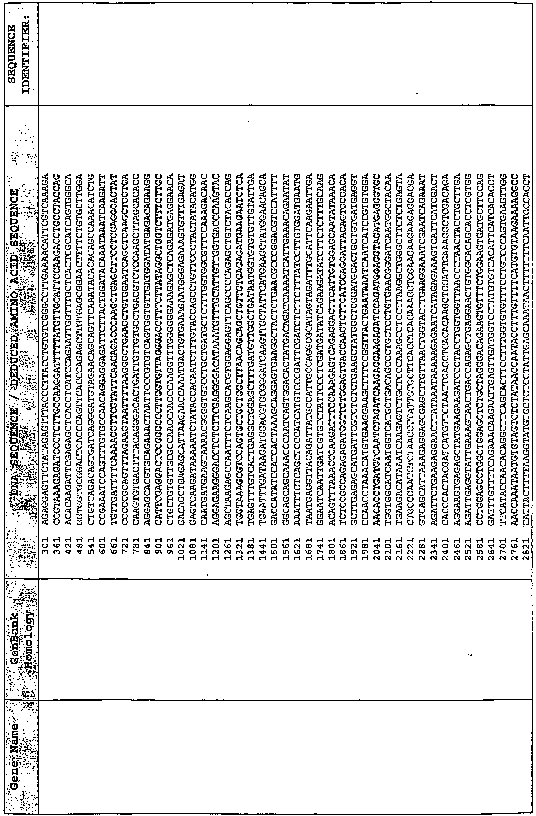


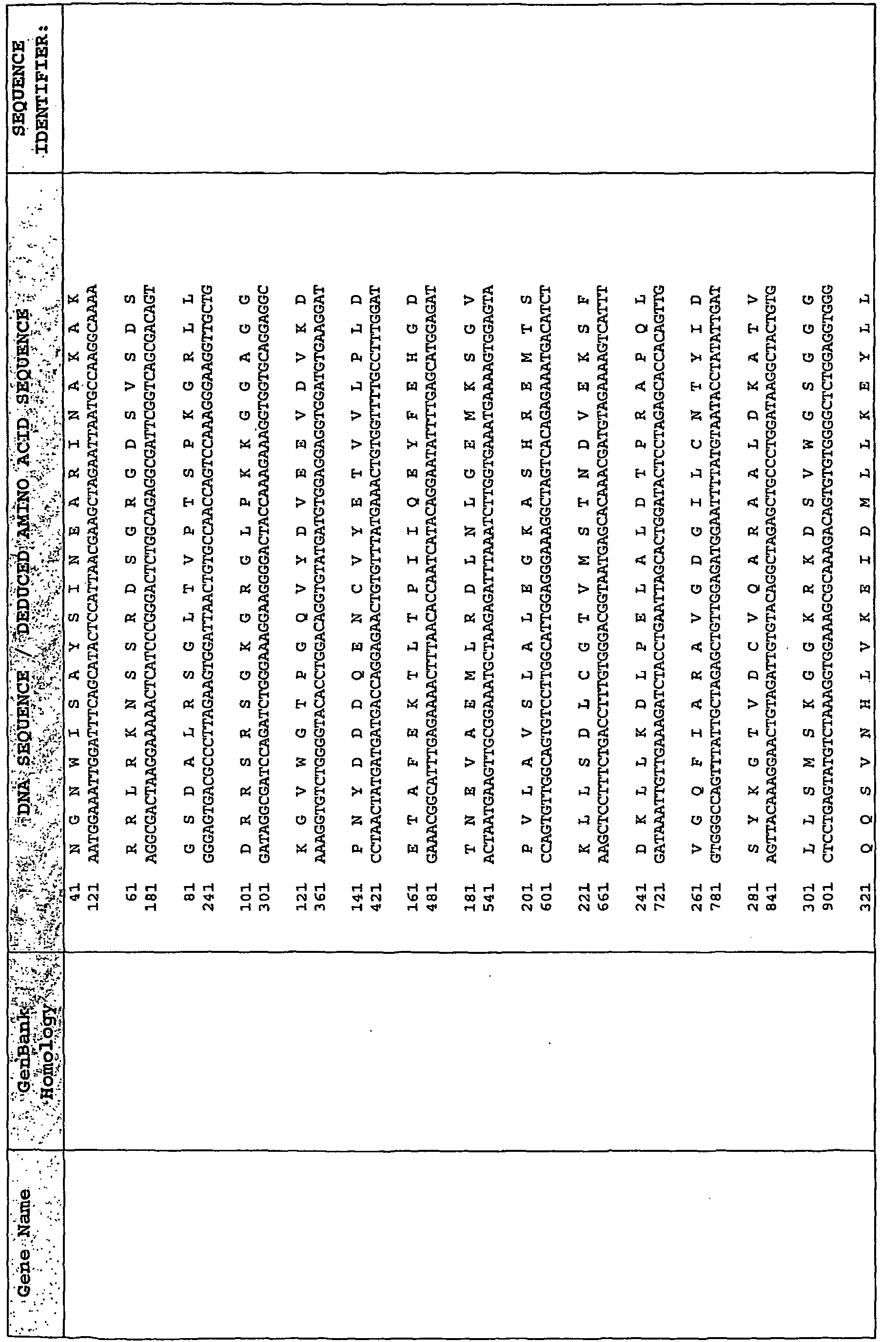


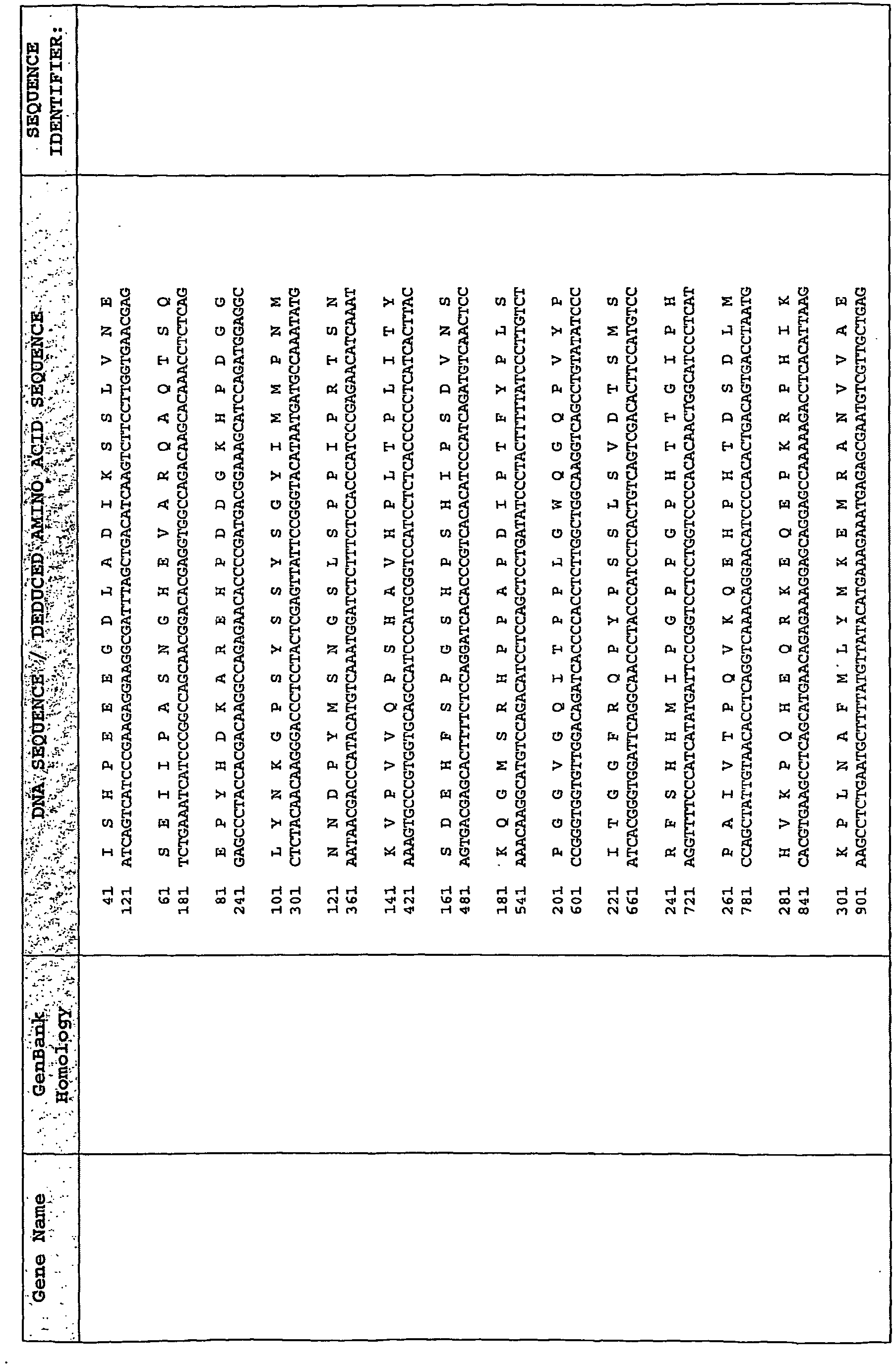


TABLE 2 Probe Set Name Sequence Sequence Identifier ILT11A mRNA for immunoglobulin-like transcript 11 protein B1961620.V1.3_at AGGACGTGACCTACGCCCAGGTGAA SEQ ID NO:327 B1961620.V1.3_at AGGGACCCCAGAAGACATAGGAGCT SEQ ID NO:328 B1961620.V1.3_at AGGTGAACCACTTGACCCTCAGAGG SEQ ID NO:329 B1961620.V1.3_at AGTGGTACGCTGCTATGGCCATCCA SEQ ID NO:330 B1961620.V1.3_at AGCAGAGCCCAGTGGTACGCTGCTA SEQ ID NO:331 B1961620.V1.3_at CATAGGAGCTGCCTCCAGAGGACAC SEQ ID NO:332 B1961620.V1.3_at GAGACTGCAGGGACCCCAGAAGACA SEQ ID NO:333 B1961620.V1.3_at GATGCCACCCTCCATGGAGGGAGAC SEQ ID NO:334 B1961620.V1.3_at GTATGCACAGGCTGCTATATCTGAA SEQ ID NO:335 B1961620.V1.3_at TGGCCATCCACTAGCCCAGGAAGGA SEQ ID NO:336 B1961620.V1.3_at TAGCCCAGGAAGGACCCGGATGCCA SEQ ID NO:337 No homology B1961856.V1.3_at ATGCTGGCAAAACCTCTAAGACTGT SEQ ID NO:338 B1961856.V1.3_at AGTTTCCCTTCTGCAGCTTGTTTTT SEQ ID NO:339 B1961856.V1.3_at ACCTGATTCTGCCTCTTTAATGAAT SEQ ID NO:340 B1961856.V1.3_at ACAGTTCCTATGCATGGTTTTTAAC SEQ ID NO:341 B1961856.V1.3_at CACGAGGGAAGAGGGTCTCCTTCCA SEQ ID NO:342 B1961856.V1.3_at GCTTGTTTTTTCTCACTATCTGTAT SEQ ID NO:343 B1961856.V1.3_at GAACAATTTAATTTCTTGGCCGTGT SEQ ID NO:344 B1961856.V1.3_at GGCCGTGTTTAGTAACAGTTCCTAT SEQ ID NO:345 B1961856.V1.3_at GAAAGTTGATTTGTCCTAGTGCAAA SEQ ID NO:346 B1961856.V1.3_at GAAACTCAATGCCTTATGTGCTCAC SEQ ID NO:347 B1961856.V1.3_at TTATGTGCTCACTCAGTTTCCCTTC SEQ ID NO:348 Equus caballus tissue inhibitor of metalloproteinase-1 (TIMP-1) mRNA complete cds B1961880.V1.3_at AGACCACCTTACAGCGGCGTTATGA SEQ ID NO:349 B1961880.V1.3_at AGCTGAAGCTTGCACAGTGTTCACC SEQ ID NO:350 B1961880.V1.3_at ACAGGCTCTGACAAGGGCTTCCAGA SEQ ID NO:351 B1961880.V1.3_at GAATCCTGTCTCCAGCAGAAGCTGA SEQ ID NO:352 B1961880.V1.3_at GAATGCTCAGTGTTTCCCTGTTCAT SEQ ID NO:353 B1961880.V1.3_at GAACCGCAGCGAGGAGTTTCTCATC SEQ ID NO:354 B1961880.V1.3_at GGACGAGAAGCTGTACATCACCACC SEQ ID NO:355 B1961880.V1.3_at TGAGTTCGTCATCAGGGCCAAGTTC SEQ ID NO:356 B1961880.V1.3_at TGCGGATACTTCCACAGGTCGGAGA SEQ ID NO:357 B1961880.V1.3_at TGATTGCTTGTGGACGGACCAGCTC SEQ ID NO:358 B1961880.V1.3_at TTCTCATCGCCGGACAACTATTGGA SEQ ID NO:359 PREDICTED Homo sapiens zinc finger, SWIM domain containing 6 (ZSWIM6), mRNA B1961900.V1.3_at GTTAAAAGGCTGACCCACGTGGCTT SEQ ID NO:360 B1961900.V1.3_at AGAACACCTCCATCCTGTGATAAGT SEQ ID NO:361 B1961900.V1.3_at ACGTGGCTTTGCAGTGCTGTTCGTC SEQ ID NO:362 B1961900.V1.3_at AAAAGTCCACACAGCTCTCCTGTTT SEQ ID NO:363 B1961900.V1.3_at GTCTACGTTCACAGCCTCAAAAAAG SEQ ID NO:364 B1961900.V1.3_at GCTGTTCGTCCAGAAGCATGGCACA SEQ ID NO:365 B1961900.V1.3_at GAACAGAGCCACACCGGTATTATAT SEQ ID NO:366 B1961900.V1.3_at GTTCTATTGCGTTTGCTGACTAAAT SEQ ID NO:367 B1961900.V1.3_at GGTTTGTTTTTACTTGAGCCTGCCT SEQ ID NO:368 B1961900.V1.3_at GGATTCAGCTTTACTCCTTTGTAAC SEQ ID NO:369 B1961900.V1.3_at TGAGCCTGCCTTTGTACCCTTTTTA SEQ ID NO:370 Equus caballus interleukin-4 receptor alpha-chain mRNA. B1961932.V1.3_at AGGATTTGCCTAGAGAGGCCCGTTC SEQ ID NO:371 B1961932.V1.3_at TGTTACCCGAGTCTGACCCAGTCCT SEQ ID NO:372 B1961932.V1.3_at GAATGACTTAGGAGGCCCCAGGAAA SEQ ID NO:373 B1961932.V1.3_at ATCACTGGATTGGATGCTGAGCCTA SEQ ID NO:374 B1961932.V1.3_at AGTCCTGGGTTAGCTGCCGCCATAT SEQ ID NO:375 B1961932.V1.3_at AGAGCTTCATCTAGCTGGCACCAGA SEQ ID NO:376 B1961932.V1.3_at GCTGCCGCCATATCACTGGATTGGA SEQ ID NO:377 B1961932.V1.3_at GATGCTGAGCCTAGAAACTGATCAA SEQ ID NO:378 B1961932.V1.3_at GTTCCTTCAACAGAGCTTCATCTAG SEQ ID NO:379 B1961932.V1.3_at TGGCACCAGAGGCAGGATTGCACCT SEQ ID NO:380 B1961932.V1.3_at TTGCACCTGTGGTGGGTGCTTAGCC SEQ ID NO:381 Equus caballus interleukin-4 receptor alpha-chain mRNA B1961932.V1.3_s_at AGGGAGTGGGTCACAGCCCATGACC SEQ ID NO:382 B1961932.V1.3_s_at ATGGGATCCCCAGAGTGGACCTGCC SEQ ID NO:383 B1961932.V1.3_s_at ATGGAGAGCGTTGCTGGCAGCCATA SEQ ID NO:384 B1961932.V1.3_s_at AGTGACAGTTTGCTGTCAGGTCCCC SEQ ID NO:385 B1961932.V1.3_s_at AGACCTTGGGTCGAGTAACGCTTGT SEQ ID NO:386 B1961932.V1.3_s_at ACGTTTTGCCTGCACATGGCTCAGA SEQ ID NO:387 B1961932.V1.3_s_at GCCAGTTTGTGCACCCATGGAGAGC SEQ ID NO:388 B1961932.V1.3_s_at GAAATTGTGATTAGCCGGTAGTGAC SEQ ID NO:389 B1961932.V1.3_s_at GAGTAACGCTTGTTTGTGTGTATCT SEQ ID NO:390 B1961932.V1.3_s_at GGCATTCCAGACAGCTGACCCGGCA SEQ ID NO:391 B1961932.V1.3_s_at TGGGCCTCCTCTTAGGCATGGGATC SEQ ID NO:392 Equus caballus adenosine A2A receptor mRNA BM734452.V1.3_at AGACTTTCCCAGGTTCGGGAGCTGC SEQ ID NO:393 BM734452.V1.3_at AAGTGGAGGCTCTAGCTGACTGCCT SEQ ID NO:394 BM734452.V1.3_at CTCAGAGCTGCCCAAGTGGAGGCTC SEQ ID NO:395 BM734452.V1.3_at CCGACTCCAGCAGCACGTAGAAGTG SEQ ID NO:396 BM734452.V1.3_at GCCGTGAGCTGCCATGTGCTTCTCA SEQ ID NO:397 BM734452.V1.3_at GCTGCTGTGTCCAGAGGTGGCATTT SEQ ID NO:398 BM734452.V1.3_at GTAAGCGCGAGGAAATCCTTTTTAT SEQ ID NO:399 BM734452.V1.3_at GGCATTTGACTATTTTTGACCAGGA SEQ ID NO:400 BM734452.V1.3_at GGCTGCTGGGTCTGTTGTCAGTCCT SEQ ID NO:401 BM734452.V1.3_at TGTCAGTCCTGCTGCTAACCTGGCA SEQ ID NO:402 BM734452.V1.3_at TTTTATTGTATTATCTTCCCTCCCT SEQ ID NO:403 No Homology BM734531.V1.3_at ACCACTTCATGTTCTCTACAGAGCT SEQ ID NO:404 BM734531.V1.3_at ACAGAGCTGTCCAGAGCCGAGGCTG SEQ ID NO:405 BM734531.V1.3_at AAACGAGTCCGAGGGCACAGCCAGG SEQ ID NO:406 BM734531.V1.3_at CGTTGCCCGCTGTTGGTCATGACAA SEQ ID NO:407 BM734531.V1.3_at GAGTCTCTGTCAGGATCCTTTTGAA SEQ ID NO:408 BM734531.V1.3_at GAGTCACCCAAGGAACTTATGCAGA SEQ ID NO:409 BM734531.V1.3_at GTCCTGTGGCTTTTGTGTGTCTCTC SEQ ID NO:410 BM734531.V1.3_at GGCCTTGCTTGAGAGAGGTCCATCC SEQ ID NO:411 BM734531.V1.3_at GGTCACTTAGCAGCGACTTCTTGGA SEQ ID NO:412 BM734531.V1.3_at GGAGCCAGGTGTCTGCATTTGAACA SEQ ID NO:413 BM734531.V1.3_at TTATGCAGATGCCATGTCCTCACTC SEQ ID NO:414 Human UbA52 adrenal RNA for ubiquitin-52 amino acid fusion protein. BM734661.V1.3_at ATTTGCCGCAAGTGTTATGCTCGCC SEQ ID NO:415 BM734661.V1.3_at ATCTTTGTGAAGACCCTGACGGGCA SEQ ID NO:416 BM734661.V1.3_at AACCTGCGCCCCAAGAAGAAGGTCA SEQ ID NO:417 BM734661.V1.3_at AGCAGCGTTTGATTTTTGCCGGCAA SEQ ID NO:418 BM734661.V1.3_at GAAGGTCAAATAAGGCCCCTCCTCT SEQ ID NO:419 BM734661.V1.3_at CACTCTCTCAGACTACAATATCCAG SEQ ID NO:420 BM734661.V1.3_at GCAAGAAGAAGTGCGGCCACACCAA SEQ ID NO:421 BM734661.V1.3_at GACGCAGACATGCAGATCTTTGTGA SEQ ID NO:422 BM734661.V1.3_at GGTTGAGCCCAGTGACACCATTGAG SEQ ID NO:423 BM734661.V1.3_at TCGTGCTGTCAACTGCCGCAAGAAG SEQ ID NO:424 BM734661.V1.3_at TTTTGCCGGCAAACAGCTGGAGGAC SEQ ID NO:425 Homo sapiens STAT4 mRNA BM734780.V1.3_at AGTCATCTCTTATTCTTCATCTTTG SEQ ID NO:426 BM734780.V1.3_at ACAGGATGACCTCTTGACACTCAAA SEQ ID NO:427 BM734780.V1.3_at AATAAAGGCCGCTTGTCTGCTTTGC SEQ ID NO:428 BM734780.V1.3_at GAAAACCCTCTGAAGTACCTGTATC SEQ ID NO:429 BM734780.V1.3_at GAGAGAAAACCTGAGTCCCACGACC SEQ ID NO:430 BM734780.V1.3_at TGGCATGACACTCCCAATTGGGTGT SEQ ID NO:431 BM734780.V1.3_at GATGAAGTCTCCATATTCTGCCGAA SEQ ID NO:432 BM734780.V1.3_at GGTTGAAGTCTGAAGCTCTGCTCTC SEQ ID NO:433 BM734780.V1.3_at TGATCTTTGCCAACAGTCATCTCTT SEQ ID NO:434 BM734780.V1.3_at TATGTTCCTTCTGTTTTTATCCCCA SEQ ID NO:435 BM734780.V1.3_at TTTGCTGACATCCTTCGAGACTACA SEQ ID NO:436 Homo sapiens phosphate cytidylyltransferase 1 choline alpha isoform, mRNA (cDNA clone MGC: 49915 IMAGE: 5744747) BM734844.V1.3_at ATGTTTGCTCCAACACAGAGGACAG SEQ ID NO:437 BM734844.V1.3_at ACATCAGACATCATCACCCGAATTG SEQ ID NO:438 BM734844.V1.3_at AAGGCTGCAGCCTATGATATCAGTG SEQ ID NO:439 BM734844.V1.3_at CATCACCTCCAGAAGCTCTTTGTTG SEQ ID NO:440 BM734844.V1.3_at GAAATACCACTTGCAGGAGCGAGTT SEQ ID NO:441 BM734844.V1.3_at GACAGAAGGTATCTCCACATCAGAC SEQ ID NO:442 BM734844.V1.3_at GATGGCCTTTCTCTGGCAAGACTTC SEQ ID NO:443 BM734844.V1.3_at GATGCTGCAGGCCATCAGTCCAAAG SEQ ID NO:444 BM734844.V1.3_at GTCCGGGACTATGATGTGTACGCGA SEQ ID NO:445 BM734844.V1.3_at GGGCTACACAGCCAAGGAGCTCAAT SEQ ID NO:446 BM734844.V1.3_at TCAGTCCAAAGCAGAGTCCCAGCAG SEQ ID NO:447 No Homology BM734891.V1.3_at ACTGGTATTCTCAGCTTCGTATGCT SEQ ID NO:448 BM734891.V1.3_at AAGGATCCTCCTTCTGGACTTGTTT SEQ ID NO:449 BM734891.V1.3_at CAGCGAGATCAGCAAGGCCTTTTCT SEQ ID NO:450 BM734891.V1.3_at CAATGTGCATGTTTCAGACCTTCTG SEQ ID NO:451 BM734891.V1.3_at GCTTGCCACTGCCTTTGGGATATAG SEQ ID NO:452 BM734891.V1.3_at TAATGGGTCCCTTCAGTCAGTGGCA SEQ ID NO:453 BM734891.V1.3_at GTGACTTTTGGTTACCTGGGCACAC SEQ ID NO:454 BM734891.V1.3_at GGTAGTGTTGGATCTCAGCCACCCA SEQ ID NO:455 BM734891.V1.3_at TCGTATGCTGCAGTACCTGTGGACA SEQ ID NO:456 BM734891.V1.3_at TATAGGGACTGCAGCCTTTGTTTCT SEQ ID NO:457 BM734891.V1.3_at TTCTGCTCTCTCTGGAATGGGTAGT SEQ ID NO:458 Homo sapiens matrix metalloproteinase 9 (gelatinase B, 92kDa gelatinase, 92kDa type IV collagenase) mRNA (cDNA clone BM734900.V1.3_at ACGACGTCTTCCAGTACCGAGAGAA SEQ ID NO:459 BM734900.V1.3_at ACTGGCGCGTGAGTTCCCGGAATGA SEQ ID NO:460 BM734900.V1.3_at AAGTGGGCTACGTGAGCTTCGACCT SEQ ID NO:461 BM734900.V1.3_at GCTGGATTCCGCCTTTGAGGAGCCA SEQ ID NO:462 BM734900.V1.3_at GAAGGAGCCAGTTTGCCGGTTTCAA SEQ ID NO:463 BM734900.V1.3_at GAAGATTTTCTTCTTCTCTGGGCGC SEQ ID NO:464 BM734900.V1.3_at GAGGAGCCACTCACCAAGAAGATTT SEQ ID NO:465 BM734900.V1.3_at GTGTCAGCCCGGTGGACCAGATGTT SEQ ID NO:466 BM734900.V1.3_at GTGTACACAGGCAAGTCGGCGCTAG SEQ ID NO:467 BM734900.V1.3_at GGTTTCAAACTGGTGGGTCTGTTCT SEQ ID NO:468 BM734900.V1.3_at TAGGCCCGAGGCGTCTGGACAAGCT SEQ ID NO:469 Homo sapiens S100 Calcium binding protein P, mRNA. BM734933.V1.3_at AGGGCAGCAAGCAGAGCCTGACCAA SEQ ID NO:470 BM734933.V1.3_at AAAGCTCTTGTTGGCAGTCGTTTCC SEQ ID NO:471 BM734933.V1.3_at GCCGAGGTGGACTTCAGCGAGTTCA SEQ ID NO:472 BM734933.V1.3_at CCTGGCGTCCAGAAAGCTCTTGTTG SEQ ID NO:473 BM734933.V1.3_at GAGCATGATCATCGACGTCTTTGCC SEQ ID NO:474 BM734933.V1.3_at GAGTTCATCGTGTTTGTGGCCGCGC SEQ ID NO:475 BM734933.V1.3_at GAGGTGGGACATTTCCTCGGCCACG SEQ ID NO:476 BM734933.V1.3_at GTGGGTCCAAATCCAGCACCATGAC SEQ ID NO:477 BM734933.V1.3_at GGTGCCGTGGACAAACTGCTCAAGG SEQ ID NO:478 BM734933.V1.3_at TCATGGAGAAGGAGCTCCCGGGCTT SEQ ID NO:479 BM734933.V1.3_at TCAAGGACCTGGACGCCAACGGAGA SEQ ID NO:480 Human mRNA for coupling protein G(s) alpha-subunit (alpha-S1) (stimulatory regulatory component Gs of adenylyl cyclase) BM734975.V1.3_at ATGATTAACAAAGCCACCTTTCCCT SEQ ID NO:481 BM734975.V1.3_at ATTGTTCACGCAGTTAATCACCCAC SEQ ID NO:482 BM734975.V1.3_at ATCTCTGTGATTCTGTTCCTCAACA SEQ ID NO:483 BM734975.V1.3_at ATGCACCTCCGTCAGTATGAGCTGC SEQ ID NO:484 BM734975.V1.3_at CTGCTCGCTGAGAAAGTCCTTGCTG SEQ ID NO:485 BM734975.V1.3_at GCTCCCTTCAGCTTGGTTAAATATA SEQ ID NO:486 BM734975.V1.3_at GAATTTCTGAGAATCAGCCCCGCTA SEQ ID NO:487 BM734975.V1.3_at GACTACTTTCCAGAATTTGCTCGCT SEQ ID NO:488 BM734975.V1.3_at GTTCCCTCTCTCTTTCAGTAAGTAA SEQ ID NO:489 BM734975.V1.3_at GGCCAAGTACTTCATCCGTGACGAA SEQ ID NO:490 BM734975.V1.3_at TCGCTACACTACTCCTGAGGATGCT SEQ ID NO:491 Human mRNA for macrophage inflammatory protein-2alpha (MIP2alpha) BM735057.V1.3_s_at AAGGGCAGCTCCGACTGACCAGAGG SEQ ID NO:492 BM735057.V1.3_s_at GCTGTAGCTGAAGTAGGCCCTGCCC SEQ ID NO:493 BM735057.V1.3_s_at GCAGCTCCGACTGACCAGAGGAAGA SEQ ID NO:494 BM735057.V1.3_s_at GAAGTAGGCCCTGCCCTTACAGGAA SEQ ID NO:495 BM735057.V1.3_s_at GAAGCTGATCAGTGGCTGTAGCTGA SEQ ID NO:496 BM735057.V1.3_s_at GACTGACCAGAGGAAGAAGCTGATC SEQ ID NO:497 BM735057.V1.3_s_at GATGCTAAACAAGGGCAGCTCCGAC SEQ ID NO:498 BM735057.V1.3_s_at GGAAGTTTGTCTCAACCCTGAAGCC SEQ ID NO:499 BM735057.V1.3_s_at GGCCCTGCCCTTACAGGAATCAAAG SEQ ID NO:500 BM735057.V1.3_s_at TGGACAGGAAGTTTGTCTCAACCCT SEQ ID NO:501 BM735057.V1.3_s_at TAAACAAGGGCAGCTCCGACTGACC SEQ ID NO:502 Horse serpin mRNA. BM735091.V1.3_at ATGCTTGCCATGCTGATGCCAGAGG SEQ ID NO:503 BM735091.V1.3_at ATCTAGCCCGCCTAGGTGTACAAGA SEQ ID NO:504 BM735091.V1.3_at CATTCATTTTCTTCATTCGGCACAA SEQ ID NO:505 BM735091.V1.3_at GCTAACATCCTGTTCTTAGGCAGAT SEQ ID NO:506 BM735091.V1.3_at GCAAAGCTGATCTGTCTGGCATGTC SEQ ID NO:507 BM735091.V1.3_at GAATCTGTATCTCGCTGAAGTCAAT SEQ ID NO:508 BM735091.V1.3_at GAAAATTTCAATGCCGACCATCCAT SEQ ID NO:509 BM735091.V1.3_at GGGCCAGAGATCTTTTCGTATCGAA SEQ ID NO:510 BM735091.V1.3_at TGAAGTCAATGTCCACTTGCCCAGG SEQ ID NO:511 BM735091.V1.3_at TTTGGCGTGGGTAGCTATTTTCCTT SEQ ID NO:512 BM735091.V1.3_at TTAGGCAGATTTTCTTCCCCTTAGA SEQ ID NO:513 Equus caballus mRNA for ferritin light chain. BM735286.V1.3_at AGGAGCCTCCGGAGTCCAGCGGCCT SEQ ID NO:514 BM735286.V1.3_at AGCTTTTTAACTAGCCTGGAGCCTT SEQ ID NO:515 BM735286.V1.3_at CGTCAGAGCTTCTGCCTGAGCCTCT SEQ ID NO:516 BM735286.V1.3_at CTGCAGCCACTAGAGATAGCTTTTT SEQ ID NO:517 BM735286.V1.3_at CTCCGGAGTCCAGCGGCCTTTGAGG SEQ ID NO:518 BM735286.V1.3_at CTGGCGTCAGAGCTTCTGCCTGAGC SEQ ID NO:519 BM735286.V1.3_at GAGATAGCTTTTTAACTAGCCTGGA SEQ ID NO:520 BM735286.V1.3_at TTTGGTATCCCCCTGGCGTCAGAGC SEQ ID NO:521 BM735286.V1.3_at TCCCTGCAGCCACTAGAGATAGCTT SEQ ID NO:522 BM735286.V1.3_at TTTTAACTAGCCTGGAGCCTTCTGC SEQ ID NO:523 BM735286.V1.3_at TAACTAGCCTGGAGCCTTCTGCCCA SEQ ID NO:524 No Homology BM735386.V1.3_at ATTTGCTGACCTTGTGGTGCTCTCA SEQ ID NO:525 BM735386.V1.3_at AAGTTGTCACGTGCAGTGTCTTTCT SEQ ID NO:526 BM735386.V1.3_at AAGGGCAATCCTGCTTCACTGGTGT SEQ ID NO:527 BM735386.V1.3_at GCTGGTGCCCCTGTAAAGAGGCTTT SEQ ID NO:528 BM735386.V1.3_at GAAGTTGTCACCATGAAGTTGTCAC SEQ ID NO:529 BM735386.V1.3_at GAGCGTTTCCTTTACCTGGAAGTTG SEQ ID NO:530 BM735386.V1.3_at GAGACCTGTTGTGCTGTGTTATTTT SEQ ID NO:531 BM735386.V1.3_at GATGGCTGTTTTCCTGTCCCAGAGA SEQ ID NO:532 BM735386.V1.3_at GATGTCTGACCTTAGTGTTCCAAAC SEQ ID NO:533 BM735386.V1.3_at GTTAAGTATGTGGTTCCTTCTCAGA SEQ ID NO:534 BM735386.V1.3_at TAAAGGATGTCCTGGTGCTGGCTCA SEQ ID NO:535 No Homology BM735452.V1.3_at CGAACCCCGGAAGTCTGCAAAATTG SEQ ID NO:536 BM735452.V1.3_at AGGATGACTCGGGTCCAGTGGCCCA SEQ ID NO:537 BM735452.V1.3_at AAGTCTGCAAAATTGGGTGCCCCAA SEQ ID NO:538 BM735452.V1.3_at CTGCTCTCTGCTCCCTAAATTATTG SEQ ID NO:539 BM735452.V1.3_at CCAGAATCTGCCCACGGAGTCGGTG SEQ ID NO:540 BM735452.V1.3_at CCCCGGTTTCATATCTGTATTTATA SEQ ID NO:541 BM735452.V1.3_at GGGTGCCCCAAGAGCTGGAAACTCA SEQ ID NO:542 BM735452.V1.3_at GAGGCCTCAGTGCAGTGCCTTCCCG SEQ ID NO:543 BM735452.V1.3_at GTGCGCAGGTCCCACTCAGGATGAC SEQ ID NO:544 BM735452.V1.3_at GGAAACTCAGGAAACCCCAGGTGCT SEQ ID NO:545 BM735452.V1.3_at TTAATATATGCAATTCCCCATCCCT SEQ ID NO:546 Homo sapiens male-specific lethal 3-like 1 (Drosophila); transcript variant 1, mRNA BM780999.V1.3_at TACTTCTTAGGTACCTGAACGGCCT SEQ ID NO:547 BM780999.V1.3_at ATTTGAGTTGCTTACCTACTGTAGT SEQ ID NO:548 BM780999.V1.3_at AGTGTGTCTAGGTTAGGCTGTTTCA SEQ ID NO:549 BM780999.V1.3_at AAAGCATTTCCTGTCTTGTTTGAGA SEQ ID NO:550 BM780999.V1.3_at GCTTGTTACAGTTCATTTTCTCTAA SEQ ID NO:551 BM780999.V1.3_at GACAGGCAGGCCCATTAATTTGAGT SEQ ID NO:552 BM780999.V1.3_at GATGGTGGGCCTTCAGACAGAGTAC SEQ ID NO:553 BM780999.V1.3_at GATGAGGCCGAGCAACTCTGTCCAA SEQ ID NO:554 BM780999.V1.3_at GGAAACCGCAAGGTAGTCCCAGAAT SEQ ID NO:555 BM780999.V1.3_at TAATGTTATGGTTCCCTTTCTGTAA SEQ ID NO:556 BM780999.V1.3_at TTGCCACTGTACACGTGATCTGTGA SEQ ID NO:557 Homo sapiens replication protein A1, 70kDa, mRNA BM781033.V1.3_at AGATAGCCTTCCTTCAGGGTTTAAT SEQ ID NO:558 BM781033.V1.3_at AAAGGCGCTCTCTTTCTAGAGGTGC SEQ ID NO:559 BM781033.V1.3_at AAAGCCTTGCTTTAGTTTCACTTCA SEQ ID NO:560 BM781033.V1.3_at AATTGACCCTAAATCTGCAGACAGT SEQ ID NO:561 BM781033.V1.3_at CAGGGACATCCACGCAACTACAAAG SEQ ID NO:562 BM781033.V1.3_at GAATGCACACTTTTTCTACCTGTAC SEQ ID NO:563 BM781033.V1.3_at GAAGAAGCCCTTTGTTTCTCTTGCT SEQ ID NO:564 BM781033.V1.3_at GACATTTCTGCAACTTGGGTTCCTT SEQ ID NO:565 BM781033.V1.3_at GTAAGCGCGTCTTCCTAGAATTCAA SEQ ID NO:566 BM781033.V1.3_at TAACTAGGTTTCTGCTCCATTGGTG SEQ ID NO:567 BM781033.V1.3_at TACCTGTACATATTTCTTGCGTTTG SEQ ID NO:568 Human mRNA for cathepsin H BM781103.V1.3_at ATCCCTCTGGTGTGAGCCAGGATGG SEQ ID NO:569 BM781103.V1.3_at AAGAATATTCTTTCCTTCCTGAAGG SEQ ID NO:570 BM781103.V1.3_at CACCCACACCTTCTGATGCAAAGGA SEQ ID NO:571 BM781103.V1.3_at CAGCCTTGTGCCTTTTAAGCATGAT SEQ ID NO:572 BM781103.V1.3_at CATTGGTGGAAGTCCTGCCCTGGAG SEQ ID NO:573 BM781103.V1.3_at CCCCGTGCTGTGTCCAGAAATCTAA SEQ ID NO:574 BM781103.V1.3_at GCAGCCTGGCCATTGGTGGAAGTCC SEQ ID NO:575 BM781103.V1.3_at GAAGAATTCCACCAAGCAACAAGCC SEQ ID NO:576 BM781103.V1.3_at GATGCAAAGGATCACCAGCCTTGTG SEQ ID NO:577 BM781103.V1.3_at TTCCTTCCTGAAGGGCTACTTTTCA SEQ ID NO:578 BM781103.V1.3_at TTAACTGACTCAAACCCACGTGGAC SEQ ID NO:579 Homo sapiens transforming acidic coiled-coil containing protein 3 (TACC3) mRNA BM781237.V1.3_s_at GTACTACAGTACAGCCAGAAGGACC SEQ ID NO:580 BM781237.V1.3_s_at AGCAGTCCTTATACCTCAAGTTCGA SEQ ID NO:581 BM781237.V1.3_s_at AAGTTCGACCCGCTCCTGATGGACA SEQ ID NO:582 BM781237.V1.3_s_at AAGAGAACCTGTCGCTCAGGAGCGA SEQ ID NO:583 BM781237.V1.3_s_at GGACCCATCGTGGACGTACTACAGT SEQ ID NO:584 BM781237.V1.3_s_at GTGGAACTTGACTTCCTAGGCCCTC SEQ ID NO:585 BM781237.V1.3_s_at TGGACCCACCTTTGTGTGTCCTCGG SEQ ID NO:586 BM781237.V1.3_s_at TGATGGACAGCCCTCAGAGACCTGC SEQ ID NO:587 BM781237.V1.3_s_at TCCGGATGTGCCTGTGCTGGACCCA SEQ ID NO:588 BM781237.V1.3_s_at TGCCGCCAATCTCGTGGAACTTGAC SEQ ID NO:589 BM781237.V1.3_s_at TCCCCGCGACAGACAGTGTGCAGGA SEQ ID NO:590 Homo sapiens, chromosome 6 open reading frame 166, mRNA BM781278.V1.3_at ACATGCATTCTTCCTCAGTGGACCA SEQ ID NO:591 BM781278.V1.3_at AAAGAACAGCCCTTATTCACTCTAC SEQ ID NO:592 BM781278.V1.3_at CTGATGTCAATACCACCCGGATGTG SEQ ID NO:593 BM781278.V1.3_at CAGCCTGCTAGTTATGTTTCATGAA SEQ ID NO:594 BM781278.V1.3_at CACGTTTTCTGCATTTGTGGGCTGC SEQ ID NO:595 BM781278.V1.3_at TCTGCAACATCCTCACCATTAAAAA SEQ ID NO:596 BM781278.V1.3_at GTGGCCTCATTTGCTGTATCATTAC SEQ ID NO:597 BM781278.V1.3_at TGGCAACCACTTTAGGCAGCAACTG SEQ ID NO:598 BM781278.V1.3_at TGTGGACCTTTGCTACCTGTATTAA SEQ ID NO:599 BM781278.V1.3_at TTCAAGCTTTCGTCAGTGGCAACCA SEQ ID NO:600 BM781278.V1.3_at TTATTCACTCTACGGCAGGTTGGGA SEQ ID NO:601 Homo sapiens KIAK0002 mRNA BM781319.V1.3_at ATAACCTGTTATTCTGACTAGCTTA SEQ ID NO:602 BM781319.V1.3_at ATGTTCATTACCCTTGTACCATGTA SEQ ID NO:603 BM781319.V1.3_at AGTGCCTTACTGACTGTAGCCATTA SEQ ID NO:604 BM781319.V1.3_at GAGTCAATGTTATTCTCTCACAATG SEQ ID NO:605 BM781319.V1.3_at GATCTCTCTCTCTCAGTTACAGTTT SEQ ID NO:606 BM781319.V1.3_at GTTCTTACTGGCCAACTCTTAAATC SEQ ID NO:607 BM781319.V1.3_at GTTTCTCATCTTGGACTTCTTTTAT SEQ ID NO:608 BM781319.V1.3_at GTGTGATGCCATATCGAGTCAATGT SEQ ID NO:609 BM781319.V1.3_at TGTACCATGTACCTCAGATCTCTCT SEQ ID NO:610 BM781319.V1.3_at TCACTGGGCCATTGGATTTTTTCCA SEQ ID NO:611 BM781319.V1.3_at TAAGAGGTCCTATCTGTTCAGTGTT SEQ ID NO:612 Homo sapiens inhibitor of Bruton a gammaglobulinemia tyrosine kinase (IBTK) mRNA BM781379.V1.3_at ATTCGGCAACCCTGATGAGTTTGTC SEQ ID NO:613 BM781379.V1.3_at AGGGACCACTGGCAGTACCTATGTG SEQ ID NO:614 BM781379.V1.3_at AGCATGGATGCTAGTTCGCCGTGGA SEQ ID NO:615 BM781379.V1.3_at AAAACCCTTGGCTTTGATTCAGATT SEQ ID NO:616 BM781379.V1.3_at GTTGAAAGGACACCGCAGGGACCAC SEQ ID NO:617 BM781379.V1.3_at GAGGAGCACGCGATACAAGATTTAT SEQ ID NO:618 BM781379.V1.3_at GATTCAGATTGAGGAGCACGCGATA SEQ ID NO:619 BM781379.V1.3_at GTTCGCCGTGGACTTGAGATGTATT SEQ ID NO:620 BM781379.V1.3_at GGTGCATTAGCTATTATTTCTGATC SEQ ID NO:621 BM781379.V1.3_at GGTTTTCTATGAGGCATTCGGCAAC SEQ ID NO:622 BM781379.V1.3_at GGATCTAATTTCTTTGCTGCAGTTT SEQ ID NO:623 No Homology BM781394.V1.3_at ACCAGTGCTGAACATTAGTGCCTTT SEQ ID NO:624 BM781394.V1.3_at GAACCTGCTGAATTTAACTTAACCG SEQ ID NO:625 BM781394.V1.3_at AAAATACAGTCAACCCTTCACCATG SEQ ID NO:626 BM781394.V1.3_at AAGAGTTCAGCTTCACGTTTGCCAA SEQ ID NO:627 BM781394.V1.3_at CAACAGGCGGAGACAGCTGTCCTTT SEQ ID NO:628 BM781394.V1.3_at CCATATCACCTGCTATTTCCAAGAA SEQ ID NO:629 BM781394.V1.3_at GCTGCATCTCATACAATGGCTCTCT SEQ ID NO:630 BM781394.V1.3_at GAAACAGCCCGATACATAACACTCC SEQ ID NO:631 BM781394.V1.3_at TCTGCCCATCAATTACACTTTCTTT SEQ ID NO:632 BM781394.V1.3_at TAACCGAGAGGATCACCGTGACAAA SEQ ID NO:633 BM781394.V1.3_at TAATGCTGGCATTTTGGATACTACC SEQ ID NO:634 No Homology BM781409.V1.3_at ATGGATCCCCTTATTATTCTTTCAC SEQ ID NO:635 BM781409.V1.3_at AGTTTTTACATCTGCCTCAAGGCCT SEQ ID NO:636 BM781409.V1.3_at AGAATGAAACTGAGTCTCCCTGGCC SEQ ID NO:637 BM781409.V1.3_at AGATCCTTCGCTTGTCTGCTTAAAA SEQ ID NO:638 BM781409.V1.3_at AATTCTGAACAACTCTCTGCAACTG SEQ ID NO:639 BM781409.V1.3_at GTTCCTCCTTTTCTTTAGGTCTCAG SEQ ID NO:640 BM781409.V1.3_at GCACGAGCTGACCTGGAGTACTACA SEQ ID NO:641 BM781409.V1.3_at GACTTCCCAGTGCACTCAGAATGAA SEQ ID NO:642 BM781409.V1.3_at GAGTTCTCTATGTCACAGTTTTTAC SEQ ID NO:643 BM781409.V1.3_at GTCTCAGCTTACATATGTCACCTTC SEQ ID NO:644 BM781409.V1.3_at TTGTTTATCACTGTATCCCTAGCAC SEQ ID NO:645 No Homology BM781416.V1.3_at ATGCTGCAGCTGTTTGTCAGGAACA SEQ ID NO:646 BM781416.V1.3_at AGTATTTGCCATCACATGCTGCAGC SEQ ID NO:647 BM781416.V1.3_at ACAATTACTGGCTTTTCTGCTGTAA SEQ ID NO:648 BM781416.V1.3_at CAGTAATCACGCTGCAGAAATTGGC SEQ ID NO:649 BM781416.V1.3_at GAACGCAGCAGCAGGGTACGTAGCA SEQ ID NO:650 BM781416.V1.3_at GAAATTGGCAGCCTGCACCTTATGC SEQ ID NO:651 BM781416.V1.3_at TGTCCGTTTTTATTTGGGCATCCAC SEQ ID NO:652 BM781416.V1.3_at GGGCATCCACCACAGACAATTTAAA SEQ ID NO:653 BM781416.V1.3_at GGTGTCATTCATTCATTCAGTAATC SEQ ID NO:654 BM781416.V1.3_at TAAATTTCGTAGATGCGCTCAGAAC SEQ ID NO:655 BM781416.V1.3_at TTATGCGTCACCATTACCTTTAGTC SEQ ID NO:656 Homo sapiens cysteine-rich motor neuron 1 (CRIM1) BM781438.V1.3_at ATATGATTTATCCTGAGTGCTGTAT SEQ ID NO:657 BM781438.V1.3_at ATCACTCTTTTACTTTGGTTCCTGT SEQ ID NO:658 BM781438.V1.3_at AGATACCCAGTATGCTTAATGTGAA SEQ ID NO:659 BM781438.V1.3_at ACTTGGACAGAGGTTGCTGAACTTT SEQ ID NO:660 BM781438.V1.3_at AAAGGTGTTCTAGCTGTTTGCATCA SEQ ID NO:661 BM781438.V1.3_at AAAATAGAGTTACATCAGCCTGATT SEQ ID NO:662 BM781438.V1.3_at GTTGACAATTAATCCATTCCTGGCA SEQ ID NO:663 BM781438.V1.3_at GTGCTGTATCTATCACTCTTTTACT SEQ ID NO:664 BM781438.V1.3_at TGGTTCCTGTTGTGCTCTTGTAAAA SEQ ID NO:665 BM781438.V1.3_at TAAATGCTTGCTTTTTGTTTTTTCA SEQ ID NO:666 BM781438.V1.3_at TTGTTTTTTCAATCATGGCCATATT SEQ ID NO:667 Homo sapiens RNF4 mRNA. Foe1216.V1.3_at ATTGCCATGAAGTTCCTGCTGGGCT SEQ ID NO:668 Foe1216.V1.3_at CTTTCCCTTCCTCAATGTACATAGT SEQ ID NO:669 Foe1216.V1.3_at TGACTTTTTGACTGTTGGGCTCACT SEQ ID NO:670 Foe1216.V1.3_at GCTGGGCTTCGTGGAAACACTGTTT SEQ ID NO:671 Foe1216.V1.3_at GATGATACCCAGACTTCAGGAGCCA SEQ ID NO:672 Foe1216.V1.3_at GGGCAGGACATCAGTTTTCATCAGT SEQ ID NO:673 Foe1216.V1.3_at GGAGTGTTCCTCCTTTATGTGAAAA SEQ ID NO:674 Foe1216.V1.3_at TGGGCTCACTTCCAGTTAGTTTGAA SEQ ID NO:675 Foe1216.V1.3_at TTCCGGTTAGACGTCATTGCCATGA SEQ ID NO:676 Foe1216.V1.3_at TATCCGTGTATGTCAGTCTGTTTTG SEQ ID NO:677 Foe1216.V1.3_at TTTGGACTCCTCTGCTAGTGTTACA SEQ ID NO:678 No Homology Foe1263.V1.3_at ATCTCTTGGAGTTTCAGTACTGTCA SEQ ID NO:679 Foe1263.V1.3_at AGAGGTGGTCTGTAATGGATTCAAA SEQ ID NO:680 Foe1263.V1.3_at ACATAGTTCCACTGTCATTTCTGAA SEQ ID NO:681 Foe1263.V1.3_at AAAAAGCTCGACAGCACTCAGGTCC SEQ ID NO:682 Foe1263.V1.3_at AACTAATAGGTTACTGCTTCCCACA SEQ ID NO:683 Foe1263.V1.3_at CCAGTCCATCTCGTTTCTTTATAAA SEQ ID NO:684 Foe1263.V1.3_at GCTTCCCACACTACATTGGTTCAAA SEQ ID NO:685 Foe1263.V1.3_at GCTCTTTAAGTCATAGCACCGGTAA SEQ ID NO:686 Foe1263.V1.3_at GAGTTCCGGTATCTAAGTCACAATT SEQ ID NO:687 Foe1263.V1.3_at GGTTGCAGCAAACTTTTTAACTGAT SEQ ID NO:688 Foe1263.V1.3_at TAAAGGCATTGTTCCTTCCAGTCCA SEQ ID NO:689 Human-3-hydroxyacyl - CoA dehydrogenase, isoform 2 mRNA Foe1317.V1.3_at ATAATTGCCTGAATTCGTTCCCTTG SEQ ID NO:690 Foe1317.V1.3_at AGGGAAGCACTTCCTCCGAGGGCAT SEQ ID NO:691 Foe1317.V1.3_at CTTCTGAACCTTGTCATTTTCGTGA SEQ ID NO:692 Foe1317.V1.3_at AGCAGTGTCGAGAGCCATCGCGTGT SEQ ID NO:693 Foe1317.V1.3_at GCAAGCTGCCTGTAAATCGACTCTA SEQ ID NO:694 Foe1317.V1.3_at GAATGATGACTGTCTGTCTCTCTTT SEQ ID NO:695 Foe1317.V1.3_at GAAATGCAGTCTTTTAACCTATGAA SEQ ID NO:696 Foe1317.V1.3_at GTAACGGCAGATTTCTCCATTGAGA SEQ ID NO:697 Foe1317.V1.3_at GTCTCTCTTTTTTCCGTGACAAATC SEQ ID NO:698 Foe1317.V1.3_at GGAAGAGGTTTCATTCACACCCGAA SEQ ID NO:699 Foe1317.V1.3_at TACACCCTTGGTGCCTTGGAGCAAA SEQ ID NO:700 Homo sapiens mRNA; cDNA DKFZp434I0612 (from clone DKFZp434I0612). Foe1324.V1.3_at AAACCTTTTGGTGTACATCCTCTGG SEQ ID NO:701 Foe1324.V1.3_at AGATATCTGTCTCATTCATTCCACT SEQ ID NO:702 Foe1324.V1.3_at ACTTGCACTGTCTCCTGGGATCTTG SEQ ID NO:703 Foe1324.V1.3_at AACAAGTGGCTTCCTTTTTGAGATA SEQ ID NO:704 Foe1324.V1.3_at CTTTCTTTCCACAGCACTTTTGATA SEQ ID NO:705 Foe1324.V1.3_at CTCCCTGTGGCACTTGACATTTTAA SEQ ID NO:706 Foe1324.V1.3_at GTAAGGCAATCTTTCCTGTTGTAAA SEQ ID NO:707 Foe1324.V1.3_at GTGTGATCCCATCATGAGTTCATTG SEQ ID NO:708 Foe1324.V1.3_at TGGGATCTTGTCTGCAGCCCGACGT SEQ ID NO:709 Foe1324.V1.3_at GGCCCCTTAAGTGCTGCCAGAAGCA SEQ ID NO:710 Foe1324.V1.3_at TTCTGGTTATCATTTATCCTCCCTG SEQ ID NO:711 No Homology Foe146.V1.3_at ATTGTGGATTTGTCAGTTTCTCTCT SEQ ID NO:712 Foe146.V1.3_at ATTCTTCATAAGAGCTGTTGCCAAA SEQ ID NO:713 Foe146.V1.3_at ATTCATTTCACTGTACTCTTGCCAG SEQ ID NO:714 Foe146.V1.3_at ATCACTCCAGTGATGGTTGTGGTGG SEQ ID NO:715 Foe146.V1.3_at: AAAGCATAGTTCAGTGCCACTCACA SEQ ID NO:716 Foe146.V1.3_at AAGTATGTTGAAGTGTCTCTCTGTC SEQ ID NO:717 Foe146.V1.3_at AAGTGTCTCTCTGTCATTGTGGATT SEQ ID NO:718 Foe146.V1.3_at AATTGCCCTTGTCTCATAATGCTTT SEQ ID NO:719 Foe146.V1.3_at CAGGTGCCTAATGTGCGGTCTTTAA SEQ ID NO:720 Foe146.V1.3_at TGTCTACTTGGTTTTACCACCTTCT SEQ ID NO:721 Foe146.V1.3_at TCAGTTTCTCTCTGCATTTCTCAGA SEQ ID NO:722 Homo sapiens fucosyltransferase 8 (alpha (1,6) fucosyltransferase) (FUT8) transcript variant 1 Foe148.V1.3_at ATCAGTTCATCCACCTCATTATTAA SEQ ID NO:723 Foe148.V1.3_at AGGTTGATTCTTAGCCAGTGGTGGT SEQ ID NO:724 Foe148.V1.3_at AGTGTTTCTGGACATTTCTCTTTGA SEQ ID NO:725 Foe148.V1.3_at CAGGTTCTTTAGTACTCAGTGTTTC SEQ ID NO:726 Foe148.V1.3_at GTGACCTAACGAGAAGACCCGCATA SEQ ID NO:727 Foe148.V1.3_at GATGCCTCATTGGTGGAATTCCTCT SEQ ID NO:728 Foe148.V1.3_at GTATTGTAACCGCTGAATTCACTCC SEQ ID NO:729 Foe148.V1.3_at GTGCCTATATCCGAGAGACCTATGT SEQ ID NO:730 Foe148.V1.3 at GTGAGACTTCAAACATGGTGCCTAT SEQ ID NO:731 Foe148.V1.3_at TCAAACCCATGCACAGTCCAATAAT SEQ ID NO:732 Foe148.V1.3_at TACTTTGCCCCTTCTTTCAAGATTA SEQ ID NO:733 Homo sapiens tripeptidyl peptidase II mRNA Foe443.V1.3_at AGGACTTTCAGTTGGGTTGCAGCTA SEQ ID NO:734 Foe443.V1.3_at CCATAACTTTTAACTGCAAGCCAGT SEQ ID NO:735 Foe443.V1.3_at AACATCCTCTTGACGTGCAGAGCAT SEQ ID NO:736 Foe443.V1.3_at AATTGCTGGAGCATAACTGCTTCAA SEQ ID NO:737 Foe443.V1.3_at CAGGGTTTTAACATGCTTGACTTTA SEQ ID NO:738 Foe443.V1.3_at GTACACCATATCACTCTGATACCTT SEQ ID NO:739 Foe443.V1.3_at GTGCCAGACAGCTAAATCTTTATCA SEQ ID NO:740 Foe443.V1.3_at GGGTTGCAGCTATTACACGCATGAT SEQ ID NO:741 Foe443.V1.3_at GGAGTTATTTCAGGCCAATCATTTT SEQ ID NO:742 Foe443.V1.3_at TGTCTGCCAGCACCAAGGACTTTCA SEQ ID NO:743 Foe443.V1.3_at GGCCAATCATTTTTAACATCCTCTT SEQ ID NO:744 Equus caballus Toll-like receptor 4 mRNA complete cds GI9717252-3_at ATCTTTGACATCTTAGCCATCCTAA SEQ ID NO:745 G19717252-3_at AGAAGGCTCCTGATTCAGATCCTCC SEQ ID NO:746 GI9717252-3_at ACATCGTCTCCCAAGTCTTTTGAAT SEQ ID NO:747 GI9717252-3_at ACAGGACTGCTAATCCCTTTGAGTT SEQ ID NO:748 GI9717252-3_at AAACATCCTGGTCATTCTTTAGCGT SEQ ID NO:749 G19717252-3_at AAGTCAGCTAAGGAGTCCGTGCCAG SEQ ID NO:750 GI9717252-3_at GCTGCAACATACCAGGCATTGTGCT SEQ ID NO:751 GI9717252-3_at GAATGGAAACCATCTCATCTTTGAC SEQ ID NO:752 GI9717252-3_at GACTGGGCCCCAGTGAGTTCAGAAA SEQ ID NO:753 GI9717252-3_at GGCAGGTGATTCTGTCGTGCACAAG SEQ ID NO:754 GI9717252-3_at TCTCTGTTCAATTTTCCCTTTTCTA SEQ ID NO:755 Equus caballus Toll-like receptor 4 mRNA, complete cds GI9717252-3M_at ATAAGTTCTATTTCCACCTGATGCT SEQ ID NO:756 GI9717252-3M_at AGAGACTTCATTCCTGGTGTGGCCA SEQ ID NO:757 GI9717252-3M_at AAAGTTATTGTCGTGGTGTCCCAGC SEQ ID NO:758 GI9717252-3M_at CAGCACTTCATTCAGAGCCGATGGT SEQ ID NO:759 GI9717252-3M_at GCGGGTCGGTTTTCAGTATACTCAT SEQ ID NO:760 GI9717252-3M_at CTGATGCTTCTTGCTGGCTGCAAAA SEQ ID NO:761 GI9717252-3M_at GCATGCCCGTGCTGGGTTTTAACAA SEQ ID NO:762 GI9717252-3M_at GATGCCTTTGTTATCTACTCAAGCC SEQ ID NO:763 GI9717252-3M_at GGTGTGTGCAATACCCTTACAGATG SEQ ID NO:764 GI9717252-3M_at TGAGTATGAGATTGCCCAGACCTGG SEQ ID NO:765 GI9717252-3M_at TTAACAATGCCACCTGTCAGATTAG SEQ ID NO:766 Homo sapiens potassium channel tetrarisation domain containing 12 (KCTD12) WBC001D02_V1.3_at ATATAGAAGCTCCATGGGCTGCCCA SEQ ID NO:767 WBC001D02_V1.3_at GAGGGAAACGACTCCTTGTCTTTCA SEQ ID NO:768 WBC001D02_V1.3_at AGGAGACCTGGTTGCCTTATACCTT SEQ ID NO:769 WBC001D02_V1.3_at AGATTCATGCGATCCAGTCTGTATA SEQ ID NO:770 WBC001D02_V1.3_at CTGTGATGACCGAGAAGCCTTTCTT SEQ ID NO:771 WBC001D02_V1.3_at GAACACGGAATAAACTCCCACTGCA SEQ ID NO:772 WBC001D02_V1.3_at GGCTGCCCATCTGTAGATAGCTGTA SEQ ID NO:773 WBC001D02_V1.3_at GGCTCCTTTGGATATTCTGTGATGA SEQ ID NO:774 WBC001D02_V1.3_at TGCACATCCTGTCTACACCATAAGT SEQ ID NO:775 WBC001D02_V1.3_at TTGCCTTATACCTTTACGCGAACAC SEQ ID NO:776 WBC001D02_V1.3_at TTGTCTTTCATGCTGTGAGGCTCCT SEQ ID NO:777 Homo sapiens protein tyrosine phosphatase type IVA, member 2 WBC001F10_V1.3_at ATCAGTGCCTGCCTGCGTTAGGAGA SEQ ID NO:778 WBC001F10_V1.3_at AGAACTGGATTTTCTTCCTCAAAAT SEQ ID NO:779 WBC001F10_V1.3_at AAACTTTTCCCTTTGACATGCTCTG SEQ ID NO:780 WBC001F10_V1.3_at CAGCATGTCCTGTAAGCTCAGATAG SEQ ID NO:781 WBC001F10_V1.3_at CATGCTCTGATTTTTGTTGTTTGGG SEQ ID NO:782 WBC001F10_V1.3_at GACTCCAAGGGTTTTGATCAGTATC SEQ ID NO:783 WBC001F10_V1.3_at GATAGACTCTGTTGACTCCAAGGGT SEQ ID NO:784 WBC001F10_V1.3_at GTGTGCAGCCTGATTTAAAACCAAC SEQ ID NO:785 WBC001F10_V1.3_at GTGTGTGCGGTGTCCACCAGTATCA SEQ ID NO:786 WBC001F10_V1.3_at GGAGAATTACATTCCTGGTTCTGTA SEQ ID NO:787 WBC001F10_V1.3_at TTGCTAGCTGAAGCCAGTTTGACAT SEQ ID NO:788 Homo sapiens retinoblastoma-like 2 (p130) mRNA WBC001F11_V1.3_at ATTAAGAGGGATCAGCTGGCTAAGT SEQ ID NO:789 WBC001F11_V1.3_at ATATCTTTGAGTGTGTTCCTGGCAG SEQ ID NO:790 WBC001F11_V1.3_at ATCTTCTTTGATGCTTTTGTACTTT SEQ ID NO:791 WBC001F11_V1.3_at TTGTTAAAGCCCCAGTAGCCACCTT SEQ ID NO:792 WBC001F11_V1.3_at AAATTATGACCTCTTCCTTTAGGAG SEQ ID NO:793 WBC001F11_V1.3_at AAACCTCTCAGATACTGCTACTGTA SEQ ID NO:794 WBC001F11_V1.3_at GCTATTGTTCCAGCAGTTTTAACGT SEQ ID NO:795 WBC001F11_V1.3_at GAAATTCTCCAGTTTTTGATTATTA SEQ ID NO:796 WBC001F11_V1.3_at GTAGCCACCTTTTGGGCATATTTGA SEQ ID NO:797 WBC001F11_V1.3_at TACTAGGTAACTTCACATTGCTCTG SEQ ID NO:798 WBC001F11_V1.3_at TATACACCTTTATTAATCGCTATTG SEQ ID NO:799 Homo sapiens TRAF-interacting protein with a forkhead associated domain mRNA (cDNA clone MGC: 20791 WBC004E04_V1.3_at ACGAAGTGGTGTGCCGGTACTGATC SEQ ID NO:800 WBC004E04_V1.3_at CAACGGTCATTTGTTTTCAAGGTCA SEQ ID NO:801 WBC004E04_V1.3_at ACTAGTGTTGTAGCCTGTTGGTACT SEQ ID NO:802 WBC004E04_V1.3_at AAGGAACTCTCTCTTGGTTTGGTCA SEQ ID NO:803 WBC004E04_V1.3_at GTTAGTCATCGATTTGGTCTCCTGT SEQ ID NO:804 WBC004E04_V1.3_at GTACTTATAGTACCAGTGTCCTGGA SEQ ID NO:805 WBC004E04_V1.3_at GGTCATATTATTTCACAACGGTCAT SEQ ID NO:806 WBC004E04_V1.3_at TGTTATTGTTATCTGCTGTGCTGGC SEQ ID NO:807 WBC004E04_V1.3_at TCTCCTGTTGCATTCCTGGATGTAT SEQ ID NO:808 WBC004E04_V1.3_at TCCCCTTCGTTACTGTGGTATGTTA SEQ ID NO:809 WBC004E04_V1.3_at TAGACGCTCCGCTTACGAAGTGGTG SEQ ID NO:810 Homo sapiens neural precursor cell expressed
developmentally down-regulated 1WBC004G01_V1.3_at ATTCTTTGTGTGCTGTGTGTATTAT SEQ ID NO:811 WBC004G01_V1.3_at AACTATACCTTGTATACCGCACTCC SEQ ID NO:812 WBC004G01_V1.3_at AAGTGTGTTTGCCTACATGCTGTAT SEQ ID NO:813 WBC004G01_V1.3_at CTTCTTGTAGTCTCTTCTTCCAAAG SEQ ID NO:814 WBC004G01_V1.3_at CTGCCCCATTTTGACTTCTGAGATG SEQ ID NO:815 WBC004G01_V1.3_at GCCTTTTTCTATACATGCACTTAAC SEQ ID NO:816 WBC004G01_V1.3_at GATCCATTATTATTTTCCTATTGCC SEQ ID NO:817 WBC004G01_V1.3_at GTGATCTGCAGCTTCAGTGGTGTAA SEQ ID NO:818 WBC004G01_V1.3_at GTGTACATCTGCTTGTGTGATCTGC SEQ ID NO:819 WBC004G01_V1.3_at GTGCTAATGTGATCTTTCTCCTGAC SEQ ID NO:820 WBC004G01_V1.3_at TACTGGCGTGTTTACCTTCTTGTAG SEQ ID NO:821 No homology WBC004G09_V1.3_at TCACCAGTCAGTTGCTCACAAAGTC SEQ ID NO:822 WBC004G09_V1.3_at ATGCAGATCATACCACCCCAAAGTA SEQ ID NO:823 WBC004G09_V1.3_at AGTATTTTAATTGCACCTTAGTGAA SEQ ID NO:824 WBC004G09_V1.3_at AAGGTAACTTGTCCAAAGCCAGAAT SEQ ID NO:825 WBC004G09_V1.3_at AAAACTAGTATGCTGGCTTGAAATA SEQ ID NO:826 WBC004G09_V1.3_at AAGAATCTAGACTTCACCAGTCAGT SEQ ID NO:827 WBC004G09_V1.3_at CTTTACACTGTTACCTGCTTAGGAA SEQ ID NO:828 WBC004G09_V1.3_at GAGGGAGATAATAGCCCTCCCACCA SEQ ID NO:829 WBC004G09_V1.3_at GTTTGAACAGTTGTGTCCAGATTGA SEQ ID NO:830 WBC004G09_V1.3_at TACCACCCCAAAGTAGCTCTTTAAT SEQ ID NO:831 WBC004G09_V1.3_at TACTGGAATCCTTTACACTGTTACC SEQ ID NO:832 No homology WBC005D09_V1.3_at AAATCACTTTTCTTTCCCACTGACT SEQ ID NO:833 WBC005D09_V1.3_at CACTGACTGTTTTTTCTGCTTGTTG SEQ ID NO:834 WBC005D09_V1.3_at CTTGTGGGTATTGCTGTCTTGTTTT SEQ ID NO:835 WBC005D09_V1.3_at CTGCCCATTGGCCTGAAAATACTCA SEQ ID NO:836 WBC005D09_V1.3_at CTCCTTATCGCTTACAGTGTTCTTA SEQ ID NO:837 WBC005D09_V1.3_at GAATGAAATTTCCTACGCACTACTG SEQ ID NO:838 WBC005D09_V1.3_at GAGAACCTCGTGTCTGTTCTGATTC SEQ ID NO:839 WBC005D09_V1.3_at GATATGTTCTTGTATTGACTGCCCA SEQ ID NO:840 WBC005D09_V1.3_at TCCTTCCCTGATGCTAAATTTCCAA SEQ ID NO:841 WBC005D09_V1.3_at TAAGGTCCCTTCTAACTTTCATGTC SEQ ID NO:842 WBC005D09_V1.3_at TTAGGCAATCTTTCTTCTCCTTATC SEQ ID NO:843 No homology WBC005E06_V1.3_at ATTAGCTCTGTGGTGTCAGGCTCTA SEQ ID NO:844 WBC005E06_V1.3_at AGTGCATCTGTGTATCCTGGTTTAA SEQ ID NO:845 WBC005E06_V1.3_at AGCCATTTAAGGGTGGACCTCCACG SEQ ID NO:846 WBC005E06_V1.3_at AGCCTTTTCAGCCTCCATTTTGAAG SEQ ID NO:847 WBC005E06_V1.3_at AAGAGGACCTCTGACTTGCTTGAGA SEQ ID NO:848 WBC005E06_V1.3_at CAGGCTCTAGAGTTACTTCTTGTCA SEQ ID NO:849 WBC005E06_V1.3_at GCATAGTTTGCAGCCGTGTGGTAGT SEQ ID NO:850 WBC005E06_V1.3_at GAAAATTAGCTCTCTGCCATGGTTT SEQ ID NO:851 WBC005E06_V1.3_at GAGTTCTGTCAGACTTTTCCTTTTT SEQ ID NO:852 WBC005E06_V1.3_at GATTGTTCCTTGCATTTCTGAGTAT SEQ ID NO:853 WBC005E06_V1.3_at GGACCTCCACGACGATTCTGAAAAT SEQ ID NO:854 Homo sapiens vaccinia related kinase 2, mRNA (cDNA clone MGC:34431 IMAGE:5221315) WBC005F01_V1.3_at AGTCCAGGACCATACAGCTTCACAG SEQ ID NO:855 WBC005F01_V1.3_at CAACCTACCAACACTGAATCATCAT SEQ ID NO:856 WBC005F01_V1.3_at GAAACCCATCCAGATTTTACCAGTG SEQ ID NO:857 WBC005F01_V1.3_at GTTCTACAGGATTTTGGCTTACTCA SEQ ID NO:858 WBC005F01_V1.3_at GTGGCATACAATTCCCAAATTCATT SEQ ID NO:859 WBC005F01_V1.3_at GGTATACATATGCTTCTACGCCTGG SEQ ID NO:860 WBC005F01_V1.3_at GGCAGATGTGTATTATTATGGCTTC SEQ ID NO:861 WBC005F01_V1.3_at GGCTTCATCATTCTTTTTCTTTTGG SEQ ID NO:862 WBC005F01_V1.3_at GGCTTACTCAGTTTGCTCTTAGTGA SEQ ID NO:863 WBC005F01_V1.3_at TGTCTTGCTTTATATTTTCTCTGAA SEQ ID NO:864 WBC005F01_V1.3_at TTCTACGCCTGGTATGGAGGTCACA SEQ ID NO:865 Polymeric immunoglobulin receptor 3 precursor (PIGR3) WBC005F10_V1.3_at TTTCGACTGGCGCAACGTTTGGTAA SEQ ID NO:866 WBC005F10_V1.3_at ATAACTTTTGTACCGTGCCCTCTAT SEQ ID NO:867 WBC005F10_V1.3_at AGTGGTTAAGGCTTCCTATCCAGAA SEQ ID NO:868 WBC005F10_V1.3_at AGCCCAGCTTCCTGAGTACCAATAA SEQ ID NO:869 WBC005F10_V1.3_at CAGTGACTGACCCATATGTAGCAAA SEQ ID NO:870 WBC005F10_V1.3_at GCCTGCAGCGTGAGAAGACTTCCCA SEQ ID NO:871 WBC005F10_V1.3_at GCTGCTCACTCACTGTGGGATGTTA SEQ ID NO:872 WBC005F10_V1.3_at GATAATACCCACTTTCTAGGATTGT SEQ ID NO:873 WBC005F10_V1.3_at GATGTTATGAAAGCGACCCTTCCAA SEQ ID NO:874 WBC005F10_V1.3_at TAACGTTTGACCCTGAGGCCCAGAG SEQ ID NO:875 WBC005F10_V1.3_at TTCCTTCTGGATCGTTTCTCCAAGA SEQ ID NO:876 No Homology WBC007A08_V1.3_at ATGTATGAGACAGCTTCCTCCATTG SEQ ID NO:877 WBC007A08_V1.3_at AGTGCCCTTTCAGTATACCGTGTTG SEQ ID NO:878 WBC007A08_V1.3_at AAATGCTTTCTCTGTGCTTTCGTTA SEQ ID NO:879 WBC007A08_V1.3_at AAACAAATCTCTTCTACCAGCTTGC SEQ ID NO:880 WBC007A08_V1.3_at AATCCAGGTCTTCTGATTCTCATTC SEQ ID NO:881 WBC007A08_V1.3_at AATCAGTCGAGGTCAGCAGTTTGTA SEQ ID NO:882 WBC007A08_V1.3_at GCTTGCACTCAGACCAACTTGGAAA SEQ ID NO:883 WBC007A08_V1.3_at GAATTGTTGTTCTTCCTCTTTCCTA SEQ ID NO:884 WBC007A08_V1.3_at GAGCCCTCTGTCTGTGTTTTCATCA SEQ ID NO:885 WBC007A08_V1.3_at GATTGCAGCTCCTTATTTACTGGAA SEQ ID NO:886 WBC007A08_V1.3_at GTATACCGTGTTGCCTCTAAAGATT SEQ ID NO:887 Homo sapiens mRNA for enolase 1 variant, clone adSE00169 WBC007F03_V1.3_at ATTGTCATCTCTGAGGTGTCCACAG SEQ ID NO:888 WBC007F03_V1.3_at AGGTCCCCAGAGGTTTTGTGTGCAA SEQ ID NO:889 WBC007F03_V1.3_at AAATCATTGTTTTTCTCACCTCGCT SEQ ID NO:890 WBC007F03_V1.3_at GAAACCCTGGCTCTGTAATCATGTG SEQ ID NO:891 WBC007F03_V1.3_at GATCAAGACTGGTGCACCTTGCCGA SEQ ID NO:892 WBC007F03_V1.3_at GTCTGGAGCCGTGTGTTGTATCTAC SEQ ID NO:893 WBC007F03_V1.3_at GGTAGTTGTTAACTGGCCTCTCCTA SEQ ID NO:894 WBC007F03_V1.3_at GGCCAAGTACAACCAGATCCTCAGA SEQ ID NO:895 WBC007F03_V1.3_at TGCAGTCACTGGTCAGCTCATTAGA SEQ ID NO:896 WBC007F03_V1.3_at TTTGCCGGCAGGAACTTCAGAAACC SEQ ID NO:897 WBC007F03_V1.3_at TACTGTTCTCACTGCTTCCTTAGAA SEQ ID NO:898 Homo sapiens matrix metalloproteinase 8 (neutrophil collegenase) (MMP8) mRNA WBC008A10_V1.3_at AAAGCTATGTGCAAGTCCCAACCTC SEQ ID NO:899 WBC008A10_V1.3_at AAATCTGGTGCTATTTTCCACTCTT SEQ ID NO:900 WBC008A10_V1.3_at AAACACGTTTTAAGGCTATCTGCTA SEQ ID NO:901 WBC008A10_V1.3_at AAGAACATGGCTCTTCTGACGCCTT SEQ ID NO:902 WBC008A10_V1.3_at AAGGAAGGATCCTCCCTAGAGCCTT SEQ ID NO:903 WBC008A10_V1.3_at CCAACCTCGGGTACCTATGAATGTA SEQ ID NO:904 WBC008A10_V1.3_at GCTCCTATATTCCAAAGTCTCTTTA SEQ ID NO:905 WBC008A10_V1.3_at GATAGTACTCCTTGCGTTTATTGAA SEQ ID NO:906 WBC008A10_V1.3_at GTGAGTTGAATTGTGTTCCCCAAAA SEQ ID NO:907 WBC008A10_V1.3_at TGACGCCTTGATTTTGCACTCACAG SEQ ID NO:908 WBC008A10_V1.3_at TTCGTCACCTCTCTATTCATTATTT SEQ ID NO:909 Homo sapiens cDNA FLJ33312 fis, clone BNGH42005968; weakly similar to DIPZ PROTEIN WBC008A12_V1.3_at ATCTGTCAGCGTGTTTCTTTACTAC SEQ ID NO:910 WBC008A12_V1.3_at AGTGGCTGCATCTCCAGGACAGACT SEQ ID NO:911 WBC008A12_V1.3_at GAAGACATTACCCAGACTACCTAAA SEQ ID NO:912 WBC008A12_V1.3_at AAGCTGAGATTAGACCTCCCATCGG SEQ ID NO:913 WBC008A12_V1.3_at GTAGCGCAGACAGCAGTGCCTGTAT SEQ ID NO:914 WBC008A12_V1.3_at GTAGATGGCCCATTCCCAGAAGAAA SEQ ID NO:915 WBC008A12_V1.3_at GGAATTTTGTTCAGTCAGCCTTTAC SEQ ID NO:916 WBC008A12_V1.3_at GGAGCACCCAGTTGCTGGTTTCTAA SEQ ID NO:917 WBC008A12_V1.3_at TGCTGAGTTCTAGCCACTGATCTTA SEQ ID NO:918 WBC008A12_V1.3_at TAGAGAGCATTTCCAATCAACCCAC SEQ ID NO:919 WBC008A12_V1.3_at TTTATTTCTGCCTCTGGATACCAAG SEQ ID NO:920 Homo sapiens cDNA: FLJ21472 fis, clone, COL04936. WBC008D09_V1.3_at ATTTCTACAAGTCAACCTCCAATTC SEQ ID NO:921 WBC008D09_V1.3_at AGTTTCACTGAGTTTGCTGGCTTTT SEQ ID NO:922 WBC008D09_V1.3_at AGCCTCCTACAGTGCTTTAGTCATA SEQ ID NO:923 WBC008D09_V1.3_at AAATGCTCATACATGTTCCCTTTTC SEQ ID NO:924 WBC008D09_V1.3_at CACATAATGCTTTCTTTTTGGTCTT SEQ ID NO:925 WBC008D09_V1.3_at CATCATCCATGCCTTATCAATTTCT SEQ ID NO:926 WBC008D09_V1.3_at GCTATGTGTGAAAGAGCCTCCTACA SEQ ID NO:927 WBC008D09_V1.3_at GAGCCTTAATTTCGTCCCTATTGAA SEQ ID NO:928 WBC008D09_V1.3_at TGCCTGCATAGACTTACCCAGATGT SEQ ID NO:929 WBC008D09_V1.3_at TATAAAACCGGTTAGTGTCCTACAG SEQ ID NO:930 WBC008D09_V1.3_at TATTTCCCAGGAGTCAGATGGCCCT SEQ ID NO:931 Homo sapiens integrin, beta 1 (fibronectin receptor, beta polypeptide, antigen CD29 includes MDF2, MSK12) transcript variant 1A, mRNA (cDNA clone MGC:17220 IMAGE 3924411) WBC009F04_V1.3_at ATCTTGGTAACTTTTATGCCTTCTT SEQ ID NO:932 WBC009F04_V1.3_at AGGCCTTAATTTCCATAGTACTTGC SEQ ID NO:933 WBC009F04_V1.3_at AGTGCCGTAATAGACCGCTGTATAG SEQ ID NO:934 WBC009F04_V1.3_at AGTTACTTCTCACTGTCAGAGCTGC SEQ ID NO:935 WBC009F04_V1.3_at ACCTGTGTGCCATTTTGAGAGTTAC SEQ ID NO:936 WBC009F04_V1.3_at AATTCGCCTCTTAGTGAACGTGTGA SEQ ID NO:937 WBC009F04_V1.3_at GCGACTCTTATTTTCAGGCAGTGCC SEQ ID NO:938 WBC009F04_V1.3_at TGAAGCCTGGTGCTTTTTACCACCT SEQ ID NO:939 WBC009F04_V1.3_at GGCTTACGCAACAGCTGTCACTTAA SEQ ID NO:940 WBC009F04_V1.3_at TTTCATACCAGTCACATCCTTGTTT SEQ ID NO:941 WBC009F04_V1.3_at TCAGAGCTGCCCATTTTGTTTCATA SEQ ID NO:942 No Homology WBC009H07_V1.3_at AGCATTTCATGTGTCTAGTGTGGCA SEQ ID NO:943 WBC009H07_V1.3_at AGCCACCCGATGTTGCGTTTGGAAG SEQ ID NO:944 WBC009H07_V1.3_at ACAGTATGGCTGTTTGCTTTGTCCT SEQ ID NO:945 WBC009H07_V1.3_at AAACTCGTTTTGCTTCTTTGTATAT SEQ ID NO:946 WBC009H07_V1.3_at CCAGGTGACATTCTTTCCAGGTACA SEQ ID NO:947 WBC009H07_V1.3_at GCCTTGGGTTTATTGTGCACAGTTT SEQ ID NO:948 WBC009H07_V1.3_at GCGAGATTTAGTACACCTGCACAGT SEQ ID NO:949 WBC009H07_V1.3_at GCACATCCTGTTCACCTGTTGGTGA SEQ ID NO:950 WBC009H07_V1.3_at GAAGTGTTTGTTGCTCCCGTTGGAG SEQ ID NO:951 WBC009H07_V1.3_at GGCGTTCTTAGAAAGTACCCTGTCT SEQ ID NO:952 WBC009H07_V1.3_at GGCATTTCTGGATGCGGCTGTCACA SEQ ID NO:953 Homo sapiens nuclear autoantigen GS2NA mRNA WBC011D03_V1.3_at AAGTCAGATTTTTGCCCATAGTGGA SEQ ID NO:954 WBC011D03_V1.3_at AAGTGAGTCCATGACAGCTTCTCTG SEQ ID NO:955 WBC011D03_V1.3_at CACCTGCCGGGATTTCATACTTAAA SEQ ID NO:956 WBC011D03_V1.3_at AATTCACTCTCAGTTTGTAAGCCCC SEQ ID NO:957 WBC011D03_V1.3_at GCAGACCCTTCATATCTTGGATACG SEQ ID NO:958 WBC011D03_V1.3_at CTGCACACCTTACATTTCACAATTT SEQ ID NO:959 WBC011D03_V1.3_at CCCACGTCAGGATCATGTTTCTTTA SEQ ID NO:960 WBC011D03_V1.3_at GAAAAGCTACATGCCAACTACCAGA SEQ ID NO:961 WBC011D03_V1.3_at GTATGTATTACCTGTCTGTCGAGGA SEQ ID NO:962 WBC011D03_V1.3_at GTGACTGTTTGCTGTGTATGTGCCA SEQ ID NO:963 WBC011D03_V1.3_at GGAGATGCTGCATAAGGCCCCACGT SEQ ID NO:964 No Homology WBC011F05_V1.3_at ATAACCTGTTTCCTCTAAGATCTTG SEQ ID NO:965 WBC011F05_V1.3_at ACAGGGTGTTTCCACTTGTGTAGCA SEQ ID NO:966 WBC011F05_V1.3_at AAAACTCTACTAGGGCTCTTGCTGA SEQ ID NO:967 WBC011F05_V1.3_at AAAAGGCATCTTTGTTCTCAACAGA SEQ ID NO:968 WBC011F05_V1.3_at AAGCAGACGTCTCTGAGCGTTTCCA SEQ ID NO:969 WBC011F05_V1.3_at GAGCGTTTCCAGTGTTTTATCCAAA SEQ ID NO:970 WBC011F05_V1.3_at GCTGACTTCAGTGCCTTTTGTAAAT SEQ ID NO:971 WBC011F05_V1.3_at GAGATTCTTTCTCCTCGTTAAGTTT SEQ ID NO:972 WBC011F05_V1.3_at GAGGATGCCCAAGACCGAGCAACAG SEQ ID NO:973 WBC011F05_V1.3_at GATTTTCCTCAACTTGCTTTTTGGG SEQ ID NO:974 WBC011F05_V1.3_at TAAGATCTTGCTATACTGTCCTGTA SEQ ID NO:975 No Homology WBC012A03_V1.3_at ATCTATTTGGTTAGGCCTAGGCCAT SEQ ID NO:976 WBC012A03_V1.3_at ATCGCCCCTTTTTATTCAACATCTG SEQ ID NO:977 WBC012A03_V1.3_at AGTGCCCTGAGTGATGTCCAGTTCC SEQ ID NO:978 WBC012A03_V1.3_at AGTCCCCATATGTGTTAATGTCTCA SEQ ID NO:979 WBC012A03_V1.3_at AGCGTGGACTGTGCCTGAGACCGAC SEQ ID NO:980 WBC012A03_V1.3_at CTAGGCCATGCCTTGGCTGAGTATA SEQ ID NO:981 WBC012A03_V1.3_at GTCCAGTTCCATTAGACACTCTGTC SEQ ID NO:982 WBC012A03_V1.3_at GGTGACTGTGAGAAATCGCCCCTTT SEQ ID NO:983 WBC012A03_V1.3_at TGTACAGAGGCATTCAGCGTGGACT SEQ ID NO:984 WBC012A03_V1.3_at TAGTCTGTACGTGAACTGCTCCAAG SEQ ID NO:985 WBC012A03_V1.3_at TTTACTCTGTTAGGAATCTTCTCGA SEQ ID NO:986 H. sapiens mRNA for inwardly rectifing potassium channel Kir4.2. WBC012C10_V1.3_at AACGTCTGACGGCAGTGGTGTCACT SEQ ID NO:987 WBC012C10_V1.3_at CTTTTCCTTTGATCTGGTGGCTAAA SEQ ID NO:988 WBC012C10_V1.3_at GCATTTCCATGTTTGAGAGGCTTCC SEQ ID NO:989 WBC012C10_V1.3_at GCACCTTTTATTGCGCAGATGCTGA SEQ ID NO:990 WBC012C10_V1.3_at GAGGACCCTTTTGTTACAGCAGAGC SEQ ID NO:991 WBC012C10_V1.3_at GAGGCTTCCTTTTAAATGCTTTGTC SEQ ID NO:992 WBC012C10_V1.3_at GTGGATGCACTCTCAAAAAACCTCA SEQ ID NO:993 WBC012C10_V1.3_at GGAAGAGCCCGGACTGCACCTTTTA SEQ ID NO:994 WBC012C10_V1.3_at GGCTACAAACTTGATCTTTTCCTTT SEQ ID NO:995 WBC012C10_V1.3_at TGGTGTCACTGCTGTTTAACTCTGC SEQ ID NO:996 WBC012C10_V1.3_at TTAACTCTGCGAGCTGTTTCCACAT SEQ ID NO:997 No homology. WBC012E05_V1.3_at ATGTACCTGCTGATCATCACTGTGT SEQ ID NO:998 WBC012E05_V1.3_at AAATCTTTCAATAACCTGCCAGTGG SEQ ID NO:999 WBC012E05_V1.3_at GACCGTCCTGACTGTATATTTTCAA SEQ ID NO:1000 WBC012E05_V1.3_at GACGTTGTCTGTGATGGTGTCCACG SEQ ID NO:1001 WBC012E05_V1.3_at GAGATCATGCTTGCGTTACTAGGAC SEQ ID NO:1002 WBC012E05_V1.3_at GTCTGAGCCTCATGTACCTATTTGA SEQ ID NO:1003 WBC012E05_V1.3_at GTATTCTGACTCTAACATGGACTTT SEQ ID NO:1004 WBC012E05_V1.3_at GGATATGCTCACTGATCTGTGCTTA SEQ ID NO:1005 WBC012E05_V1.3_at TCAGCCTTGCTGGATTTGGAGTCTT SEQ ID NO:1006 WBC012E05_V1.3_at TTGTACATCTCACTGCTTAGCTTAG SEQ ID NO:1007 WBC012E05_V1.3_at TTCTCTTCAGGTGCAGCCTTACTGG SEQ ID NO:1008 Homo sapiens sialyltransferase 1 (beta-galactoside alpha-2,6-sialyltransferase), transcript variant 2 mRNA WBC013A09_V1.3_at AGCTCAGGCCCGATCTGCTTGGGAA SEQ ID NO:1009 WBC013A09_V1.3_at AGCCCATCTCTCTGAAGGGATTTAT SEQ ID NO:1010 WBC013A09_V1.3_at AATTCCTGGCTCTCTTTATGTTCTG SEQ ID NO:1011 WBC013A09_V1.3_at GCCGCTTACTGCTGCGTGAACTAAT SEQ ID NO:1012 WBC013A09_V1.3_at GAATTGCAGGAAAGCCCATCTCTCT SEQ ID NO:1013 WBC013A09_V1.3_at GAAGGTGGCATTCCATCAGCAGAAG SEQ ID NO:1014 WBC013A09_V1.3_at GTAGCCATCTATTCAGCCTATATCA SEQ ID NO:1015 WBC013A09_V1.3_at GTGAACTAATCTTTTCCACCTCTCT SEQ ID NO:1016 WBC013A09_V1.3_at TGGTGTGGGACCCTGAGACTACACT SEQ ID NO:1017 WBC013A09_V1.3_at TCATGATATCCTCTAATCCTTCCAA SEQ ID NO:1018 WBC013A09_V1.3_at TTAATTCTGTGCTCTCTCTATATGG SEQ ID NO:1019 Homo sapiens nucleosome assembly protein 1 like 2 mRNA WBC013A11_V1.3_at ATCCTTGCTGCAGACTTTGAAATTG SEQ ID NO:1020 WBC013A11_V1.3_at AACAGTTTCCAATGACTCTTTCTTT SEQ ID NO:1021 WBC013A11_V1.3_at TACGTGAGCGTATAATCCCAAGATC SEQ ID NO:1022 WBC013A11_V1.3_at GATGCTGAAGCTATCCTTGCTGCAG SEQ ID NO:1023 WBC013A11_V1.3_at GATTCTGATCCCTTTTCTTTTGATG SEQ ID NO:1024 WBC013A11_V1.3_at GTTGTTGTTTGTTTGTTTGCTTGCT SEQ ID NO:1025 WBC013A11_V1.3_at GTGGGACAGTTCGTACTGTGACCAA SEQ ID NO:1026 WBC013A11_V1.3_at GGTCTTCTGCTTCCCTGGAAAGGAT SEQ ID NO:1027 WBC013A11_V1.3_at TGAGGATAACCTGCACTGGTCTTCT SEQ ID NO:1028 WBC013A11_V1.3_at TGCTTGCTTTTGTTTTTGCAGCCTA SEQ ID NO:1029 WBC013A11_V1.3_at TTGCCCCTCCTGAAGTTCCTGAGAG SEQ ID NO:1030 Homo sapiens nucleosome assembly protein 1-like 2 mRNA WBC013E05_V1.3_at ATCCGAGCCCTACGCTCAGGTGGTG SEQ ID NO:1031 WBC013E05_V1.3_at AGGTGGTGCCTTAAGACGCCAAGGA SEQ ID NO:1032 WBC013E05_V1.3_at AGCTCTCATACTCTTCAGAATTCTC SEQ ID NO:1033 WBC013E05_V1.3_at AAGACTGATGATTCCTTCCTGAAGA SEQ ID NO:1034 WBC013E05_V1.3_at GATGATTCCTTCCTGAAGATCAGTT SEQ ID NO:1035 WBC013E05_V1.3_at TGTCTCCATGGAAGCAATAATTCTC SEQ ID NO:1036 WBC013E05_V1.3_at TGCTAGATGACCTCGCACTATTTAC SEQ ID NO:1037 WBC013E05_V1.3_at TAGGGACCTGATCCATGTCTCCATG SEQ ID NO:1038 MBC013E05_V1.3_at TAATCTCTAGGGACCTGATCCATGT SEQ ID NO:1039 WBC013E05_V1.3_at TACGCTCAGGTGGTGCCTTAAGACG SEQ ID NO:1040 WBC013E05_V1.3_at TTTTCCTCCCCAAGACTGATGATTC SEQ ID NO:1041 Homo sapiens cDNA FLJ45679 fis, clone ERLTF2001835 WBC013E10_V1.3_at AGAGGATCTCTCTTTTGAAAAGCTA SEQ ID NO:1042 WBC013E10_V1.3_at AAGAACACTTACAGCTCATATACAG SEQ ID NO:1043 WBC013E10_V1.3_at AAGGAGTTTCCTGTGGTTTCTGCCG SEQ ID NO:1044 WBC013E10_V1.3_at CGACTATTACTTTCTCTCAAGTGCC SEQ ID NO:1045 WBC013E10_V1.3_at GAATATGTTCTCCTTTGTAGCCTCT SEQ ID NO:1046 WBC013E10_V1.3_at GAAAAACACAGCTTAGCCCCTTCCT SEQ ID NO:1047 WBC013E10_V1.3_at GTAGAATGACACCAAAGCTGCAACT SEQ ID NO:1048 WBC013E10_V1.3_at GGAAATCAGTCAGTACCATCGTTAA SEQ ID NO:1049 WBC013E10_V1.3_at TTGTAGCCTCTTGACAAAGCAGGAA SEQ ID NO.1050 WBC013E10_V1.3_at TTGTCCACTGAAACTTTGCTAACGT SEQ ID NO:1051 WBC013E10_V1.3_at TTCCTCTAGCCTTCAGTCAATTTTT SEQ ID NO:1052 No Homology WBC014E06_V1.3_at ATGTATGCGTGAGAAGGCCCCGTGA SEQ ID NO:1053 WBC014E06_V1.3_at ATTTATTTTTGGTTCTCTGCTGTAA SEQ ID NO:1054 WBC014E06_V1.3_at AGGCCCCGTGATATAATAGTGCTTA SEQ ID NO:1055 WBC014E06_V1.3_at ACCACACTTCTGTTGTCTTTTCAAA SEQ ID NO:1056 WBC014E06_V1.3_at GTTACCTGCCTCCAGAGTTCTAAAA SEQ ID NO:1057 WBC014E06_V1.3_at GTATGCTCAGCACCTAATGCACATA SEQ ID NO:1058 WBC014E06_V1.3_at GTATTTTCAAACAGCTTTTCAGGAT SEQ ID NO:1059 WBC014E06_V1.3_at TCTGTTGTAATTGCGTGTCTGTCCT SEQ ID NO:1060 WBC014E06_V1.3_at TAAAATCACCTTTACTGACGGTTGT SEQ ID NO:1061 WBC014E06_V1.3_at TAGTGCTTAGCATTCTCTGTTGTAA SEQ ID NO:1062 WBC014E06_V1.3_at TTTCCTCTTGTCCTTAAACTCCATA SEQ ID NO:1063 Homo sapiens purinegic receptor P2Y, G-protein coupled, 13, transcript variant 2 NBC014H06_V1.3_at CTAACATTTGTATGGACCCCTTCAT SEQ ID NO:1064 WBC014H06_V1.3_at CAGCAGGACTGATTGCCGGGTGCAA SEQ ID NO:1065 WBC014H06_V1.3_at ACCGTTTTTGTCCTAATGCTTCTGT SEQ ID NO:1066 WBC014H06_V1.3_at ACCTCTCTGCCAAACGTGATCTTAA SEQ ID NO:1067 WBC014H06_V1.3_at AAATCACACCATCCAGACTGACAAT SEQ ID NO:1068 WBC014H06_V1.3_at AACAACTCTCTTTTTGGCAGCAACT SEQ ID NO:1069 WBC014H06_V1.3_at AAGCCACACCGTCGTCTGTGAGAAA SEQ ID NO:1070 WBC014H06_V1.3_at CTTTGCTCCATTTCATTTTGCCAGA SEQ ID NO:1071 WBC014H06_V1.3_at GAGAAAGTGCGCCTCCTTAAAGGGT SEQ ID NO:1072 WBC014H06_V1.3_at GCTTCTGTTTTATGTGGTGATCGCA SEQ ID NO:1073 WBC014H06_V1.3_at TAAAGTGTTCGTTGTGGTGGCCGTC SEQ ID NO:1074 Homo sapiens copine III WBC015B08_V1.3_at ATAGTGTGCAGTTAGCGTGGTCCCC SEQ ID NO:1075 WBC015B08_V1.3_at AGGCTCTTCTGTGCT'PCTATGATAT SEQ ID NO:1076 WBC015B08_V1.3_at AGTGCCTTGTTCTGATTTTTGTTCT SEQ ID NO:1077 WBC015B08_V1.3_at AGAGGCAGCCTTTCCAGAGCAAATC SEQ ID NO:1078 WBC015B08_V1.3_at AAGTCGTCACGATCGCGAGAGTTGA SEQ ID NO:1079 WBC015B08_V1.3_at CTGTCTCTTTATGTGTGCGGTTTAT SEQ ID NO:1080 WBC015B08_V1.3_at CCCGCTGCCTGTACTGAATAGTGTG SEQ ID NO:1081 WBC015B08_V1.3_at GTATCTTCCGTTTGAATTCCCTTTG SEQ ID NO:1082 WBC015B08_V1.3_at GTGCCTGGGCAGATGTAGTTTCTCC SEQ ID NO:1083 WBC015B08_V1.3_at TGCTTTGCCTTTTTCGTTCTGTAAA SEQ ID NO:1084 WBC015B08_V1.3_at TTTAATAAACTCCTCCTGTCTCTTT SEQ ID NO:1085 No Homology WBC016A12_V1.3_at ATGCAGATCAGATGGCGTTTCTCCC SEQ ID NO:1086 WBC016A12_V1.3_at ACGTGTTCAGACTTGTGTTTTCATA SEQ ID NO:1087 WBC016A12_V1.3_at AAGTATGGGCTGTTTCACGTGTTCA SEQ ID NO:1088 WBC016A12_V1.3_at GACTATGTACTGTCCACCTGCATTA SEQ ID NO:1089 WBC016A12_V1.3_at GATTGATTTCCTACTAGTCTTTGAC SEQ ID NO:1090 WBC016A12_V1.3_at GTTTCTCCCAGCTCAGTGGATTTTC SEQ ID NO:1091 WBC016A12_V1.3_at GGAGCCTGGCTTATTTGATTGATTT SEQ ID NO:1092 WBC016A12_V1.3_at GGAAGTCCCTGTTCTGACTATGTAC SEQ ID NO:1093 WBC016A12_V1.3_at TCCCTTCATTCTGGTTTCCATGTGA SEQ ID NO:1094 WBC016A12_V1.3_at TCACCAACTCCCTTTTGGATTTCAT SEQ ID NO:1095 WBC016A12_V1.3_at TAAACAACCATGTCTGCATCTAGGG SEQ ID NO:1096 Homo sapiens mRNA for HsMcm6 WBC016G11_V1.3_at ATGATGTGTGTATCTTCCTGGGAAA SEQ ID NO:1097 WBC016G11_V1.3_at ACTGTGCTGCTTTTCATGTGTAGGA SEQ ID NO:1098 WBC016G11_V1.3_at GAATGCTTCTGGGAGTGATGCTTCA SEQ ID NO:1099 WBC016G11_V1.3_at GAAGAAGATCCCTACCTGGTGGTTA SEQ ID NO:1100 WBC016G11_V1.3_at GAGTCTGCATGGAGCTCTCCTAGAG SEQ ID NO:1101 WBC016G11_V1.3_at GTTAACCCTAACTACCTGCTTGAAG SEQ ID NO:1102 WBC016G11_V1.3_at GTTTCTGTCATACGCTTTTATACCA SEQ ID NO:1103 WBC016G11_V1.3_at GGATGGTTGTGTCATGTTTCTGTCA SEQ ID NO:1104 WBC016G11_V1.3_at GGCTTTAGCCCTTTGTTTACTGTGC SEQ ID NO:1105 WBC016G11_V1.3_at TCACCCACTACGATCATGTTCTAAT SEQ ID NO:1106 WBC016G11_V1.3_at TAACCCGAGAAACTGCAGCCTGCTG SEQ ID NO:1107 Homo sapiens G protien-coupled receptor 155 WBC017B09_V1.3_at AGGAGACCTTCCGAGTTACAGCAAC SEQ ID NO:1108 WBC017B09_V1.3_at ACACACGTCCTGTTATGTGCATGAA SEQ ID NO:1109 WBC017B09_V1.3_at AAAGCCTCGTTGCTGTACCAGGAGA SEQ ID NO:1110 WBC017B09_V1.3_at AAACCTGCATGGAAGCCTAGTGTAT SEQ ID NO:1111 WBC017B09_V1.3_at GAATCGGAACATCTTTTCCCATGGG SEQ ID NO:1112 WBC017B09_V1.3_at GATGCTTTCAGCAGAACTGTCTTGC SEQ ID NO:1113 WBC017B09_V1.3_at GTAACTTTTACTCCTCTTGACAAGC SEQ ID NO:1114 WBC017B09_V1.3_at TGTCCACACTGTAACTCTCATTATG SEQ ID NO:1115 WBC017B09_V1.3_at TGCAGGCACCTCAGACTCAAGTGGA SEQ ID NO:1116 WBC017B09_V1.3_at TTGATTCACATGCATGCAGGCACCT SEQ ID NO:1117 WBC017B09_V1.3_at TTGCTGCGGCACTTTGTTTTAACAT SEQ ID NO:1118 No homology WBC017C08_V1.3_at ATGCTACCTTTTTAGGTTGTCCTGA SEQ ID NO: 1119 WBC017C08_V1.3_at AAGTTGGTACAGTTATTTTCCTCAT SEQ ID NO:1120 WBC017C08_V1.3_at AATACCCGTATTTTAGTTGTCATAG SEQ ID NO:1121 WBC017C08_V1.3_at CGGTGTGACTTTGCTTAGGTTTCTC SEQ ID NO:1122 WBC017C08_V1.3_at GCCATCTTTGGCCAAGTGTTAGGAG SEQ ID NO:1123 WBC017C08_V1.3_at GAGTTTTGGCTTTGTAGTCAGGCAA SEQ ID NO:1124 WBC017C08_V1.3_at GTCAGGCAAGCCTATGTTGGGTCCT SEQ ID NO:1125 WBC017C08_V1.3_at GTGTATTCTCTTACTCTTGAGCTTT SEQ ID NO:1126 WBC017C08_V1.3_at GGCTGTTGCCACTTAATAGCGGTGT SEQ ID NO:1127 WBC017C08_V1.3_at TGAGCCTCATTATCCACATCTGTAA SEQ ID NO:1128 WBC017C08_V1.3_at TTAGGTTTCTCAACTTCGCTGAGCC SEQ ID NO:1129 Human transcriptional repressor (CTCF) mRNA WBC018B05_V1.3_at ATTATGAGCGGGTCACAGCAGCCCT SEQ ID NO:1130 WBC018B05_V1.3_at ATCATGTAGCAGAACGGCACCCAGA SEQ ID NO:1131 WBC018B05_V1.3_at AGAGCCTCAGTCTTGCTGATTTCAA SEQ ID NO:1132 WBC018B05_V1.3_at GCCATCAGTTAGCTATCAGACTCTA SEQ ID NO:1133 WBC018B05_V1.3_at GCGGAGCCAGCATTTGAACCTTGTA SEQ ID NO:1134 WBC018B05_V1.3_at GACATTTTCTTTGCTCTGTTTGTAG SEQ ID NO:1135 WBC018B05_V1.3_at GAGATCGTGAATTCCAGCCCGCGGA SEQ ID NO:1136 WBC018B05_V1.3_at GATTCTTGCACATGAACTGTCACAC SEQ ID NO:1137 WBC018B05_V1.3_at GTTTGTGTTCTGTAAATCCTCTGTT SEQ ID NO:1138 WBC018B05_V1.3_at GGAGGATCTACTGCTGTACAGCTAA SEQ ID NO:1139 WBC018B05_V1.3_at TGATTTCCCATCGACATTTTCTTTG SEQ ID NO:1140 No homology WBC018D03_V1.3_at ATGTCTTGGATTGCACTTTCGTGGA SEQ ID NO:1141 WBC018D03_V1.3_at AGTTGGTGGCTCCAGTTTGCTTTTC SEQ ID NO:1142 WBC018D03_V1.3_at AGCAGCCTTGGTCGTTTTCGTTGGA SEQ ID NO:1143 WBC018D03_V1.3_at AGCTCATCTCAATGTCCTGTTGACT SEQ ID NO:1144 WBC018D03_V1.3_at GGTTTTGATTCCTCTGCTAGGACGC SEQ ID NO:1145 WBC018D03_V1.3_at GAAATCTTTGGTTTTGTCCTGTGAG SEQ ID NO:1146 WBC018D03_V1.3_at GTAGATCACCGAAACTAACTTGCTG SEQ ID NO:1147 WBC018D03_V1.3_at GTCCTGTGAGACCAGTGCTTGACAT SEQ ID NO:1148 WBC018D03_V1.3_at TGCTTGACATCGTCTGTTCGCCTGA SEQ ID NO:1149 WBC018D03_V1.3_at TAGGACGCTGCCTAAACCAGCTCAT SEQ ID NO:1150 WBC018D03_V1.3_at TTTCTCTTCGGTCTCGTGGTAGCAG SEQ ID NO:1151 Human mRNA for integrin alpha-4 subunit WBC018F03_V1.3_at AATTTAACCTCACTGGATTGGCACC SEQ ID NO:1152 WBC018F03_V1.3_at ATCAGGGCATTCTTATGTGAACTTG SEQ ID NO:1153 WBC018F03_V1.3_at ATTAGGCCAGATTCTGCCCAGTTAG SEQ ID NO:1154 WBC018F03_V1.3_at AGATTGCTGGATAACATGACTTTCA SEQ ID NO:1155 WBC018F03_V1.3_at AAATGATGGCATTTCAGATTGCTGG SEQ ID NO:1156 WBC018F03_V1.3_at GCCCAGTTAGATATTTTGCACAGTT SEQ ID NO:1157 WBC018F03_V1.3_at GAACACAGCTCCCAATTAGGCCAGA SEQ ID NO:1158 WBC018F03_V1.3_at GTACCTGCAATGTTTACTTTCAGTT SEQ ID NO:1159 WBC018F03_V1.3_at GGAACATTAGTTCTTTCTTATTCAG SEQ ID NO:1160 WBC018F03_V1.3_at TTGGCACCTCCCCAGGAAAATCAGG SEQ ID NO:1161 WBC018F03_V1.3_at TTGTGGTTTGTCTGTACCAGGCCAT SEQ ID NO:1162 No Homology WBC019B05_V1.3_at ATATATGCTGTCTGTTTTGTGTACA SEQ ID NO:1163 WBC019B05_V1.3_at AACTCACTCTTACAGTAGTCTGCTT SEQ ID NO:1164 WBC019B05_V1.3_at AATGTTGTCTTTTAAATACTCCGAT SEQ ID NO:1165 WBC019B05_V1.3_at CAAGCCATTTGTGGGAATCCTAAAA SEQ ID NO:1166 WBC019B05_V1.3_at CCAGCTGTTTATTTTATTGAGTCCT SEQ ID NO:1167 WBC019B05_V1.3_at GAGTAACAAACTCAGCCTTCTGTAA SEQ ID NO:1168 WBC019B05_V1.3_at GAGGATCATCTGACTTTTCACTTAC SEQ ID NO:1169 WBC019B05_V1.3_at GTAGTCTGCTTATTTCCAGCTGTTT SEQ ID NO:1170 WBC019B05_V1.3_at GTGCTTTATGTACTGTTATCCTATA SEQ ID NO:1171 WBC019B05_V1.3_at TGTAAAAACTCCTTTTCTGCCACAC SEQ ID NO:1172 WBC019B05_V1.3_at TTGGGTTTTCTTTTCAACTCACTCT SEQ ID NO:1173 Human proline-rich protein (PRP) mRNA WBC019C11_V1.3_at ATGTAAAGCTCTGTGTCCGAAACCA SEQ ID NO:1174 WBC019C11_V1.3_at AGTTTATCCACTTTACCATCTCTTT SEQ ID NO:1175 WBC019C11_V1.3_at AAAAATCATGCAGTGTCTCCCAAAG SEQ ID NO:1176 WBC019C11_V1.3_at GCTGTATAAGCTGTCTCTGGAGATC SEQ ID NO:1177 WBC019C11_V1.3_at GAAAAATGTACCTTGCTGCCTTCAA SEQ ID NO:1178 WBC019C11_V1.3_at GACGGCTGTCTGTGGTTAAGGATCA SEQ ID NO:1179 WBC019C11_V1.3_at GTGACATGTTTTTGCTTTCCTGGAC SEQ ID NO:1180 WBC019C11_V1.3_at GGTTGGTCTCCAAAGTATCACTTGC SEQ ID NO:1181 WBC019C11_V1.3_at TGAGAACGGCCCTGGAGCTGTATAA SEQ ID NO:1182 WBC019C11_V1.3_at TGACCCTCACTATCGTTTGGTTGGT SEQ ID NO:1183 WBC019C11_V1.3_at TTTTGTGCCATCACTCCTGGTAATA SEQ ID NO:1184 Human translation initiation factor 3 large subunit mRNA. WBC020F05_V1.3_at ATGACCGTCGTCGTGAGCGAGATGA SEQ ID NO:1185 WBC020F05_V1.3_at ATGGATGGACCACGGTACGACGTTA SEQ ID NO:1186 WBC020F05_V1.3_at AGATTGATCATGCACCGTATCCACA SEQ ID NO:1187 WBC020F05_V1.3_at AGTTCTTGGAGACGTGCTGATGACA SEQ ID NO:1188 WBC020F05_V1.3_at ACATAGGTTTGATCACATTCGCGGA SEQ ID NO:1189 WBC020F05_V1.3_at AAGAGGACCTCCTTTGAGATCAGAA SEQ ID NO:1190 WBC020F05_V1.3_at CGCGGATTATTAGACTTGTGCTTCA SEQ ID NO:1191 WBC020F05_V1.3_at GAGAGATTCGAGTCGCCGTGACGAT SEQ ID NO:1192 WBC020F05_V1.3_at GATAGGGAATCTCTTCGTCGTACTA SEQ ID NO:1193 WBC020F05_V1.3_at GGATTCTTTAATGTTGTCTCACCAT SEQ ID NO:1194 WBC020F05_V1.3_at TGGGTTCGAACTCATGTGTCACATA SEQ ID NO:1195 Human' CSaids binding protein (CSBP1) mRNA WBC020H01_V1.3_at ATGGCAATCACAGCTCGCCTGGGAT SEQ ID NO:1196 WBC020H01_V1.3_at AGGTCAAATCCTCACGATGCACAGC SEQ ID NO:1197 WBC020H01_V1.3_at ACAGTGCGGTGATGCATGCGCTCCA SEQ ID NO:1198 WBC020H01_V1.3_at AAAGCTACTTCCAGTGTTGGCTATG SEQ ID NO:1199 WBC020H01_V1.3_at AAGGCCTCATCTCCTTTATATGCAG SEQ ID NO:1200 WBC020H01_V1.3_at AAGGTGTTTCCATTTCTCCATCACA SEQ ID NO:1201 WBC020H01_V1.3_at CATCTCCTGATTTCTCTGAACGGAA SEQ ID NO:1202 WBC020H01_V1.3_at CTGAACTCAGCTGGAGCCTGTGGAA SEQ ID NO:1203 WBC020H01_V1.3_at GAAGAGCTCCTTGGTTTCTGAGCAT SEQ ID NO:1204 WBC020H01_V1.3_at GACCCTGGGTCAACTGGAGTGAGAA SEQ ID NO:1205 WBC020H01_V1.3_at GAGGGCCCACATATTTAAATCTTCT SEQ ID NO:1206 No homology WBC021E02_V1.3_at TGATCGTTCACAAGCGTACTCACGC SEQ ID NO:1207 WBC021E02_V1.3_at GCAGTTTTTTGTACTTGATCGTTCA SEQ ID NO:1208 WBC021E02_V1.3_at GAATGTGGTAAAGCCCTCAGTTGTC SEQ ID NO:1209 WBC021E02_V1.3_at GTACTCACGCTGGAGAGAAACCTTA SEQ ID NO:1210 WBC021E02_V1.3_at GAACCACACTGGAGAGATGCTCTAG SEQ ID NO:1211 WBC021E02_V1.3_at GAAACATGGGAAGCCCTTCACTGTT SEQ ID NO:1212 WBC021E02_V1.3_at GAGAATCTCATCACAACCTGGCATG SEQ ID NO:1213 WBC021E02_V1.3_at GTACCATGAAAGCACTTACCTGCAG SEQ ID NO:1214 WBC021E02_V1.3_at TCAGTTGTCCCAATTCACTTTGAAG SEQ ID NO:1215 WBC021E02_V1.3_at TTCCCATTTCCTTTTGGTACCATGA SEQ ID NO:1216 WBC021E02_V1.3_at TTACCTGCAGAGAAATCCTGTCAGT SEQ ID NO:1217 No homology WBC021E07_V1.3_at AGTTCCCCTTCAAGGAGCCAGAGAG SEQ ID NO:1218 WBC021E07_V1.3_at ACCAGCAGCTGGAAGCACCAAGTAT SEQ ID NO:1219 WBC021E07_V1.3_at AACACACTGATTTCTTCTTGGGTCC SEQ ID NO:1220 WBC021E07_V1.3_at CTGAGCTTCAGCACACTGGCAGAGA SEQ ID NO:1221 WBC021E07_V1.3_at CGATCAGAGCTGTCCCTCAAGGAGG SEQ ID NO:1222 WBC021E07_V1.3_at GAGTTTTATCAGCAGAATTCCCTAA SEQ ID NO:1223 WBC021E07_V1.3_at GAGGAGAAACTCCAGGCTCTGAGCC SEQ ID NO:1224 WBC021E07_V1.3_at GATTATTCTGAATACTGCTGAGCTT SEQ ID NO:1225 WBC021E07_V1.3_at GATTGTTTCAAGTCGCTGGACACCA SEQ ID NO:1226 WBC021E07_V1.3_at GGGTGATGCCTTGTCATGGTTAATT SEQ ID NO:1227 WBC021E07_V1.3_at GGAGAGCCATTAGCAAACCAGCAGC SEQ ID NO:1228 Homo sapiens T-cell receptor interacting molecule, mRNA (cDNA clone MGC:3435.4 IMAGE:5227396). WBC022B03_V1.3_at AGAGTGTCCGTTTGTACACGCCAAA SEQ ID NO:1229 NBC022B03_V1.3_at AGATGAGCAGTTACACGCGACGGAT SEQ ID NO:1230 WBC022B03_V1.3_at CTGGACTCTGATCTAGCTCAATGAT SEQ ID NO:1231 WBC022B03_V1.3_at GCCTTTCTATGACTGCTTTGGTTGA SEQ ID NO:1232 WBC022B03_V1.3_at GAACCTATGAACTAGCTGGACTCTG SEQ ID NO:1233 WBC022B03_V1.3_at GAAACACGATCCTTATTTGCCAGAC SEQ ID NO:1234 WBC022B03_V1.3_at GAAATTCCCATATGACACACTGTGA SEQ ID NO:1235 WBC022B03_V1.3_at GACAGCCTGATCGTTTGTTGGTGAG SEQ ID NO:1236 WBC022B03_V1.3_at GTTGATAGTTTTGCACCTGAGGACC SEQ ID NO:1237 WBC022B03_V1.3_at TGAGATGGTGACTTTCCTCTTATCC SEQ ID NO:1238 WBC022B03_V1.3_at TGATGATCCCATCAGGCTGTTCGGA SEQ ID NO:1239 PREDICTED Bos taurus similar to Caspase recruitment domain protein 8. (Apoptotic protein NDPP1) (DACAR) (CARD-inhibitor of NF-kappaB activating ligand) (CARDINAL) (Tumor-upregulated CARD-containing antagonist of CASP9) (TUCAN) (LOC511229), partial mRNA WBC022C04_V1.3_at AGCCCATTGGCTCTTATTAATCAGT SEQ ID NO:1240 WBC022C04_V1.3_at AGAACGGTGATCTCTTCTTGAGGAA SEQ ID NO:1241 WBC022C04_V1.3_at AACTATAGCTTTGCCTGTGAAGAAC SEQ ID NO:1242 WBC022C04_V1.3_at TAGCCTTAGCCGCACCAAGGGAGAA SEQ ID NO:1243 WBC022C04_V1.3_at CCATTAGTCTTCTGTAGGTGTTAGC SEQ ID NO:1244 WBC022C04_V1.3_at CCAGGGAATATCGAGCCCATTGGCT SEQ ID NO:1245 WBC022C04_V1.3_at TAAGCTGGTTTCCTAACATTTTCAA SEQ ID NO:1246 WBC022C04_V1.3_at GTCAGGGATCCTGTGGAGTTCAAAT SEQ ID NO:1247 WBC022C04_V1.3_at GATCATTTGCTCCACTCAGAGGTAG SEQ ID NO:1248 WBC022C04_V1.3_at GTAGATGTTATCATTCTCCCAGAAT SEQ ID NO:1249 WBC022C04_V1.3_at TTTTCAACCTTGCTCATACTCTGGA SEQ ID NO:1250 No-homology WBC022D03_V1.3_at AGGGACGATACTTCTTACTGTTGTT SEQ ID NO:1251 WBC022D03_V1.3_at AGCAGTTTATGTGGTTGATTCTCAA SEQ ID NO:1252 WBC022D03_V1.3_at ACGCTGGATTGTAGTCTGTTTACTT SEQ ID NO:1253 WBC022D03_V1.3_at AACAGTGGTCTCTTTGACAAGGCAT SEQ ID NO:1254 WBC022D03_V1.3_at CTTCTTACTGTTGTTGACGCTGGAT SEQ ID NO:1255 WBC022D03_V1.3_at GCAGGGTGATGCTGTCTGGTCTTCA SEQ ID NO:1256 WBC022D03_V1.3_at GACTGTATCTTATCCACCATTGTAT SEQ ID NO:1257 WBC022D03_V1.3_at TTTATCCTAACAGTGGCCCTTTGCA SEQ ID NO:1258 WBC022D03_V1.3_at TGTTTACTTGTCTTTCTCTTTCGTA SEQ ID NO:1259 WBC022D03_V1.3_at TCTTCAGCTCTTCACACTTACTTAA SEQ ID NO:1260 WBC022D03_V1.3_at TTAAGACCTGGCCTTGACTGAATTT SEQ ID NO:1261 No homology WBC023F12_V1.3_at GACCGTAAAACTATGTTCAGGCACT SEQ ID NO:1262 WBC023F12_V1.3_at AATACCAGCAAGTCATGTCTTGTGT SEQ ID NO:1263 WBC023F12_V1.3_at AATTGATTGCTGACTTGACCTGCCT SEQ ID NO:1264 WBC023F12_V1.3_at GACCTGCCTGGTCAAATATCTGAAT SEQ ID NO:1265 WBC023F12_V1.3_at GATGGCATTACTGGGTAGATTCTAA SEQ ID NO:1266 WBC023F12_V1.3_at GATTCTAACCCATGATTCTAACTGT SEQ ID NO:1267 WBC023F12_V1.3_at GTTACAGTTCAAATTGTCCCACTTC SEQ ID NO:1268 WBC023F12_V1.3_at GTATTACCCTCTATCATAACACACT SEQ ID NO:1269 WBC023F12_V1.3_at GTAACTATTGTTGCTATTCATGTAA SEQ ID NO:1270 WBC023F12_V1.3_at TACTGTATCAGCTGTAGTTGGGTGA SEQ ID NO:1271 WBC023F12_V1.3_at TTCAGGCACTTACTCTTCGTTTATA SEQ ID NO:1272 Adducin 3 (gamma) (ADD3), transcript variant 2 WBC024B05_V1.3_at ATGAACCTCTGTGTCCTGTGGAAAA SEQ ID NO:1273 WBC024B05_V1.3_at ATGAGCCAATGAACCTCTGTGTCCT SEQ ID NO:1274 WBC024B05_V1.3_at AGTGAACTATTTGCACCTTTTGCTA SEQ ID NO:1275 WBC024B05_V1.3_at CACCTTTTGCTAATGCCTCTATTTA SEQ ID NO:1276 WBC024B05_V1.3_at CAGTGTTTTAATCTCTTAGTGGAAA SEQ ID NO:1277 WBC024B05_V1.3_at CTGGTTCTGTTTGGCGTATGTGTAT SEQ ID NO:1278 WBC024B05_V1.3_at GCCTCTATTTACTTGCTTTGGCATA SEQ ID NO:1279 WBC024B05_V1.3_at GATCTCACTAACTACTGGAATCAGT SEQ ID NO:1280 WBC024B05_V1.3_at GTAACCTGTGAACTATGCTTTTCCA SEQ ID NO:1281 WBC024B05_V1.3_at GGAAACTCTCAGTTGCTTAATTCTG SEQ ID NO:1282 WBC024B05_V1.3_at GGAAATTTCATTTTAGATCTCACTA SEQ ID NO:1283 No homology WBC024F08_V1.3_at ATGTGTGTCTCTATGTACCCAAGCC SEQ ID NO:1284 WBC024F08_V1.3_at AGCTCTGCAAGTCACTTACCTGAAG SEQ ID NO:1285 WBC024F08_V1.3_at AGACTCAGGGATGCTGTTTCCAGCT SEQ ID NO:1286 WBC024F08_V1.3_at AGCGTGTGTCTACATGTGTGTCTCT SEQ ID NO:1287 WBC024F08_V1.3_at AAGCACAGTGTCTCTCGAATTTCGG SEQ ID NO:1288 WBC024F08_V1.3_at GAGAGGCGAGCATCTGGCTGTACTT SEQ ID NO:1289 WBC024F08_V1.3_at GTACCCTCCTCACATTTTTGCATAT SEQ ID NO:1290 WBC024F08_V1.3_at GGTGGCCACCTGCATGAGTGTATTA SEQ ID NO:1291 WBC024F08_V1.3_at GGATGCATTCTCTTGTTTTGCTTGA SEQ ID NO:1292 WBC024F08_V1.3_at TGAGTCCTGTGAGATGCCCTTGTTA SEQ ID NO:1293 WBC024F08_V1.3_at TCTGGTTCTCTCTCTCAGGAATAAG SEQ ID NO:1294 Horse serpin mRNA WBC024G03_V1.3_at AGTAAAGCACCCCAATTAAGGCCTG SEQ ID NO:1295 WBC024G03_V1.3_at AAAATGCTGCATTTTCTTGCTCCCA SEQ ID NO:1296 WBC024G03_V1.3_at CCAGCTTCTTGCGTTTTCTGATGAA SEQ ID NO:1297 WBC024G03_V1.3_at GACAAACTCGTCTTGACCGTTGTTT SEQ ID NO:1298 WBC024G03_V1.3_at GTTGTTACACCAGTGCGATGGCTTT SEQ ID NO:1299 WBC024G03_V1.3_at GGCACAAAGATCTCACTGTTACCCA SEQ ID NO:1300 WBC024G03_V1.3_at TAGGGCAGCACTCCAATTTCAAACT SEQ ID NO:1301 WBC024G03_V1.3_at TTTTGTTCTCCTTTATCCTACCTGA SEQ ID NO:1302 WBC024G03_V1.3_at TTCAAACTGTTAGCCACCTGGTGAT SEQ ID NO:1303 WBC024G03_V1.3_at TTGTTTCTAACCCTTTGTTCCACAG SEQ ID NO:1304 WBC024G03_V1.3_at TTGCTCCCACAAAGCTGGTATGCTG SEQ ID NO:1305 No homology WBC024G08_V1.3_at AGTGAACTTGTCCTTGCCAGATGGG SEQ ID NO:1306 WBC024G08_V1.3_at ACTCAGCAGTGAGTTGCCTGTGGCT SEQ ID NO:1307 WBC024G08_V1.3_at AAATAAGTGCATTCTCCTCAGAGGA SEQ ID NO:1308 WBC024G0B_V1.3_at CTCCATGCCTTCAACTCAACAAATG SEQ ID NO:1309 WBC024G08_V1.3_at GCTGGAAACAAGCTGTGCCTTCAAA SEQ ID NO:1310 WBC024G08_V1.3_at GCCTATGTAGGCCTTTTCTGCAGAA SEQ ID NO:1311 WBC024G08_V1.3_at GTCTTTAAGCATCTTCAGCTCTGAT SEQ ID NO:1312 WBC024G08_V1.3_at GGGCTTTTTGTTCAGTCTGTATCAG SEQ ID NO:1313 WBC024G08_V1.3_at GGAAACTATTTCTAACATCTGGTGA SEQ ID NO:1314 WBC024G08_V1.3_at TGGCTGTCTTTTCTTGTATTCTGTA SEQ ID NO:1315 WBC024G08_V1.3_at TCAGAACAGCGTTTGTGCTCCATGC SEQ ID NO:1316 Homo sapiens interferon gamma receptor 2 (interferon gamma transducer 1) mRNA WBC024H07_V1.3_at ATTTTGTTCATTGTCGGTTGGGCCA SEQ ID NO:1317 WBC024H07_V1.3_at ATTTTCTCAATCATACACGTCGTTA SEQ ID NO:1318 WBC024H07_V1.3_at AGGCAGGTCACCTAATCTGTCTCAG SEQ ID NO:1319 WBC024H07_V1.3_at AGGACTGAGCTCCTGAAGCTGCTGA SEQ ID NO:1320 WBC024H07_V1.3_at CCAGTCTCCCAGCTTAAAACGTGAT SEQ ID NO:1321 WBC024H07_V1.3_at AAATGGCTCTGGCACACCTGTGATA SEQ ID NO:1322 WBC024H07_V1.3_at GAAGCTGCTGATGTCCCTGTCAGGA SEQ ID NO:1323 WBC024H07_V1.3_at GAAGATGTTCTCCAAAGCACTTTGA SEQ ID NO:1324 WBC024H07_V1.3_at GAAGCAAAGCATTGGCCTGCTCCAC SEQ ID NO:1325 WBC024H07_V1.3_at GTGTCTGTCGTTGTGTTTCCAGAGA SEQ ID NO:1326 WBC024H07_V1.3_at TGTCAGGACTTTACGGAGCCCACTA SEQ ID NO:1327 No homology WBC025A06_V1.3_at ATCGTGTCAGCACAGAAACCTTCTC SEQ ID NO:1328 WBC025A06_V1.3_at AGAGAATTAGTCACTGGGCCAGGCG SEQ ID NO:1329 WBC025A06_V1.3_at ACAATTCAGAAGTGTCCAGCCCAGC SEQ ID NO:1330 WBC025A06_V1.3_at AACCTTCTCAGGTACAGGCTTTGTT SEQ ID NO:1331 WBC025A06_V1.3_at GCTTCCCACGGAACTGAGCAGAGAA SEQ ID NO:1332 WBC025A06_V1.3_at GAAGGGCAGCCCACTGGCATCGTGT SEQ ID NO:1333 WBC025A06_V1.3_at GGTGTGAAAGACTCCTGCTATTATA SEQ ID NO:1334 WBC025A06_V1.3_at TGTGTCGGTTACCACTCAAGACGCA SEQ ID NO:1335 WBC025A06_V1.3_at TCCAGTGTGGTGGTTAGTCTGCCTT SEQ ID NO:1336 WBC025A06_V1.3_at TATTATAAGCACTCAGTCCGCTTCC SEQ ID NO:1337 WBC025A06_V1.3_at TTGCGGTTCTGGACTTTGTTTTTAT SEQ ID NO:1338 Homo sapiens TBP-interacting protein, mRNA WBC025C03_V1.3_at AGTTCCAGTCGCAGATCAGCTCCAA SEQ ID NO:1339 WBC025C03_V1.3_at AGATGTTTGCTCCATACGGGACCAT SEQ ID NO:1340 WBC025C03_V1.3_at AGAGCCCGCTGATGAGCGAGTTCCA SEQ ID NO:1341 WBC025C03_V1.3_at AGACAGTTTGGCTTTCTTCCATTGT SEQ ID NO:1342 WBC02SC03_V1.3_at AACTGAAGCGATCGGCCATGAGAGC SEQ ID NO:1343 WBC025C03_V1.3_at GAAAGACTCATCGTCCACTAACTTG SEQ ID NO:1344 WBC025C03_V1.3_at GGGACCATTACGTTGACCATACGAT SEQ ID NO:1345 WBC025C03_V1.3_at GTGTATTTCCATAATCCAGAGGTTG SEQ ID NO:1346 WBC025C03_V1.3_at GGTCCAGTATCTATTTGCCCTGTAA SEQ ID NO:1347 WBC025C03_V1.3_at GGCAGCCATCTTTGAGAGCATCCAG SEQ ID NO:1348 WBC025C03_V1.3_at TGACCATTCCCGAGGCCGAGAAGAG SEQ ID NO:1349 No Homology WBC027B01_V1.3_at ATCTTGGCACCTTGCTCTTTTAGAA SEQ ID NO:1350 WBC027B01_V1.3_at AAAATACATTGCTCACCTTTCCTGT SEQ ID NO:1351 WBC027B01_V1.3_at AACTCTAGAACTCTCAGACGCTGCG SEQ ID NO:1352 WBC027B01_V1.3_at GATTCTCAGGGAAACTCCGACTGGA SEQ ID NO:1353 WBC027B01_V1.3_at GCTAGATTTCAAAGGCTCGCTTCGC SEQ ID NO:1354 WBC027B01_V1.3_at CTGGATTTATTTTTCTACCTGTGTA SEQ ID NO:1355 WBC027B01_V1.3_at GCTGGTGCTCTATAGTTTTGTTCTC SEQ ID NO:1356 WBC027B01_V1.3_at GTAAAATCCTGTCAGACCACCGATA SEQ ID NO:1357 WBC027B01_V1.3_at GTACATCTAGTTCCTCACCATAGAA SEQ ID NO:1358 WBC027B01-V1.3_at TCGCTTCGCAAAAATACTCCATCTT SEQ ID NO:1359 WBC027B01_V1.3_at TTCAGCCCCTGAAGCTTGCTAATAT SEQ ID NO:1360 No homology WBC027D09_V1.3_at ATTTTTGCTGCCATACACTAACTCA SEQ ID NO:1361 WBC027D09_V1.3_at ATTTTGCCTGCACTCTTCATTCTTA SEQ ID NO:1362 WBC027D09_V1.3_at GAAACTTATGGAATGTGACCCTGAT SEQ ID NO:1363 WBC027D09_V1.3_at GAGGATGCCACAGAGGTTTGCAAGA SEQ ID NO:1364 WBC027D09_V1.3_at GATGTGTGGCAAATACTCAATGTTA SEQ ID NO:1365 WBC027D09_V1.3_at GTGTTATTGCATTAGCTCCCTTCGA SEQ ID NO:1366 WBC027D09_V1.3_at GGAGCTACTTCACAATTTTGCCTGC SEQ ID NO:1367 WBC027D09_V1.3_at TGGAAGCACCAAATACTGTCTTATT SEQ ID NO:1368 WBC027D09_V1.3_at TTCGATGGGCATCTCTATGAATTAG SEQ ID NO:1369 WBC027D09_V1.3_at TAACCCCTTGCACCATGACTGATGT SEQ ID NO:1370 WBC027D09_V1.3_at TTGTGATCACTCTTTCTGACGCATA SEQ ID NO:1371 Homo sapiens collagenase mRNA. WBC027F05_V1.3_at ATACTGGGCTCTGAATGGCTATGAC SEQ ID NO:1372 WBC027F05_V1.3_at ATGACATTCAGCAAGGTTATCCCAA SEQ ID NO:1373 WBC027F05_V1.3_at AGATGACTTCTTTCTTTTCTTCAGC SEQ ID NO:1374 WBC027F05_V1.3_at AGCATAGCAAGTACCTTTCCAGGAA SEQ ID NO:1375 WBC027F05_V1.3_at AATTTCATTTCTCTGTTCTGGCCAT SEQ ID NO:1376 WBC027F05_V1.3_at CTTCCCAAGCAGTGTCCAAGCAATT SEQ ID NO:1377 WBC027F05_V1.3_at TCAGCGGACCAGTTTATTATGCATT SEQ ID NO:1378 WBC027F05_V1.3_at GAAGCAGGTTATCCCCAAAGCATAG SEQ ID NO:1379 WBC027F05_V1.3_at GACATATCAAACTACGGCTTCCCAA SEQ ID NO:1380 WBC027F05_V1.3_at TGATGCAGCTGTTTCCTATAGGAGT SEQ ID NO:1381 WBC027F05_V1.3_at TTTCTCTCCTGTGTTCCATTAGGTA SEQ ID NO:1382 Homo sapiens ribosomal protein S8, mRNA (cDNA clone MGC:90249 IMAGE:6527505) WBC27H11_V1.3_at ATGTATCGCTTCCAGACCAGGCCAG SEQ ID NO:1383 WBC027H11_V1.3_at AGGCAAATAAGTCCCTCTGGTCCTA SEQ ID NO:1384 WBC027H11_V1.3_at AGGGCAAGCTTCTCGCATGTATCGC SEQ ID NO:1385 WBC027H11_V1.3_at AAAATCAGCAGTCTCCTGGAGGAGC SEQ ID NO:1386 WBC027H11_V1.3_at AAGACCCTGGTGAAGAACTGCATCG SEQ ID NO:1387 WBC027H11_V1.3_at CAGACGGCTATGTGCTCGAGGGCAA SEQ ID NO:1388 WBC027H11_V1.3_at GAAAGCCCTACCACAAGAAGCGGAA SEQ ID NO:1389 WBC027H11_V1.3_at GAACTGCATCGTGCTGGTTGACAGC SEQ ID NO:1390 WBC027H11_V1.3_at GGCAAGGAGCTGGAGTTCTATCTGA SEQ ID NO:1391 WBC027H11_V1.3_at GGAGCAGTTCCAGCAGGGCAAGCTT SEQ ID NO:1392 WBC027H11_V1.3_at GGCTCCGAGTGCTGTACGCGCAAAA SEQ ID NO:1393 Homo sapiens hypothetical protein FLJ25421, mRNA (cDNA clone MGC: 33348 IMAGE 4831324) WBC028D04_V1.3_at GTGGTAAGACCTCAAACTGCAAGTG SEQ ID NO:1394 WBC028D04_V1.3_at ACAAGCCTTTGCCTTGACTGAGGAG SEQ ID NO:1395 WBC028D04_V1.3_at AAATCTTTCTTTTCTTGTGGATGAC SEQ ID NO:1396 WBC028D04_V1.3_at AAGCCCTTTATTTTACAACTCTGTG SEQ ID NO:1397 WBC028D04_V1.3_at AACTCTGTGGAGCTTCATTATTAAC SEQ ID NO:1398 WBC02BD04_V1.3_at GATGGTTTGATTTCTCTTTGTCAGG SEQ ID NO:1399 WBC028D04_V1.3_at GTACGTGTCACATGTTCTCGTGTCG SEQ ID NO:1400 WBC02SD04_V1.3_at GTGACTATTGCAGAGCCTTTGGATA SEQ ID NO:1401 WBC028D04_V1.3_at TCGTGTCGGGTTTCAGGCTCTGCAA SEQ ID NO:1402 WBC028D04_V1.3_at TCATGCTCTGTACTTGACCTCAAAA SEQ ID NO:1403 WBC028D04_V1.3_at TATAGATTCTCATCACTGTGGCGAA SEQ ID NO:1404 Home sapiens regulator of G-protein signaling 13 mRNA WBC028E11_V1.3_at ATTCAGGAAGTATTCCGCCAGCCAT SEQ ID NO:1405 WBC028E11_V1.3_at ATCACAGTCTTCTTCCGAATCTGAT SEQ ID NO:1406 WBC028E11_V1.3_at AAAGATGCCATACATTTGCCTTTGG SEQ ID NO:1407 WBC028E11_V1.3_at GAGGCATAGCTCCACGTTAGGACTG SEQ ID NO:1408 WEC028E11_V1.3_at GAGTAATTAGCCCTCGTCTTTGGAC SEQ ID NO:1409 WBC028E11_V1.3_at GAGATAGCGGCAGTCAGTCATTTGA SEQ ID NO:1410 WBC028E11_V1.3_at GTCCACATTTGGGTTCATTCCTAAG SEQ ID NO:1411 WBC028E11_V1.3_at GGACTTACCCAGTTATACGGAGCAC SEQ ID NO:1412 WBC028E11_V1.3_at TGGTCACGGAAACATGCCCAGATGA SEQ ID NO:1413 WBC028E11_V1.3_at TGCCGCATTGGCTCATATTCAGGAA SEQ ID NO:1414 WBC028E11_V1.3_at TTTGAGGTCCTAGCGATCAGGCATC SEQ ID NO:1415 Homo sapiens mRNA for KIAA1689 protein, partial cds WBC028G09_V1.3_at CTCCTGAACACTTACGTCTTAGCAA SEQ ID NO:1416 WBC028G09_V1.3_at ACACCTGTGTTTAATCTGATTCATG SEQ ID NO:1417 WBC028G09_V1.3_at AACTTTGTTAACATCCGTCTCTTGC SEQ ID NO:1418 WBC028G09_V1.3_at AATCATGTTATCTTTGGGCAGTCGT SEQ ID NO:1419 WBC028G09_V1.3_at CGTCTCTTGCACATCTACAACAAAT SEQ ID NO:1420 WBC028G09_V1.3_at GCATTCGAGAATCAAGTCAGCCAGA SEQ ID NO:1421 WBC028G09_V1.3_at GACCATGTATACTCTCTTGCTTGAA SEQ ID NO:1422 WBC028G09_V1.3_at TGGAGAGACCTGTTCACACCTGTGT SEQ ID NO:1423 WBC028G09_V1.3_at TGGGCAGTCGTTGCATTCGAGAATC SEQ ID NO:1424 WBC028G09_V1.3_at TGAATATCCACTGTTTACCACCATG SEQ ID NO:1425 WBC028G09_V1.3_at TTTTCTTGGCTTTTCCTCCTGAACA SEQ ID NO:1426 Homo sapiens protein associated with Myc mRNA. WBC030C11_V1.3_at ATTACCGTCATTGGCCTGTGAAGCA SEQ ID NO:1427 WBC030C11_V1.3_at AGTGCAATACTGAGTGTGCCTCATT SEQ ID NO:1428 WBC030C11_V1.3_at AATCCTGTACAATTGCATCACGGGT SEQ ID NO:1429 WBC030C11_V1.3_at CGGGACCATTTTGACATCCATGAAA SEQ ID NO:1430 WBC030C11_V1.3_at GAATATTCTGGTTTATTTCCTAGAC SEQ ID NO:1431 WBC030C11_V1.3_at GAAGGATGCCAAATACCATGTACAT SEQ ID NO:1432 WBC030C11_V1.3_at GAATGATGCTGGCTTTCAAGCAAAA SEQ ID NO:1433 WBC030C11_V1.3_at GTATACTACCTTTTTGTAATCCTGT SEQ ID NO:1434 WBC030C11_V1.3_at GTGCCTCATTATCTTGCAGCTGTAA SEQ ID NO:1435 WBC030C11_V1.3_at GTGTTTGCATGTGGCCATTACCGTC SEQ ID NO:1436 WBC030C11_V1.3_at TCCCCATTATTTCGGTTTATCTTCT SEQ ID NO:1437 Homo sapiens progestin and adipoO receptor family member XI (PAOR11) WBC031B06_V1.3_at ATAAATGAGCAGACCCTGCCTTTGG SEQ ID NO:1438 WBC031B06_V1.3_at AGATGCCTGTTGTTTTATTGTGTAT SEQ ID NO:1439 WBC031B06_V1.3_at AGAAACTCATGCATCTGTGGTCCAT SEQ ID NO:1440 WBC031B06_V1.3_at CACCGCTATCTGCATGATTCTGAAC SEQ ID NO:1441 WBC031B06_V1.3_at CTGCCTTTGGATTGGGTGCTGATAA SEQ ID NO:1442 WBC031B06_V1.3_at GAAGTCTGCTCAAAGTAACCTGTCA SEQ ID NO:1443 WBC031B06_V1.3_at GATTAATCTCCTCTTGGTGTTTTGA SEQ ID NO:1444 WBC031B06_V1.3_at GTTTCCCCTCAGTGTCTTATATTAA SEQ ID NO:1445 WBC031B06_V1.3_at GGGTGCTGATAACGACTATTTTCTA SEQ ID NO:1446 WBC031B06_V1.3_at GGTCCATGGGTTATATCCTGGCTCA SEQ ID NO:1447 WBC031B06_V1.3_at TATTATTTTTGGTTTCCCCTCAGTG SEQ ID NO:1448 Homo sapiens ribosomal protein L4, mRNA (cDNA clone MGC:87190 IMAGE:5259747) WBC031C11_V1.3_at CGTGGGACGTTTTTGCATTTGGACT SEQ ID NO:1449 WBC031C11_V1.3_at GCTAAACCCGTATGCGAAGACCATG SEQ ID NO:1450 WBC031C11_V1.3_at AGGCCAAGAATCACAAACTCCGGGT SEQIDNO:1451 WBC031C11_V1.3_at AGATGATCTTTATGGCACTTGGCGT SEQ ID NO:1452 WBC031C11_V1.3_at AGCCTGCTGCGTAAACTTGTTTGTT SEQ ID NO:1453 WBC031C11_V1.3_at AAGAGTAACTACAACCTTCCCATGC SEQ ID NO:1454 WBC031C11_V1.3_at AAGCGCTTTCCGCAAGTTAGATGAT SEQ ID NO:1455 WBC031C11_V1.3_at GAAGACCATGCGCAGGAACACCATT SEQ ID NO:1456 WBC031C11_V1.3_at GAAGATTCATCGCAGGGTCCTGAAG SEQ ID NO:1457 WBC031C11_V1.3_at GTAAAGCTGCTTCCCTCAAGAGTAA SEQ ID NO:1458 WBC031C11_V1.3_at GGTCACATCATTTTGGACAGCTCCT SEQ ID NO:1459 Homo sapiens syntenin (syc1) mRNA. WBC031D02_V1.3_at ATAGGACAAACTTCTGACTCCCTTC SEQ ID NO:1460 WBC031D02_V1.3_at AACCGTGCTACTTTGGCATGGTCAT SEQ ID NO:1461 WBC031D02_V1.3_at AAATGTAACTACTGCTGCCTTTCTG SEQ ID NO:1462 WBC031D02_V1.3_at AAATGTTCAGCAGCCCAGAGCAGTT SEQ ID NO:1463 WBC031D02_V1.3_at GAAACCCACCATGCAGTTCTGTATT SEQ ID NO:1464 WBC031D02_V1.3_at GACTCCCTTCTTTCATTTTTAGATT SEQ ID NO:1465 WBC031D02_V1.3_at GTTACTGGTATCACTTTCTGAGAAA SEQ ID NO:1466 WBC031D02_V1.3_at GGTAATTTGTTCCATGTCCTGACTC SEQ ID NO:1467 WBC031D02_V1.3_at GGCAGTGACACTTAACCTGATTTTT SEQ ID NO:1468 WBC031D02_V1.3_at. TAATTTGGGCATTCTACTCTTAGCA SEQ ID NO:1469 WBC031D02_V1.3_at TAAACTGTCGCCTTAACTATGCTAA SEQ ID NO:1470 Homo sapiens solute carrier family 2 (facilitated glucose transporter), member 14, mRNA WBC031G11_V1.3_at AAATGGAGATTGGACCCCGATCTTC SEQ ID NO:1471 WBC031G11_V1.3_at CCTCTGGTCATTGAGCCTTTGGATA SEQ ID NO:1472 WBC031G11_V1.3_at GTATTTTAGTTCTCAGTTCTCACCA SEQ ID NO:1473 WBC031G11_V1.3_at GAACAGGTGACTTTCCTCTAACTGT SEQ ID NO:1474 WBC031G11_V1.3_at GATCTTCCCTTTCTTGTATGTTGCT SEQ ID NO:1475 WBC031G11_V1.3_at GTGTGAGCTCAGGACATGTGGCCTC SEQ ID NO:1476 WBC031G11_V1.3_at GGTCATCTACTTTCCCAGTATTTTT SEQ ID NO:1477 WBC031G11_V1.3_at TTTTCCCGTTTCTCAGTGACCAGAT SEQ ID NO:1478 WBC031G11_V1.3_at TGCTTACACATTTCAGGCCCTTTAG SEQ ID NO:1479 WBC031G11_V1.3_at TTTACACTTGCTACAGGTCACGGAC SEQ ID NO:1480 WBC031G11_V1.3_at TTCTGGCTCCTCAAAAGCAGTGGGT SEQ ID NO:1481 Homo sapiens hypothetical protein MGC12966, mRNA WBC032D03_V1.3_at AGATACCGTGTCTTCAGACTGTCTC SEQ ID NO:1482 WBC032D03_V1.3_at AACCCTAGCAGCGTTGCCTGAGATA SEQ ID NO:1483 WBC032D03_V1.3_at GAGGCCCTACGGTGAAAGTTGCAAT SEQ ID NO:1484 WBC032D03_V1.3_at GCTGAATATTACTACACCGTGTGCT SEQ ID NO:1485 WBC032D03_V1.3_at GTATTCGTAAAAGTCAGCCCCTCGG SEQ ID NO:1486 WBC032D03_V1.3_at GTAGGCTGTTACCATTTCTGCGTTT SEQ ID NO:1487 WBC032D03_V1.3_at TGAGATTTTTCTGAGGTGTCCCCTT SEQ ID NO:1488 WBC032D03_V1.3_at TCCCCTTTTGTTGATACCTTCGATA SEQ ID NO:1489 WBC032D03_V1.3_at TCTGACCATCTACCGGAAGTGCGTA SEQ ID NO:1490 WBC032D03_V1.3_at TAAAAACGAGCTTGCGACTCTGCCG SEQ ID NO:1491 WBC032D03_V1.3_at TACTTGCTCTGAACCTTGCTGAGTT SEQ ID NO:1492 Homo sapiens mRNA; cDNA DKFZp686H1588 WBC032H02_V1.3_at ATACGAGTCAGCTTCTGTAGCATAA SEQ ID NO:1493 WBC032H02_V1.3_at AGTGTACAAGCGTCATTTATAGTTT SEQ ID NO:1494 WBC032H02_V1.3_at AGTGGCCTCACTTCAAGCAGTTTGC SEQ ID NO:1495 WBC032H02_V1.3_at AGCAGTTTGCCTTTGTTCTTTAAAT SEQ ID NO:1496 WBC032H02_V1.3_at AACTTTTGCTTACTGTGCTCACATC SEQ ID NO:1497 WBC032H02_V1.3_at AATTCTTCCACTTGTTTTACATCAC SEQ ID NO:1498 WBC032H02_V1.3_at GTATTATGCTTTTCTGTTGAGGGAG SEQ ID NO:1499 WBC032H02_V1.3_at GTGGTTTGTCACTGTAATACGAGTC SEQ ID NO:1500 WBC032H02_V1.3_at. TTATTTGGGTTAAAGTGGCCTCACT SEQ ID NO:1501 WBC032H02_V1.3_at GGTTTGATTTCCTATAATTCTTCCA SEQ ID NO:1502 WBC032H02_V1.3_at GGCTTTGAGGATTTTTCTGGCATTA SEQ ID NO:1503 No Homology WBC033D12_V1.3_at TTTTCCCATAACTGAGCCTACGTGA SEQ ID NO:1504 WBC033D12_V1.3_at AAAATGTGTCACTCGCTTTCTTCTG SEQ ID NO:1505 WBC033D12_V1.3_at CAGCGTGAACGTACCATAATTTTAA SEQ ID NO:1506 WBC033D12_V1.3_at GCTCATGTCTTCATTTGCTTATCCA SEQ ID NO:1507 WBC033D12_V1.3_at TTTTCTTTCAGGGAGTTTGCCCCAG SEQ ID NO:1508 WBC033D12_V1.3_at GCAACTCTCTAGTGTCAGCGTGTAG SEQ ID NO:1509 WBC033D12_V1.3_at GTTGTCCCTTCGGTGGTGTATGAAA SEQ ID NO:1510 WBC033D12_V1.3_at TGTAGAGCCCAATACTTCCCAGTAT SEQ ID NO:1511 WBC033D12_V1.3_at TATGCATTCCTTTGGTTACTTGTGA SEQ ID NO:1512 WBC033D12_V1.3_at TAGTCTCTTGTTTCATTATGCATTC SEQ ID NO:1513 WBC033D12_V1.3_at TTGGCAGATTGCTTCAGAGACATGT SEQ ID NO:1514 Homo sapiens splicing factor, arginine/serine-rich 2, mRNA (cDNA clone MGC: 5480 IMAGE 3452024) WBC035E01_V1.3_at ATAACCTAACTTATGCCCCATTTTG SEQ ID NO:1515 WBC035E01_V1.3_at. ATGGCTATGTAACCCACGAGTGCTA SEQ ID NO:1516 WBC035E01_V1.3_at ACGAGTGCTATCTAGGTGACCTTCC SEQ ID NO:1517 WBC035E01_V1.3_at ACAATCTCAGTCCTTTTTCTTACAG SEQ ID NO:1518 WBC035E01_V1.3_at AACAGTATGTCTTCCTGGGAGGGCA SEQ ID NO:1519 WBC035E01_V1.3_at AAGTGAAGTGTCTAGTTGCTGCCCT SEQ ID NO:1520 WBC035E01_V1.3_at CAGATGGTACTGCTTGTGCTCTCAA SEQ ID NO:1521 WBC035E01_V1.3_at GCAGACTCTGCTATGTTTTCTTTGT SEQ ID NO:1522 WBC035E01_V1.3_at GACACATTTCATATCCCACTTGTAG SEQ ID NO:1523 WBC035E01_V1.3_at GTTGCTGCCCTTTTTACTGAGTTAA SEQ ID NO:1524 WBC035E01_V1.3_at TGAGTCCTAGTTGCCATACCAGGTT SEQ ID NO:1525 Homo sapiens annexin A2, mRNA WBC036E08_V1.3_at GAAAATGCTTTCCTGAACCTGGTCC SEQ ID NO:1526 WBC036E08_V1.3_at GAACAAGCCCCTGTATTTTGCTGAC SEQ ID NO:1527 WBC036E08_V1.3_at GAGCTACAGCCCTTATGACATGTTG SEQ ID NO:1528 WBC036E08_V1.3_at GAGGATGATGTTAGCCTTGTGCCCC SEQ ID NO:1529 WBC036E08_V1.3_at GGCACGCGCGATAAGGTCCTGATCA SEQ ID NO:1530 WBC036E08_V1.3_at TGTGCTTCCTGCTAACAGTTCTAGA SEQ ID NO:1531 WBC036E08_V1.3_at TGATCAGAATCATGGTCTCCCGCAG SEQ ID NO:1532 WBC036E08_V1.3_at TGACCGACTGTACGACTCCATGAAG SEQ ID NO:1533 WBC036E08_V1.3_at TGCCTCTTGTGTACTGTGTCGTAAA SEQ ID NO:1534 WBC036E08_V1.3_at TCTAGTCTCTCCTTTTAGCCAAAGA SEQ ID NO:1535 WBC036E08_V1.3_at TATTTTAGATGCCTACGCGTTACCC SEQ ID NO:1536 Homo sapiens programmed cell death 4 (neoplastic transformation inhibiutor), transcript variant 1, mRNAWBC037C09_V1.3_at AGGGAGCATACTGATTGGTCTTAAA SEQ ID NO:1537 WBC037C09_V1.3_at AAGCTACCTTTTGTAAGTGCCATGT SEQ ID NO:1538 WBC037C09_V1.3_at AAGTGCCATGTTTATGACCTAATCA SEQ ID NO:1539 WBC037C09_V1.3_at CCATGTTGGCTGCTGCTGTTGAAAT SEQ ID NO:1540 WBC037C09_V1.3_at GACCTAATCATTCCAAGTTTTGCAT SEQ ID NO:1541 WBC037C09_V1.3_at GACTTATTCATCTTTCCAGGGAGCA SEQ ID NO:1542 WBC037C09_V1.3_at GTGGAATATTCTCATAAGCTACCTT SEQ ID NO:1543 WBC037C09_V1.3_at GGTGGCCGGAACATCTATTTTTCTA SEQ ID NO:1544 WBC037C09_V1.3_at TAAGCTGCTAAAACCCCATGTTGGC SEQ ID NO:1545 WBC037C09_V1.3_at TGCCACTCCTTTCTTTCAAGGACAG SEQ ID NO:1546 WBC037C09_V1.3_at TTGCATTGATGTCTGACTGCCACTC SEQ ID NO:1547 Homo sapiens ribosomal protein- L3, mRNA (cDNA clone MGC: 70878 IMAGE; 3852576) WBC038A02_V1.3_at ATCTACAAGATTGGCCAGGGCTACC SEQ ID NO:1548 WBC038A02_V1.3_at ATGGCCGCTTCCAGACTGTGGAGGA SEQ ID NO:1549 WBC038A02_V1.3_at AGGGCTACCTCATCAAGGACGGCAA SEQ ID NO:1550 WBC038A02_V1.3_at AGGTTGACCTCAAGTTCATCGACAC SEQ ID NO:1551 WBC038A02_V1.3_at AGAAGCGAGTGCTGACCCTTCGCAA SEQ ID NO:1552 WBC038A02_V1.3_at AGAACAATGCCTCCACTGATTACGA SEQ ID NO:1553 WBC038A02_V1.3_at AAGTCCCTGCTGGTGCAGACGAAGC SEQ ID NO:1554 WBC038A02_V1.3_at GACAAGAGCATCAACCCTCTGGGTG SEQ ID NO:1555 WBC038A02_V1.3_at GACCAACGACTTTGTCATGCTGAAA SEQ ID NO:1556 WBC038A02_V1.3_at GATGCCAGCATCAGATCGTGCAGCT SEQ ID NO:1557 WBC038A02_V1.3_at GTGGCTTTGTCCACTACGGTGAAGT SEQ ID NO:1558 Homo sapiens ribosomal protein L15, mRNA.. WBC038B01_V1.3_at ATATATCGCATTCGTGTGCGCCGTG SEQ ID NO:1559 WBC038B01_V1.3_at ATAAGGCGCGCAGACTGGGCTACAA SEQ ID NO:1560 WBC038B01_V1.3_at AGAGGGCTGACATCTGCAGGCCGCA SEQ ID NO:1561 WBC038B01_V1.3_at AGCTCAAGTTTGCTCGAAGCCTTCA SEQ ID NO:1562 WBC038B01_V1.3_at ACTATTGGTGGTTCTCGCCGTGCAG SEQ ID NO:1563 WBC038B01_V1.3_at AAGCCTTCAGTCTGTTGCCGAGGAA SEQ ID NO:1564 WBC038B01_V1.3_at GCTTCACCGTTACCGCTAATATAAG SEQ ID NO:1565 WBC038B01_V1.3_at GCAAGCCTGTGCATCATGGTGTTAA SEQ ID NO:1566 WBC038B01_V1.3_at GACGTGATGCGATTTCTTCTCAGGG SEQ ID NO:1567 WBC038B01_V1.3_at GAGAAGGCGCAATACTCTGCAGCTT SEQ ID NO:1568 WBC038B01_V1.3_at GAGGTGATCCTCATTGATCCATTCC SEQ ID NO:1569 Homo sapiens NPF/calponin-like protein, mRNA WBC039D09_V1.3_at ATTACTTCTAGCTTGGTTCCTACAA SEQ ID NO:1570 WBC039D09_V1.3_at AGGATAATAGAGCTCCTCTCCTCTG SEQ ID NO:1571 WBC039D09_V1.3_at AAAGTCTGTCTTGCCACTATGAGTA SEQ ID NO:1572 WBC039D09_V1.3_at AATATTTTCTTCTCTAGCATCGCAC SEQ ID NO:1573 WBC039D09_V1.3_at CTAGCATCGCACTGTCATTTTTTGT SEQ ID NO:1574 WBC039D09_V1.3_at GTTGGGTCTCAACATTGGCTCAAGA SEQ ID NO:1575 WBC039D09_V1.3_at GTAACCTAGCCTGAGACAAGTCCTG SEQ ID NO:1576 WBC039D09_V1.3_at GGCTTATCATCTGAGAATGCGCCAT SEQ ID NO:1577 WBC039D09_V1.3_at TGCGCCATCTTTTTCTCTGGATAAT SEQ ID NO:1578 WBC039D09_V1.3_at TTTTTTCCAGTCCTGCCATATCTAA SEQ ID NO:1579 WBC039D09_V1.3_at TTCTGCAGTTGTTTTGCGTTTCACT SEQ ID NO:1580 Leu-8 pan leukocyte antigen WBC039F12_V1.3_at ACATGCACCTTCAACTGCTCAGAAG SEQ ID NO:1581 WBC039F12_V1.3_at AAACATTTGGCTGATTTTTGCTTTT SEQ ID NO:1582 WBC039F12_V1.3_at GCAGGATACCAAGCTCTATGTTTTA SEQ ID NO:1583 WBC039F12_V1.3_at GATCATCAGGAATCTGGTCCAGCAC SEQ ID NO:1584 WBC039F12_V1.3_at GTTGGCATTTATCATTTGGCTGGCA SEQ ID NO:1585 WBC039F12_V1.3_at GTTTTATAGACATCAGTCCCTGGAG SEQ ID NO:1586 WBC039F12_V1.3_at GTGAAGCAGCCCAGTACATACTTTC SEQ ID NO:1587 WBC039F12_V1.3_at GTCATGGTTACTGCATTATCTGGGT SEQ ID NO:1588 WBC039F12_V1.3_at GGTCCAGCACTAGTCCAATGTGTCA SEQ ID NO:1589 WBC039F12_V1.3_at TGGACAGGAGTTTCACGGCGATCAA SEQ ID NO:1590 WBC039F12_V1.3_at TCATCCCTGTGGCAGTCATGGTTAC SEQ ID NO:1591 Human lymphocyte dihydropyrimidine dehydrogenase WBC044E01_V1.3_at ATATATTTTCTATTACTGCCAAACA SEQ ID NO:1592 WBC044E01_V1.3_at ATCTCTTTTTTGTGGCATATTTTAA SEQ ID NO:1593 WBC044E01_V1.3_at ATGCAACACCAGTTTGACTATTCTT SEQ ID NO:1594 WBC044E01_V1.3_at AATGTGCACTTACCAAAAAATCTGT SEQ ID NO:1595 WBC044E01_V1.3_at CCAGTTTGACTATTCTTTCTTTTAG SEQ ID NO:1596 WBC044E01_V1.3_at GTTTGCATGCTCTGCTTCGGCGGCC SEQ ID NO:1597 WBC044E01_V1.3_at GTAACAATCGGAAGCATATTTCTAT SEQ ID NO:1598 WBC044E01_V1.3_at GTATGTCTCTATTTTCATAATCAGC SEQ ID NO:1599 WBC044E01_V1.3_at GTACTTTTAGAAATGTGCACTTACC SEQ ID NO:1600 WBC044E01_V1.3_at TGGGCACAGACCAGGCCACGCTGTG SEQ ID NO:1601 WBC044E01_V1.3_at TTCATTTATCTCTTTTTTGTGGCAT SEQ ID NO:1602 H.sapiens mRNA for BS69 protein. WBC047A01_V1.3_at CTCCTCGTTGCTGTGTGCTTAATTA SEQ ID NO:1603 WBC047A01_V1.3_at GAACACTTACCTTCTCTGATTGTTT SEQ ID NO:1604 WBC047A01_V1.3_at GTTGAGCTTAGCCACTATGCACATT SEQ ID NO:1605 WBC047A01_V1.3_at GTTGACACGGTTGCACATTTCCAAT SEQ ID NO:1606 WBC047A01_V1.3_at GTAATTATTCATTGTGGCTGTCCAG SEQ ID NO:1607 WBC047A01_V1.3_at GGCTGTCCAGTTCTATGATGCATTC SEQ ID NO:1608 WBC047A01_V1.3_at TGTTACTTTGGCTTTTTTGTCTCCG SEQ ID NO:1609 WBC047A01_V1.3_at TGAGGCGTCGTAACCAAGAACACTT SEQ ID NO:1610 WBC047A01_V1.3_at TCGATCCACCCCTATGAGAGCAAGT SEQ ID NO:1611 WBC047A01_V1.3_at TGAACCGGGCAGTGACTTGGCGTCT SEQ ID NO:1612 WBC047A01_V1.3_at TTAATTAGTGTTGCCACGGTGCTGT SEQ ID NO:1613 No homology WBC107.V1.3_s_at ATTCTTTTTCTTTTCCAGTATGAAG SEQ ID NO:1614 WBC107.V1.3_s_at ATAGCTTTCATACCTACATTTCAAG SEQ ID NO:1615 WBC107.V1.3_s_at ATACCTATCTCAGTTATTCTTTTTC SEQ ID NO:1616 WBC107.V1.3_s_at CCAGTATGAAGTTTGCTGAATGTAC SEQ ID NO:1617 WBC107.V1.3_s_at GCTAGACTTATTACTGTGCACGAGA SEQ ID NO:1618 WBC107.V1.3_s_at GAAGCTGTTCTTGAACTAACACGGA SEQ ID NO:1619 WBC107.V1.3_s_at GATTGTCTTGATTTGATCCGAAACA SEQ ID NO:1620 WBC107.V1.3_s_at GGGCCGTGTCAGGAAATTACCTATA SEQ ID NO:1621 WBC107.V1.3_s_at GGTGTAGAAGCTCAGCTAGACTTAT SEQ ID NO:1622 WBC107.V1.3_s_at GGATAATATCTTCATGTTTCTGAAA SEQ ID NO:1623 WBC107.V1.3_s_at TGTGCACGAGAGTAAATACCTATCT SEQ ID NO:1624 No Homology WBC117.gRSP.V1.3_at AAATAATCTTGCAGTACCGGAGTAT SEQ ID NO:1625 WBC117.gRSP.V1.3_at GAAACTTTTCCTGTAGAGGGCCGCT SEQ ID NO:1626 WBC117.gRSP.V1.3_at GCAAAAGCATTCATCAGCAGCGTGT SEQ ID NO:1627 WBC117.gRSP.V1.3_at GAAAGTTTCCCACCTGCTAGGAAAG SEQ ID NO:1628 WBC117.gRSP.V1.3_at GAAATGGCCTCAGGAAAGTTTCCCA SEQ ID NO:1629 WBC117.gRSP.V1.3_at: GACCCTGCCGTCATCATGCAAAAGC SEQ ID NO:1630 WBC117.gRSP.V1.3_at GAGGGCCGCTTTGTAAATATTTTAG SEQ ID NO:1631 WBC117.gRSP.V1.3_at GAGGGCAACACAATTTCTGTCGCAA SEQ ID NO:1632 WBC117.gRSP.V1.3_at TGGCCACTTTTGTCATTCAGCACTT SEQ ID NO:1633 WBC117.gRSP.V1.3_at TGAATTTTACCTGTTTTCTACTCTA SEQ ID NO:1634 WBC117.gRSP.V1.3_at TACCTATCTAATACAGCAGCCACTG SEQ ID NO:1635 Homo sapiens CDC14 cell division cycle 14 homolog A (S cerevisiae) (CDC14A) transcript variant 1 mRNAWBC215.gRSP.V1.3_at AACACTAATTTGTGTGCAGCTATTA SEQ ID NO:1636 WBC215.gRSP.V1.3_at AATTGACTACTGTAAATTTTCCACA SEQ ID NO:1637 WBC215.gRSP.V1.3_at AATTGTGACCTTTTCTAAAGCTGAA SEQ ID NO:1638 WBC215.gRSP.V1.3_at GAAAGGGAACCATATTGACAGTTTT SEQ ID NO:1639 WBC215.gRSP.V1.3_at GACAGTTTTAGTTCTGTGAACACTA SEQ ID NO:1640 WBC215.gRSP.V1.3_at GAGACTATTGTACGTGCTTTCAGCA SEQ ID NO:1641 WBC215.gRSP.V1.3_at GTTGACTTCAATAACTACAGACCAA SEQ ID NO:1642 WBC215.gRSP.V1.3_at GTTTGACTCAGGTTTGTTGACTTCA SEQ ID NO:1643 WBC215.gRSP.V1.3_at GTATAGCATAGTAATTCTACTCCAA SEQ ID NO:1644 WBC215.gRSP.V1.3_at GTGCTTTCAGCAAAACATCTTATTC SEQ ID NO:1645 WBC215.gRSP.V1.3_at GGAGGGCTACCTTGAGACTATTGTA SEQ ID NO:1646 Homo sapiens hypothetical protein MGC5576, mRNA WBC309.V1.3_at ATTCAGATCTTTTCAGCTGTTCTTG SEQ ID NO:1647 WBC309.V1.3_at ATCATACTGTGGTTCTAGGTTATCA SEQ ID NO:1648 WBC309.V1.3_at AGGAAAACTCCTCTCTGTAGCACCA SEQ ID NO:1649 WBC309.V1.3_at AGTGCCTGGCCATATGCCCAGCAAA SEQ ID NO:1650 WBC309.V1.3_at AAAGCAAACTTTTTCCTGTAGAGAG SEQ ID NO:1651 WBC309.V1.3_at AAGCAAGCACAGACCGTTCAGCGTA SEQ ID NO:1652 WBC309.V1.3_at CTGTAGCACCATTTCTGTTTCTGAG SEQ ID NO:1653 WBC309.V1.3_at GAAGTTTTTTCATGTGTTCCCAAAT SEQ ID NO:1654 WBC309.V1.3_at GTTAGTGCAGCTCCAGAACTGTCAT SEQ ID NO:1655 WBC309.V1.3_at TCAGCTGTTCTTGGGCCGTTATAAA SEQ ID NO:1656 WBC309.V1.3_at TTTGATTTACCCTTCATCCATTGGC SEQ ID NO:1657 Homo sapiens lymphoid enhancer-binding factor 1 (LEF1) mRNA (cDNA clone MGC : 60112 IMAGE. 6054813) WBC310.V1.3_at AGCCGACCCGTTTGCAGTGAATTTT SEQ ID NO:1658 WBC310.V1.3_at ATTTTCTGCCGATCTCTTCTTTTTT SEQ ID NO:1659 WBC310.V1.3_at AAAGCCCAGCACTTGAATTGTTCTT SEQ ID NO:1660 WBC310.V1.3_at AAACTTTCCCAGATATTCTCAGCCG SEQ ID NO:1661 WBC310.V1.3_at GAGTCCTGTTTCGTTTTATTTTCTG SEQ ID NO:1662 WBC310.V1.3_at GATGAGTGGCTTCTCTGTGATCACC SEQ ID NO:1663 WBC310.V1.3_at GTCATCTAACGTTTGTCATTCCAGT SEQ ID NO:1664 WBC310.V1.3_at TGTGATCACCTGTCACGATTCGTGG SEQ ID NO:1665 WBC310.V1.3_at TGAATTTTCATTTCCTTCCTTCTTG SEQ ID NO:1666 WBC310.V1.3_at TTGCCCCTTGTTGTAAAAAGCCCAG SEQ ID NO:1667 WBC310.V1.3_at TTGGTCGAGTGTTCGGCAGAGCCAC SEQ ID NO:1668 Homo sapiens lymphoid enhancer- binding factor 1, mRNA (cDNA clone MGC : 60112 IMAGE : 6054813)WBC311.V1.3_at GGTCTTGGAACATAGGAGGTGCTCA SEQ ID NO:1669 WBC311.V1.3_at ATTTTTGGCCATATTTCACTTAGTT SEQ ID NO:1670 WBC311.V1.3_at AGTATTCCCGTATTGCATTTTGCTA SEQ ID NO:1671 WBC311.V1.3_at ACTCACTTTTTAACTGTACACCTTG SEQ ID NO:1672 WBC311.V1.3_at AAATGTATTGTTTCAACTCACTTTT SEQ ID NO:1673 WBC311.V1.3_at AAATACTAGTTGACTGCACAGCGAG SEQ ID NO:1674 WBC311.V1.3_at AATGCATGATGATTATTTTTGGCCA SEQ ID NO:1675 WBC311.V1.3_at GACTGCACAGCGAGTAGTGTACTAA SEQ ID NO:1676 WBC311.V1.3_at GAGATGTCCTTATTGGGTAACCATG SEQ ID NO:1677 WBC311.V1.3_at GTACACCTTGTAGTTATTTTCTATT SEQ ID NO:1678 WBC311.V1.3_at TATTTCACTTAGTTTTTTCAGGCTA SEQ ID NO:1679 Homo sapiens maltase-glucoamylase mRNA. WBC327.V1.3_at ATTGCCAGGACAGTTCCAGGCTCTG SEQ ID NO:1680 WBC327.V1.3_at ATAACTTCTTTTTGCAGTGGTGTGC SEQ ID NO:1681 WBC327.V1.3_at AGCGTAATCAGGGATTGGGCCCCAC SEQ ID NO:1682 WBC327.V1.3_at CAGTGGTGTGCTTTGAAATTTGTCA SEQ ID NO:1683 WBC327.V1.3_at GCTCTGGAGCTAACTAAGCCATCAA SEQ ID NO:1684 WBC327.V1.3_at GAAATGCTTAGCTATAGGACTGGGA SEQ ID NO:1685 WBC327.V1.3_at GATGTATATAACCACAGTTCCCATA SEQ ID NO:1686 WBC327.V1.3_at GTCTTATTTCTATACTCTGCCTGAT SEQ ID NO:1687 WBC327.V1.3_at GTGTGCATGTCTATGTGAGCGTAAT SEQ ID NO:1688 WBC327.V1.3_at GGAAGAGCCAACTCACCTGAAAGAA SEQ ID NO:1689 WBC327.V1.3_at TTGGGCCCCACTCTTGGTAAATACA SEQ ID NO:1690 Homo sapiens radixin, mRNA (cDNA clone MGC : 48283 IMAGE : 52844-38), complete cds WBC335.V1.3_at AATTCCAACAGGCTTTTACCATTAG SEQ ID NO:1691 WBC335.V1.3_at ATGCTTTCAAGCCTGTGTTCACTTT SEQ ID NO:1692 WBC335.V1.3_at ACTAGAGGGTGAACGCTGCTGTTGA SEQ ID NO:1693 WBC335.V1.3_at AACGCTGCTGTTGATGCTTTCAAGC SEQ ID NO:1694 WBC335.V1.3_at AATCCATTTTTAAAGCTGTGTGAAT SEQ ID NO:1695 WBC335.V1.3_at GAGCGTTAAAACAATTGCCATAAAG SEQ ID NO:1696 WBC335.V1.3_at GAGAGCACATGTTCATATAGCAATG SEQ ID NO:1697 WBC335.V1.3_at GTCACATCAGTTTGATATCAGTGTT SEQ ID NO:1698 WBC335.V1.3_at GTGCACTACCATTGTCACATCAGTT SEQ ID NO:1699 WBC335.V1.3_at GTGTTCACTTTTCCAGAGCGTTAAA SEQ ID NO:1700 WBC335.V1.3_at TTCATTTTGAATTGTGTGCACTACC SEQ ID NO:1701 No Homology WBC374.V1.3_at ATTTGGGCTTCTAACTGCAGCGTTT SEQ ID NO:1702 WBC374.V1.3_at ATTTTGATTATGTTGCTTACCTTGA SEQ ID NO:1703 WBC374.V1.3_at ATAATTTAGTGTCTCTGGCACACAC SEQ ID NO:1704 WBC374.V1.3_at ATGTTTTCACATTCCTTTGTCCGGA SEQ ID NO:1705 WBC374.V1.3_at ACTTCAGTTGTGATCTTGGTGACAT SEQ ID NO:1706 WBC374.V1.3_at AACTCAGCATCCTTGTTTTCTGCAA SEQ ID NO:1707 WBC374.V1.3_at CCATGTCTTTGTTGTACTTGTGCAG SEQ ID NO:1708 WBC374.V1.3_at GACATATCTGTTGGCTTAGGTTTTA SEQ ID NO:1709 WBC374.V1.3_at GATGCTTTTGTTTTGGGTTATGGTC SEQ ID NO:1710 WBC374.V1.3 at TTATCGCAGATGTTCGGTTGACCAA SEQ ID NO:1711 WBC374.V1.3_at TTGTAATTACCTTCCCATGTCTTTG SEQ ID NO:1712 No Homology WBC964.gRSP.V1.3_at AGTGATTCAGTTTTCTATAGCTTAC SEQ ID NO:1713 WBC964.gRSP.V1.3_at ACCACTGCAAGAGGACGCTGAGATT SEQ ID NO:1714 WBC964.gRSP.V1.3_at ACCCTATTTCTCACTGTCTTCATTA SEQ ID NO:1715 WBC964.gRSP.V1.3_at ACTGTCTTCATTATTTCACGTAGGA SEQ ID NO:1716 WBC964.gRSP.V1.3_at AAAGCCTTGTTATTATTTGATCCTA SEQ ID NO:1717 WBC964.gRSP.V1.3_at CACTTCTAACCTGAAAGCTGCTTGG SEQ ID NO:1718 WBC964.gRSP.V1.3_at CAAAATGGATCTGTGCGCTGTGGCA SEQ ID NO:1719 WBC964.gRSP.V1.3_at GATTTTCTATGTCTTCCACTTCTAA SEQ ID NO:1720 WBC964.gRSP.V1.3_at GTGCGCTGTGGCAGAACTGTTATTT SEQ ID NO:1721 WBC964.gRSP.V1.3_at GGATTTTACAGCTCAACTAACAGTG SEQ ID NO:1722 WBC964.gRSP.V1.3_at TACAGTTTATTTTTTGCACACCCTA SEQ ID NO:1723 Homo sapiens AT rich interactive domain 5A (MRF1-like) mRNA (cDNA clone MGC:75508 IMAGE : 30409017) BM734632.V1.3_at ACCGACTGACGCCTCAGGAGGAAAT SEQ ID NO:1724 BM734632.V1.3_at GCTGAGAACTCCAGGCACCGACTGA SEQ ID NO:1725 BM734632.V1.3_at AGGGCAGGCTGAGAACTCCAGGCAC SEQ ID NO:1726 BM734632.V1.3_at AGTCTTAAAGATGGGCTGCTATTGG SEQ ID NO:1727 BM734632.V1.3_at AGAAAGGGCAGCCTCTACCCTAAGC SEQ ID NO:1728 BM734632.V1.3_at ACGCCTACCCCACTGAGGTACTGAA SEQ ID NO:1729 BM734632.V1.3_at CTGGTCAAAGAACCCCAGCTGGTGT SEQ ID NO:1730 BM734632.V1.3_at GGACTTCTTCCCCAGTCTTAAAGAT SEQ ID NO:1731 BM734632.V1.3_at TGCATTCCACAAAGGCGGCTCCAGA SEQ ID NO:1732 BM734632.V1.3_at TGAGGTACTGAAGCCCATCAGCCAG SEQ ID NO:1733 BM734632.V1.3_at TAAGCCTAAAGCCTGCTGGGTGTCC SEQ ID NO:1734 Homo sapiens sorting nexin 3 (SNX3), transcript variant 1 mRNAWBC028C09_V1.3_at AATGTGGTGCACACTATAGCCTCAC SEQ ID NO:1735 WBC02BC09_V1.3_at GAAATAGCTCTCTGTGCTGTTTGAA SEQ ID NO:1736 WBC028C09_V1.3_at GAGTAGCTCCCTTCATCTGGAAGAT SEQ ID NO:1737 WBC028C09_V1.3_at GTATAACCTGCAGTGTCCTAACTAA SEQ ID NO:1738 WBC028C09_V1.3_at GTAGGAGCACTACACCATTGGTTCC SEQ ID NO:1739 WBC028C09_V1.3_at GGTTCCCTACCCTACATGTAAGAGA SEQ ID NO:1740 WBC028C09_V1.3_at GTGTCATGTTTGCTTCTTGAATCTG SEQ ID NO:1741 WBC028C09_V1.3_at TAGAATATTTCTCTTATCCCCTTTT SEQ ID NO:1742 WBC028C09_V1.3_at TAGCCTCACAAACCTGTTATTCCAG SEQ ID NO:1743 WBC028C09_V1.3_at TTTAGTGAAAATCCCTCATTCTGGC SEQ ID NO:1744 WBC028C09_V1.3_at TTACTGGCTTGGTCTTATCTGCACA SEQ ID NO:1745 Human Lyn B protein mRNA. WBC039G08_V1.3_at ATGCCTATCATGACACTTCTTTCTA SEQ ID NO:1746 WBC039G08_V1.3_at AAACTGCTCTCTCTTCCAAAGGGAT SEQ ID NO:1747 WBC039G08_V1.3_at AAACATTTTCCTTCCTTGGAGGCTG SEQ ID NO:1748 WBC039G08_V1.3_at AACGCGTCACTTGTGAGCCTGCAGA SEQ ID NO:1749 WBC039G08_V1.3_at AATGGTCCAGCTTTTCAATTCCACC SEQ ID NO:1750 WBC039G08_V1.3_at CTGCAGAACCTTCTAGGGCCTTATA SEQ ID NO:1751 WBC039G08_V1.3_at GAATTTTCTTTGTTGCTTCAGATGA SEQ ID NO:1752 WBC039G08_V1.3_at GGAAGCAGCAGCACTTTCCTGGTCT SEQ ID NO:1753 WBC039G08_V1.3_at TCCTACCTACAGACTTTTTTGACAG SEQ ID NO:1754 WBC039G08_V1.3_at TCCAAGATTTCAGTGATTTCCCCTC SEQ ID NO:1755 WBC039G08_V1.3_at TTTCTATTTTCTCTGTGCTTTGGCT SEQ ID NO:1756 No homology WBC21.V1.3_at ATTCCTCTGTGTTTTATGTTACTGT SEQ ID NO:1757 WBC21.V1.3_at ATCTGATTCCCCTAAATGCGTAGAA SEQ ID NO:1758 WBC21.V1.3_at ATCACTATTTTCACTTTGACTGACA SEQ ID NO:1759 WBC21.V1.3_at AGAGGCATTAGTGGCTTTGGCTAAA SEQ ID NO:1760 WBC21.V1.3_at CAAACTACTTACCTGATTGTCAAGA SEQ ID NO:1761 WBC21.V1.3_at AAGCATTGCTGTATCTATTTTTGAA SEQ ID NO:1762 WBC21.V1.3_at CAAGTTATGCTGTTTCCTCTATTCT SEQ ID NO:1763 WBC21.V1.3_at GACCTGTTAGGCAAAATCACTATTT SEQ ID NO:1764 WBC21.V1.3_at TAAGACACTTGATGTATTCCTCTGT SEQ ID NO:1765 WBC21.V1.3_at TAGAGACTCTTTCATCCATAAGCCC SEQ ID NO:1766 WBC21.V1.3_at TTTAACAAAGCTAACCACATCCCCT SEQ ID NO:1767 Homo sapiens arachidonate 5-lipoxygenase-activating protein (ALOX5AP) WBC040E12_V1.3_at AAATCATCATAACTCAACTCCTACG SEQ ID NO:1768 WBC040E12_V1.3_at AACTTTGTGTCAGCGATACCCTTTA SEQ ID NO:1769 WBC040E12_V1.3_at AACTATTATCTCATCCTCTTTTTCG SEQ ID NO:1770 WBC040E12_V1.3_at AATACAGGGTTGGTGCTCTCATCTA SEQ ID NO:1771 WBC040E12_V1.3_at GAAGTACTTCGTGGGCTATCTGGGA SEQ ID NO:1772 WBC040E1.2_V1.3_at GAAACGCATCATACTGTTCCTGTTC SEQ ID NO:1773 WBC040E12_V1.3_at GAGTTAACCTTGCTTTTCCTGGAAG SEQ ID NO:1774 WBC040E12_V1.3_at GATCCATTCTGTGTGTTTCTTGACT SEQ ID NO:1775 WBC040E12_V1.3_at TGGGCTTCGCGTCTTGAGTTAACCT SEQ ID NO:1776 WBC040E12_V1.3_at TACGGTCCTCTTTAGATTGCTGTAA SEQ ID NO:1777 WBC040E12_V1.3_at TTGACTTACCCTGCTTTCTGAAGAT SEQ ID NO:1778 No homology BM780906.V1.3_at AGCTGAAACACATCTCTTGGGTCCT SEQ ID NO:1779 BM780906.V1.3_at GCTTCCCATCATAGTTTTGCCGTTA SEQ ID NO:1780 BM780906.V1.3_at GATTCCCAGAATGCCATCGATGACC SEQ ID NO:1781 BM780906.V1.3_at GTTCCGTTTTCAAGGACCAGTCAGC SEQ ID NO:1782 BM780906.V1.3_at GTAACAGTGACTCCTGATTCCCAGA SEQ ID NO:1783 BM780906.V1.3_at GTGTGAGCAGCTCCTCCTGTATAGT SEQ ID NO:1784 BM780906.V1.3_at TGGAGCTTTTGCCTGTAGCTTGAGA SEQ ID NO:1785 BM780906.V1.3_at TGTATAGTCCTCTTCTTCACTGAAT SEQ ID NO:1786 BM780906.V1.3_at TCAGTACGTCAGTGGTGGAGCTTTT SEQ ID NO:1787 BM780906.V1.3_at TTCACTGAATGCTGGAACCTCCAAC SEQ ID NO:1788 BM780906.V1.3_at TTTACCCGCAGTATCAAGCACAAGA SEQ ID NO:1789 No homology B1961872.V1.3_at AGATTTGACCTGTTCATGCGTCTGT SEQ ID NO:1790 B1961872.V1.3_at TCCGTCTCCCGGCTATATAGTAATC SEQ ID NO:1791 B1961872.V1.3_at GAGTTGTCATTTTCTCTTCACTGGA SEQ ID NO:1792 B1961872.V1.3_at GTTCATGCGTCTGTGGAGCAGCCCT SEQ ID NO:1793 B1961872.V1.3_at GTAATCTTAGGTAGAGTGTTGCCTT SEQ ID NO:1794 B1961872.V1.3_at GTCATTTTCTCTTCACTGGATGTTT SEQ ID NO:1795 B1961872.V1.3_at TAGAGTGTTGCCTTGTGGGTTACCG SEQ ID NO:1796 B1961872.V1.3_at TGGGTTACCGTTTGCTCTGAGACTT SEQ ID NO:1797 B1961872.V1.3_at TCTGAGACTTCTCGGATGGACCCAC SEQ ID NO:1798 B1961872.V1.3_at TCTCTTCACTGGATGTTTATTTATA SEQ ID NO:1799 B1961872.V1.3_at TATGCTCTGTGTATAAGGATGAATT SEQ ID NO:1800 Equus caballus IL-1RII mRNA for interleukin-1 receptor type II BM781269.V1.3_at ATGGCACACGAGTCAACATCACTAG SEQ ID NO:1801 BM781269.V1.3_at ACCGATCTGTGGTGGATGGCCAACA SEQ ID NO:1802 BM781269.V1.3_at ACTAGTTTGCCCTGAGCTGAGTGAA SEQ ID NO:1803 BM781269.V1.3_at AAGACAGAAGCTGCCCTGACTTACA SEQ ID NO:1804 BM781269.V1.3_at GAAACGCCTCGTACTGCTATGAAAT SEQ ID NO:1805 BM781269.V1.3_at GAAATATCGAACTCCGTGTCCAGAA SEQ ID NO:1806 BM781269.V1.3_at GATGCGGGCTATTACAGCTGTGTCA SEQ ID NO:1807 BM781269.V1.3_at GTGTCATGACCTTGATCCACAATGG SEQ ID NO:1808 BM781269.V1.3_at GGACAATGCTCTCTGGATTTTGCCG SEQ ID NO:1809 BM781269.V1.3_at TAAGGTGTTTCTGGGAGCCGGCACA SEQ ID NO:1810 BM781269.V1.3_at TTGTCAACCTCTGGCTTACTAGTTT SEQ ID NO:1811 No Homology Foe1092.V1.3_at ACTGTTTCTGGCACCCACTGAAAAA SEQ ID NO:1812 Foe1092.V1.3_at ACATGGTCTGACCTGCACTGTTTCT SEQ ID NO:1813 Foe1092.V1.3_at ACAACTGCCTTCTCCATGTTTATAC SEQ ID NO:1814 Foe1092.V1.3_at GCTTTTCCAGATTTTAGTCTCCTTT SEQ ID NO:1815 Foe1092.V1.3_at GAAAACTACTGTGGCCTTTCTGCTT SEQ ID NO:1816 Foe1092.V1.3_at GAGTTGGTTCATCCCATTTGAGGAC SEQ ID NO:1817 Foe1092.V1.3_at GAGGGAGTCTATTTTCACTCATTCT SEQ ID NO:1818 Foe1092.V1.3_at GTGGCTTTTGCCTAACACTTTTGCA SEQ ID NO:1819 Foe1092.V1.3_at TGTCTTCCCATATTCATACCTGCAT SEQ ID NO:1820 FoelO92.V1.3_at TGCCTTTCTCACCATTTTGGTTTGA SEQ ID NO:1821 Foe1092.V1.3_at TAAACTTCTCTGCACTCCATGATGG SEQ ID NO:1822 No Homology WBC001E10_V1.3_at AACATTAAGCACTGTTGGCCTCCTG SEQ ID NO:1823 WBC001E10_V1.3_at AAGATGCTCATCCAGGTCATCCTGA SEQ ID NO:1824 WBC001E10_V1.3_at CTGGTGCCCAGTGATCGGAGGTCCA SEQ ID NO:1825 WBC001E10_V1.3_at GCCGTGATCATCTCTTGCCTAAGAT SEQ ID NO:1826 WBC001E10_V1.3_at GACAAATTCACAGCTCTCACAGGAA SEQ ID NO:1827 WBC001E10_V1.3_at GAAACAGTGTTCTCAACCTTGACAA SEQ ID NO:1828 WBC001E10_V1.3_at GAGGGCCCCATTTGAGAAAGTCCGC SEQ ID NO:1829 WBC001E10_V1.3_at GTCCAGGTGGAGATGCCGTGATCAT SEQ ID NO:1830 WBC001E10_V1.3_at GGTCCTAAATTATTTTCTCTCTCCT SEQ ID NO:1831 WBC001E10_V1.3_at GGATCCCTTACTGTGATGTTTGTTC SEQ ID NO:1832 WBC001E10_V1.3_at TTCCTTCTGCAGTCTTTAGGGTGAA SEQ ID NO:1833 No Homology WBC038D08_V1.3_at ATCCGTACCTACCAAGAGCACAGAG SEQ ID NO:1834 WBC03BD08_V1.3_at ATGAGCAGGTAAAGCCAACGTTTTT SEQ ID NO:1835 WBC038D08_V1.3_at AGGGACCTTGCTGCCTTTGATAAAT SEQ ID NO:1836 WBC038D08_V1.3_at CTGTGACTGCCATGCTGGTGGGAAA SEQ ID NO:1837 WBC038D08_V1.3_at CTGATTCCACACATTTCTCTTTTTT SEQ ID NO:1838 WBC038D08_V1.3_at GACTATGGGTTTGGCAGGACTTCTG SEQ ID NO:1839 WBC038D08_V1.3_at GAGCAAGTATGCACCCTGTGACTGC SEQ ID NO:1840 WBC038D08_V1.3_at GTCGTTATCCATCTTTCTTTTTCTT SEQ ID NO:1841 WBC038D08_V1.3_at GGTTAATCTGTTTGCTTTTCACTTA SEQ ID NO:1842 WBC038D08_V1.3_at TAAATCCCATGATCAAGCCATCCGT SEQ ID NO:1843 WBC038D08_V1.3_at TAAGGACCTGCCTATTGCTGATTCC SEQ ID NO:1844 Homo sapiens nuclear factor (erythroid-derived. 2) like 2, mRNA WBC020H11_V1.3_at AGCGAATACTCCCTGCAGCAGACGA SEQ ID NO:1845 WBC020H11_V1.3_at AATTTCTTAGAACACCATTTGGGCT SEQ ID NO:1846 WBC020H11_V1.3_at ACGAGAGACGGCAGCGTGTTCCTTG SEQ ID NO:1847 WBC020H11_V1.3_at ACCATTTGGGCTAGTTTCTGTGTAA SEQ ID NO:1848 WBC020H11_V1.3_at AAATTGTCTAGCTCTGGCAAAACAT SEQ ID NO:1849 WBC020H11_V1.3_at CTAAAAGCTCCTACTGTGATGTGAA SEQ ID NO:1850 WBC020H11_V1.3_at GAAGTCTTCAGCATGCTACGTGACG SEQ ID NO:1851 WBC020H11_V1.3_at GACGAAGATGGAAGACCTTACTCCC SEQ ID NO:1852 WBC020H11_V1.3_at GTTCTTATCTCATTTGTTACGTTCC SEQ ID NO:1853 WBC020H11_V1.3_at GTGTTCCTTGTTCCCAAGAGCAGGA SEQ ID NO:1854 WBC020H11_V1.3_at GGAAGATGACCTTTTGAGCTAGTGT SEQ ID NO:1855 Homo sapiens mRNA; cDNA DKFZp686D0662 (from clone DKFZp686D0662) WBC033A01_V1.3_at ATCTCTATCAGGAATACTGCCTCAG SEQ ID NO:1856 WBC033A01_V1.3_at ATGCTGGCACATCATTTTGCTGGAG SEQ ID NO:1857 WBC033A01_V1.3_at AGAGCATTAACCTGCCTAATCTTCA SEQ ID NO:1858 WBC033A01_V1.3_at AACCTGCCTAATCTTCATGGTGAAA SEQ ID NO:1859 WBC033A01_V1.3_at AATTACACCTTCTCTATTCTAGTCA SEQ ID NO:1860 WBC033A01_V1.3_at GAGTTTTTTATATACTGTAGCCTGA SEQ ID NO:1861 WBC033A01_V1.3_at GTGTTTGACTATTTTCCAACTTGTA SEQ ID NO:1862 WBC033A01_V1.3_at TGTGCATGCTGCTGTCTCTGTTGGG SEQ ID NO:1863 WBC033A01_V1.3_at TATTTTAGTAGCTGATGCTGGCACA SEQ ID NO:1864 WBC033A01_V1.3_at TCTCTATTCTAGTCATGCTGTGCAT SEQ ID NO:1865 WBC033A01_V1.3_at TACTGCCTCAGTTATTGAACTTTTT SEQ ID NO:1866 Homo sapiens membrane-spanning- 4-domains, subfamily A, member 6A, transcript variant 1, mRNA (cDNA clone MGC:22650 IMAGE:3934920)BM781186.V1.3_at ATAGGAGCCTTGTGTTTTGTCATCT SEQ ID NO:1867 BM781186.V1.3_at AAGCCTTTGGTTCAGAGCAGCCTAG SEQ ID NO:1868 BM781186.V1.3_at AAGTTCTCGGGACTATCCAGATCCT SEQ ID NO:1869 BM781186.V1.3_at CAGCATTTTACCCAAGCGTTTTCTA SEQ ID NO:1870 BM781186.V1.3_at CTGCAAACATTCTGAGCTCTCTATC SEQ ID NO:1871 BM781186.V1.3_at GAAGGCTGCTTACCCATTCATAGGA SEQ ID NO:1872 BM781186.V1.3_at GAATTATTTTGGCATCGGCTTCCTT SEQ ID NO:1873 BM781186.V1.3_at GTCATACTGGCTTCTTTGGGTCCTG SEQ ID NO:1874 BM781186.V1.3_at GTCATCTCTGGATCTCTATCAATCA SEQ ID NO:1875 BM781186.V1.3_at TAGCTGGAACCAACGGGCTGATCCT SEQ ID NO:1876 BM781186.V1.3_at TATCAGCTCTGGTGGGTTTCATCCT SEQ ID NO:1877 No Homology BM734511.V1.3_at ATCTTCTCAATCTTTCTTTACCGGG SEQ ID NO:1878 BM734511.V1.3_at ATGCCTCATCTTAGCAGTGATTTTG SEQ ID NO:1879 BM734511.V1.3_at ATCTCTTTCCTCATTTTCCTGATAG SEQ ID NO:1880 BM734511.V1.3_at AGGCTTATCTTTTTGTCTAGTGCAA SEQ ID NO:1881 BM734511.V1.3_at AAACCTGACTGAGTTTTCCTGTCTA SEQ ID NO:1882 BM734511.V1.3_at AAGCTCAGTACACTAATGCCTCATC SEQ ID NO:1883 BM734511.V1.3_at AATACGCTTGGCACTGATGGGCACT SEQ ID NO:1884 BM734511.V1.3_at CAGACCCCGGATGCTATTTATTCAA SEQ ID NO:1885 BM734511.V1.3_at GATGAGGATGGCCTTCCTGTTTCAC SEQ ID NO:1886 BM734511.V1.3_at GTTTGAATGAGTCCTTCGTGGGCCA SEQ ID NO:1887 BM734511.V1.3_at TTTACCTAAATACTTCCAGCTCCTT SEQ ID NO:1888 No Homology BM734719.V1.3_at ACGCCAATGGGTCAAACTAACTCTG SEQ ID NO:1889 BM734719.V1.3_at AGAAGTCCTCTCTGAGACTCAAGGG SEQ ID NO:1890 BM734719.V1.3_at AAAGCCCATGAGCTGCTTCTTTGTT SEQ ID NO:1891 BM734719.V1.3_at AAAAATCTCTCATCCTATTCTGCTT SEQ ID NO:1892 BM734719.V1.3_at AAGCCTTCCTAAAAGCACACTTGCC SEQ ID NO:1893 BM734719.V1.3_at AAGGGCTAAGGCAAGGTCTTCCAGA SEQ ID NO:1894 BM734719.V1.3_at CAAGAAATGACAGCCTCCAAGCCTT SEQ ID NO:1895 BM734719.V1.3_at GAAGCTTCTTCCCACCTAGAAAGAA SEQ ID NO:1896 BM734719.V1.3_at GGTGACAACGCTGGCTGCTGAAAGC SEQ ID NO:1897 BM734719.V1.3_at TTTCCACTGTCGTCAGAGCCAACAA SEQ ID NO:1898 BM734719.V1.3_at TTCTTTGTTCTCTGTCACGGGACAA SEQ ID NO:1899 Human mRNA for T3 epsilon chain (20K) of T-cell receptor (from peripheral blood lymphocytes) BM781067.V1.3_at ATCCAGATTATGAGCCCATCCGGAA SEQ ID NO:1900 BM781067.V1.3_at AGGTAAATGAGACAGCCCTTCGCTC SEQ ID NO:1901 BM781067.V1.3_at AGAGCCGTCTGACAGTTCCTGAGGA SEQ ID NO:1902 BM781067.V1.3_at CAGTGGCCACAATCATCGTAGTCGA SEQ ID NO:1903 BM781067.V1.3_at CCTTTGGCAACCGTCTGTGGGAATG SEQ ID NO:1904 BM781067.V1.3_at GCTAGGCTCCTTGTCAGCTGAGAGA SEQ ID NO:1905 BM781067.V1.3_at GAAGACCCATTATCTCTACCTGAAA SEQ ID NO:1906 BM781067.V1.3_at GACCTGTACGCTGGCCTGAATCAGA SEQ ID NO:1907 BM781067.V1.3_at GTAGGCCCCTGGATGTTCGTTTGCA SEQ ID NO:1908 BM781067.V1.3_at GTTCGTTTGCATCTCCATGAATGTG SEQ ID NO:1909 BM781067.V1.3_at GGGCTTACTGCTGCTGGTGTATTAC SEQ ID NO:1910 H. sapiens mRNA for elongation factor 1-beta. BM781233.V1.3_at ATAGATCTCTTTGGGTCTGATGACG SEQ ID NO:1911 BM781233.V1.3_at ATGTGCAGTCCATGGATGTGGCCGC SEQ ID NO:1912 BM781233.V1.3_at AGAAGGCCAGTCTGCCAGGAGTGAA SEQ ID NO:1913 BM781233.V1.3_at AGAACGCCTTGCACAGTATGAGTCA SEQ ID NO:1914 BM781233.V1.3_at AAAGCTTTGGGCAAGTATGGTCCTG SEQ ID NO:1915 BM781233.V1.3_at AAGAAACCTGCACTTGTTGCCAAGT SEQ ID NO:1916 BM781233.V1.3_at GCGGATCACTGCTTTTGAGGACTAT SEQ ID NO:1917 BM781233.V1.3_at GGCTCCTCTAAACTAGTTCCAGTGG SEQ ID NO:1918 BM781233.V1.3_at GGTCCTGCGGATGTGGAAGACACCA SEQ ID NO:1919 BM781233.V1.3_at TGGATGTGGCCGCTTTCAACAAGAT SEQ ID NO:1920 BM781233.V1.3_at TGCCAAGTCTTCCATCTTACTAGAT SEQ ID NO:1921 Homo sapiens EPK2 mRNA for serine/threonine kinase. BM781375.V1.3_at ATCACTGCTCCAAATATCTCCAAAG SEQ ID NO:1922 BM781375.V1.3_at AGTCTGTTTATATCAACGCGCAAAA SEQ ID NO:1923 BM781375.V1.3_at AGAAGACGTTTCCACAACATCTCTC SEQ ID NO:1924 BM781375.V1.3_at AATGATGTTTCTTTGCTATACTTAA SEQ ID NO:1925 BM781375.V1.3_at CCAGTGATTCTGCTGTACTGCCCAG SEQ ID NO:1926 BM781375.V1.3_at GAAATGTATTTTCCCTGTGATTGAT SEQ ID NO:1927 BM781375.V1.3_at GATACTGTATAACTCGCATAACCTT SEQ ID NO:1928 BM781375.V1.3_at GTAATTCTGAGTCACTAGGCACCCT SEQ ID NO:1929 BM781375.V1.3_at GGCACCCTATTATGGTTTTACTGCA SEQ ID NO:1930 BM781375.V1.3_at TGCCCAGCCATAAAGATCTCCAGAA. SEQ ID NO:1931 BM781375.V1.3_at TCTCTCTCTACTTCATTCTCATATA SEQ ID NO:1932 No Homology WBC003D11_V1.3_at ATCTTCATTTCCTCTTAGCTGTCAG SEQ ID NO:1933 WBC003D11_V1.3_at AGACAGAGTGTGCATTCCTTCTTGC SEQ ID NO:1934 WBC003D11_V1.3_at CATCGGACCAACTTGTAGCTGACTA SEQ ID NO:1935 WBC003D11_V1.3_at AATGTGCCCAGGCTGAACTGCTGGA SEQ ID NO:1936 WBC003D11_V1.3_at CGATAAGTCTCGCTTGTTCTTGCAC SEQ ID NO:1937 WBC003D11_V1.3_at GAGGTTGTCCTTAATCAGCCATCGG SEQ ID NO:1938 WBC003D11_V1.3_at GAGGGTGATTCGCATCTTCATTTCC SEQ ID NO:1939 WBC003D11_V1.3_at GTCACATCCATGAGCCCAGTCAAGA SEQ ID NO:1940 wBC003D11_V1.3_at GTCATTCCATTATACCAGCTGAGGT SEQ ID NO:1941 WBC003D11_V1.3_at TATGGCTTGGCCTTGAGACTTGCTT SEQ ID NO:1942 WBC003D11_V1.3_at TTCTTGCCCGTTACACGATAAGTCT SEQ ID NO:1943 Human lymphoid restricted membrane protein (Jaw1) mRNA WBC013C09_V1.3_at AGACTTAGACACAATGGGCCACCAC SEQ ID NO:1944 WBC013C09_V1.3_at ACAAGGCACTCTGTTTGTCTGTTGC SEQ ID NO:1945 WBC013C09_V1.3_at AATGGGATGTCTCTTCAGCTTATGA SEQ ID NO:1946 WBC013C09_V1.3_at CTCACAGGCCTGTTTTTCCAAAAGT SEQ ID NO:1947 WBC013C09_V1.3_at GCAGCTTTGATGAGCTTCCTCACAG SEQ ID NO:1948 WBC013C09_V1.3_at GAGGAAGCCTGCCTATGATCTTTTA SEQ ID NO:1949 WBC013C09_V1.3_at TGACACAATAGCTTCCTGGACAACA SEQ ID NO:1950 WBC013C09_V1.3_at GGGACTCCTGGATGTCTCTAGAACA SEQ ID NO:1951 WBC013C09_V1.3_at TCTGTTGCCTTCATTGTACTGTTTG SEQ ID NO:1952 WBC013C09_V1.3_at TATCTTGTGGCCATTTACCAGACTT SEQ ID NO:1953 WBC013C09_V1.3_at TTGTGATTATCTCCCCAACTGAAAT SEQ ID NO:1954 Homo sapiens pro-oncosis receptor inducing membrane injury gene mRNA WBC025B03_V1.3_at AGTCAGTTTCTTTGGTTCTCTGCAA SEQ ID NO:1955 WBC025B03_V1.3_at ACTTTCTTCTATACTTGCTTCTTTA SEQ ID NO:1956 WBC025B03_V1.3_at AATTTGCCTTTCAAGTTTCCTGTGC SEQ ID NO:1957 WBC025B03_V1.3_at AATTTTGTTTGCAGTCCTTGAGAAT SEQ ID NO:1958 WBC025B03_V1.3_at CAAGTTTCCTGTGCTTTGGGTTTGA SEQ ID NO:1959 WBC025B03_V1.3_at GACAGGCCTGAAACTTTCTTCTATA SEQ ID NO:1960 WBC025B03_V1.3_at GGTGATGCAAACCAATATTCTGCTT SEQ ID NO:1961 WBC025B03_V1.3_at GGATCAGCTTTTGGTATTTCATTGT SEQ ID NO:1962 WBC025B03_V1.3_at TACCAGTGCTTCAATGCAGTCAGTT SEQ ID NO:1963 WBC025B03_V1.3_at TAGTTATGTCTTTTCTCTGTATGTA SEQ ID NO:1964 WBC025B03_V1.3_at TTCTGCTTTGGAGCACATTTCCTAA SEQ ID NO:1965 No Homology WBC33.gRSP.V1.3_at ATGGATATACCACAAGCTGCTTTAT SEQ ID NO:1966 WBC33.gRSP.V1.3_at AGCAGTTTGGCTATTCTCAGTCCTT SEQ ID NO:1967 WBC33.gRSP.V1.3_at AAAGGTCTCCTGGAATCATGATCAG SEQ ID NO:1968 WBC33.gRSP.V1.3_at AATCCATGAACACATCTTCTGTCAG SEQ ID NO:1969 WBC33.gRSP.V1.3_at GAGCGTATGCTTTCATTTCTATTGG SEQ ID NO:1970 WBC33.gRSP.V1.3_at GATCTTGTACCCTGAAAACTTCCTA SEQ ID NO:1971 WBC33.gRSP.V1.3_at GTTGTCTCCTGTTTTGGTTATTACA SEQ ID NO:1972 WBC33.gRSP.V1.3_at GTACATCCTGTTAGACATCCTACGT SEQ ID NO:1973 WBC33.gRSP.V1.3_at TTTGCCCTCCCAAACGAATTGACTT SEQ ID NO:1974 WBC33.gRSP.V1.3_at TGCTTTATCCATTCACCTGTTGACA SEQ ID NO:1975 WBC33.gRSP.V1.3_at TTGACAGTCTTTTGGGTTGTCTCCT SEQ ID NO:1976 Homo sapiens neuronal apoptosis inhibitory protein mRNA WBC014B05_V1.3_at CAGCTAAATTTGTGTTTTCCGGAAT SEQ ID NO:1977 WBC014B05_V1.3_at AACATGTGTTGTCTGCTGGTGACTC SEQ ID NO:1978 WBC014B05_V1.3_at AACGGCCTTGGAATTCTCCTGTGAA SEQ ID NO:1979 WBC014B05_V1.3_at AACCGCTCTGTACCCTAAATGTTAT SEQ ID NO:1980 WBC014B05_V1.3_at AAGGCATACTCGCATGTTGCTAGAT SEQ ID NO:1981 WBC014B05_V1.3_at GTTAGATAACCACGCGTCTGAGACT SEQ ID NO:1982 WBC014B05_V1.3_at GGTGACTCCATGTTGCTGGATTCTA SEQ ID NO:1983 WBC014B05_V1.3_at GGATTCTACATCTGCTCGTCCAGAA SEQ ID NO;1984 WBC014B05_V1.3_at GGCTAACTCTAGGAAGGCATTCTTT SEQ ID NO:1985 WBC014B05_V1.3_at TACTTTCTCCCTTTTTATACTGCTT SEQ ID NO:1986 WBC014B05_V1.3_at TTCCAGAGCCTTTTGAGTCAACACT SEQ ID NO:1987 No Homology BM734943.V1.3_at ATTTCCTGGTAATCTCAATTCCTGC SEQ ID NO:1988 BM734943.V1.3_at ATACGATGCCGTATGATCTGGGCCA SEQ ID NO:1989 BM734943.V1.3_at AGACCTTTGCTGTGCGTCCTGGAGC SEQ ID NO:1990 BM734943.V1.3_at AGCCATGGTCATTTGTGTTACGTAT SEQ ID NO:1991 BM734943.V1.3_at AAACTTTCCTGAGCTTGTCTGAGTC SEQ ID NO:1992 BM734943.V1.3_at CAAGGAACCCTTTCTGCAAGTCAGT SEQ ID NO:1993 BM734943.V1.3_at CTGTGTGTACTATCTTTGCTGTTGT SEQ ID NO:1994 BM734943.V1.3_at GAGGTCCCTGGGAGCAGTTGCACAT SEQ ID NO:1995 BM734943.V1.3_at GTCTGAGTCTTGAATCGTTACCACG SEQ ID NO:1996 BM734943.V1.3_at GGAGGGTGACATTGCTCAAGCCATG SEQ ID NO:1997 BM734943.V1.3_at TCAGCATTCCTGTTTCCCTTATATC SEQ ID NO:1998 H. sapiens initiation factor 4B cDNA Foe1075.V1.3_at GTAAAATCCCATCCTTTCGGTTGTG SEQ ID NO:1999 Foe1075.V1.3_at AAAGGTAGGGTTTGCCCTCTTCATG SEQ ID NO:2000 Foe1075.V1.3_at ATGGCTTGGATTGGCCTAGTAGCTA SEQ ID NO:2001 Foe1075.V1.3_at AATACAGTAGCTTGCCTGCTGGGAT SEQ ID NO:2002 Foe1075.V1.3_at GAATGAATCATTTACCCAGTGGACA SEQ ID NO:2003 Foe1075.V1.3_at GTCCCACTCCTCAATACGTGTAAAA SEQ ID NO:2004 Foe1075.V1.3_at GGAAGTGACTTTTAGAGATGCCCAA SEQ ID NO:2005 Foe1075.V1.3_at TCAGTCCTAGGTTAGACCCTGTCCA SEQ ID NO:2006 Foe1075.V1.3_at TGACACCTTGGTCTTTTGTTTGCAA SEQ ID NO:2007 Foe1075.V1.3_at TCTGGATGTATCCTTGTGGGCACAT SEQ ID NO:2008 Foe1075.V1.3_at TTCATGTCTACTCCTTCCAAATAGT SEQ ID NO:2009 No Homology WBC038G11_V1.3_at ATCTGTGGCGGATGTTTCTTCTCTG SEQ ID NO:2010 WBC038G11_V1.3_at AGGAGTTCTCCTGAGCTCAGCCGAG SEQ ID NO:2011 WBC038G11_V1.3_at AGCAGTTCTTCCTGAACGGCTTTGA SEQ ID NO:2012 WBC038G11_V1.3_at AGACTGACAGCTGACTCCCAGGAGT SEQ ID NO:2013 WBC038G11_V1.3_at ACGTGCATGTGTCTTCCAGGAGCAT SEQ ID NO:2014 WBC03BG11_V1.3_at AAACAAGCGACATCAGCACCTGGGA SEQ ID NO:2015 WBC038G11_V1.3_at AAGGAAGGGCTCTGCCTGAGCAGTT SEQ ID NO:2016 WBC038G11_V1.3_at GAACTTGGTCGCATTTGGTCTGAAA SEQ ID NO:2017 WBC038G11_V1.3_at GAAGGAGTTCCCTGATCGGCTACAG SEQ ID NO:2018 WBC038G11_V1.3_at GTGAACTGCCTGTTGAACTTGGTCG SEQ ID NO:2019 WBC038G11_V1.3_at GGAGCATTGCACGTTGCCTGTAGAA SEQ ID NO:2020 Homo sapiens mRNA; cDNA DKFZp686K06110 (from clone DKFZp686K06110) WBC031F04_V1.3_at ATGTGAAACACCTCTTTGCTGCATT SEQ ID NO:2021 WBC031F04_V1.3_at ATGTGATTCTGAAGCCTGGCCAATG SEQ ID NO:2022 WBC031F04_V1.3_at ACTGAGGACGGCTTTTCCTGGACAA SEQ ID NO:2023 WBC031F04_V1.3_at AAAAGTCTTGTAGGTCACTGCGCCA SEQ ID NO:2024 WBC031F04_V1.3_at CTGCATTCTTTTTCTTCGCTTACAA SEQ ID NO:2025 WBC031F04_V1.3_at GTCTTTAATCATTGGTTCGGCTGCT SEQ ID NO:2026 WBC031F04_V1.3_at GGTCTACTTTTTAATGGCTTTCATA SEQ ID NO:2027 WBC031F04_V1.3_at TGGAAACGGTTTCACTTGCCCTTTG SEQ ID NO:2028 WBC031F04_V1.3_at TGCGCCATCTCAGCCATAGTAGTTG SEQ ID NO:2029 WBC031F04_V1.3_at TTCGGCTGCTTTTTGTACTTTGTTT SEQ ID NO:2030 WBC031F04-V1.3_at TTGCAGTTTGCTTCCATGTGATTCT SEQ ID NO:2031 Homo sapiens triggering receptor expressed on myeloid cells 1, mRNABM734862.V1.3_at ATCTACATCCATCTGGCAGTTGTGC SEQ ID NO:2032 BM734862.V1.3_at ATGAGGATGACCTCTGATCTCCATC SEQ ID NO:2033 BM734862.V1.3_at AGCATTGTCATTCCTGTGGCGTGCG SEQ ID NO:2034 BM734862.V1.3_at ACAAAGGTTATTTCTGAGGCTCAGG SEQ ID NO:2035 BM734862.V1.3_at CTCGTGACTAAGAGCCTGGTCCTTA SEQ ID NO:2036 BM734862.V1.3_at CCCTCATTTCACTGATGACCGTGGG SEQ ID NO:2037 BM734862.V1.3_at GAAGTCATTTGGATCCTAGGCCCAT SEQ ID NO:2038 BM734862.V1.3_at TGGCGTGCGCACTCGTGACTAAGAG SEQ ID NO:2039 BM734862.V1.3_at GGCAGCGACATGAGTTGGATCCGTT SEQ ID NO:2040 BM734862.V1.3_at TAAAAGAGCAGACACGGCCCCAAAC SEQ ID NO:2041 BM734862.V1.3_at TTACTGTCCTGTTTGCTGTCACACA SEQ ID NO:2042 No Homology BM735402.V1.3_at ATAACCCCACAGCTTGAGTACTTAC SEQ ID NO:2043 BM735402.V1.3_at ATTTGCTGAATAACCAGTCCTCTCC SEQ ID NO:2044 BM735402.V1.3_at AATTCCGTTTTCACAGAAGGGCTCA SEQ ID NO:2045 BM735402.V1.3_at AGAATCTTTTCTCCATGCATGTAAA SEQ ID NO:2046 BM735402.V1.3_at GAGTACTTACTGCTTGGCGGGTCAC SEQ ID NO:2047 BM735402.V1.3_at GTCCTCTCCTTTTCTGTGAAACTAA SEQ ID NO:2048 BM735402.V1.3_at GGGCTCAATTTGTTCCTTATCGTCT SEQ ID NO:2049 BM735402.V1.3_at TGGTACTCACCCATGGAGATGCTTA SEQ ID NO:2050 BM735402.V1.3_at TCCCACCCAGTTGAGTTATTTGCTT SEQ ID NO:2051 BM735402.V1.3_at TTCTCACTTACTTTCCCTTTAGGAG SEQ ID NO:2052 BM735402.V1.3_at TTATCGTCTCCCCATTGAAGTGTCG SEQ ID NO:2053 Homo sapiens v-ets erythroblastosis virus E26 oncogene homolog 2 (avian) (ETS2), mRNA. WBC025E03_V1.3_at AGACGATTTCCAGTATCTCAGCACA SEQ ID NO:2054 WBC025E03_V1.3_at CAGTGTATTCCTGGCCTTTATACGC SEQ ID NO:2055 WBC025E03_V1.3_at AAGTCAGACACGCTTCACTAGCAGT SEQ ID NO:2056 WBC025E03_V1.3_at CATGCGACTGGGAACATCTTGTTGC SEQ ID NO:2057 WBC025E03_V1.3_at CGGTGGCTGCCATCTGTACAGAAGA SEQ ID NO:2058 WBC025E03_V1.3_at GCTCGAGCCACATGAAATTGCCAAT SEQ ID NO:2059 WBC025E03_V1.3_at GACAATTTGTCTCAATACCCACACG SEQ ID NO:2060 WBC025E03_V1.3_at GTTTATCTCTCGGACATCTGTTTAT SEQ ID NO:2061 WBC025E03_V1.3_at GGATGTTTTTCTCAAGCAGGCCCTG SEQ ID NO:2062 WBC025E03_V1.3_at TGGCAGTTTTGTGTGGTCCAACCTA SEQ ID NO:2063 WBC025E03_V1.3_at TTGGGCCTCTTCTTTCTCAAATAAT SEQ ID NO:2064 No homology B1961438.V1.3_at ATCCAGGGTGGAGTCTATGCAGAAA SEQ ID NO:2065 B1961438.V1.3_at CAGAAAAGGGCCTCACCTGCCAGGG SEQ ID NO:2066 B1961438.V1.3_at AAGGAGCCTTCTTGAGGTCACTGCA SEQ ID NO:2067 B1961438.V1.3_at GAAATGACCCATAATCCAGCAACGC SEQ ID NO:2068 B1961438.V1.3_at GACATGAGCGTTTGTGTTTGCTTTA SEQ ID NO:2069 B1961438.V1.3_at GAGGTCACTGCATTTGATCTTCATG SEQ ID NO:2070 B1961438.V1.3_at GTGGGCAGAGACTTGGCATCATTGT SEQ ID NO:2071 B1961438.V1.3_at GGCATCATTGTCATCCAGCAAATAA SEQ ID NO:2072 B1961438.V1.3_at TCAGTCTCCCCTAGTCCAGAAAAGG SEQ ID NO:2073 B1961438.V1.3_at TGCGCATTGCTTTCCAAGGAGCCTT SEQ ID NO:2074 B1961438.V1.3_at TTTGATCTTCATGGCGGCACCTGGA SEQ ID NO:2075 No homology BM780914.V1.3_at GGGATGCCCAATCCCGGGACCTACA SEQ ID NO:2076 BM780914.V1.3_at AAGAACTTTGTGTGCGACCCCGAGG SEQ ID NO:2077 BM780914.V1.3_at GAGGTACAGGACTTTGAAACGTTCT SEQ ID NO:2078 BM780914.V1.3_at ATAAGAGCCCGTGGGTACTTCACCG SEQ ID NO:2079 BM780914.V1.3_at CTACACCGTAAGGATAAGAGCCCGT SEQ ID NO:2080 BM780914.V1.3_at TTGAAACGTTCTACGGGATGCCCAA SEQ ID NO:2081 BM780914.V1.3_at GGTACTTCACCGGAATCGTGAGCGA SEQ ID NO:2082 BM780914.V1.3_at TGATGCGGAAGATCTTCCCGCCCAT SEQ ID NO:2083 BM780914.V1.3_at TGCAGAAAGTACTCGCTGATGCGGA SEQ ID NO:2084 BM780914.V1.3_at TGAGTGACAGCCTGCAGAACAAGCT SEQ ID NO:2085 BM780914.V1.3_at TCCTCGTCCTGATCTGCAGAAAGTA SEQ ID NO:2086 Equus caballus matrix metalloproteinase 1 precursor (MMP1)WBC030E04_V1.3_at ATAATTTACTTCCACCAATGAGGGT SEQ ID NO:2087 WBC030E04_V1.3_at ATTGATACTGGAGAGCAGCCTCAGA SEQ ID NO:2088 WBC030E04_V1.3_at ATGACTTTCCTGGAATTGGCGACAA SEQ ID NO:2089 WBC030E04_V1.3_at AAGCCAATAGCTGGTTCAACTGCAG SEQ ID NO:2090 WBC030E04_V1.3_at GAATTTTGACTCTCCAGAAAGCCAA SEQ ID NO:2091 WBC030E04_V1.3_at GAATGCAATGTTTCGTGTACTGCTA SEQ ID NO:2092 WBC030E04_V1.3_at GAAGATGTTTTCCTGAAGAGCCATA SEQ ID NO:2093 WBC030E04_V1.3_at GAGATACGAGTCTTCACCAGAGGAA SEQ ID NO:2094 WBC030E04_V1.3_at GAGCTGCTACTAGTATTATCCTAAT SEQ ID NO:2095 WBC030E04_V1.3_at GATTTACGGATTCATATGGGCCAAT SEQ ID NO:2096 WBC030E04_V1.3_at TGGATTTTTCTATTTCTTTCACGGA SEQ ID NO:2097 No homology BM734654.V1.3_at ACCTAGACGACCTCTCGGGATTGAC SEQ ID NO:2098 BM734654.V1.3_at CGATTCCGTTATGCGGTCCAAGCAA SEQ ID NO:2099 BM734654.V1.3_at TGAACGGGACCAGCCAAACGACGGG SEQ ID NO:2100 BM734654.V1.3_at AGAACTTCTCATGCTTCATCTACAT SEQ ID NO:2101 BM734654.V1.3_at GACCGTGATGGTCAACACCAGCCAA SEQ ID NO:2102 BM734654.V1.3_at GACGACGTCCAGTATTTCCTGTATA SEQ ID NO:2103 BM734654.V1.3_at GGGATCCAGTTCTTCGATTCCGTTA SEQ ID NO:2104 BM734654.V1.3_at GGAATGTCCCCGTTACATGAGCGAC SEQ ID NO:2105 BM734654.V1.3_at TCTACATCGTGCACTTCATGATCTG SEQ ID NO:2106 BM734654.V1.3_at TTTCGGAGAAGCTCGTCTACACCAA SEQ ID NO:2107 BM734654.V1.3_at TTGACCTCTTACAATTACTTCGTGG SEQ ID NO:2108 No Homology BM734722.V1.3_at CGGGCGACTCGCAGAATCAATACAT SEQ ID NO:2109 BM734722.V1.3_at ACAGAGCCCCGGTCAGCGGGTGAAA SEQ ID NO:2110 BM734722.V1.3_at AACTGAACGATAACCATCCGACCGA SEQ ID NO:2111 BM734722.V1.3_at AATCAATACATTTTCCCGAGTCTGG SEQ ID NO:2112 BM734722.V1.3_at TCGGCTGCCTGGTGAAGAGGTTCCT SEQ ID NO:2113 BM734722.V1.3_at GCGTTTCTGCAGCTATTTTTCTACT SEQ ID NO:2114 BM734722.V1.3_at GGTCCCGCGCATCAAAGACAAACTG SEQ ID NO:2115 BM734722.V1.3_at GCGACTTCCAGTACGAGCTGGTCAT SEQ ID NO:2116 BM734722.V1.3_at GAAGAGGTTCCTCCGGAGACACAGT SEQ ID NO:2117 BM734722.V1.3_at GGAGACACAGTCTGTTCCAGCCGGT SEQ ID NO:2118 BM734722.V1.3_at TGAAGTTTGGCCAAGAGGCTTCCCG SEQ ID NO:2119 Homo sapiens SUI1 isolog B1961499.V1.3_at AGGCCACCCTTCTAGCAGAGTTAAA SEQ ID NO:2120 B1961499.V1.3_at AGAACTTGCGGCTTGACTAGCAGCA SEQ ID NO:2121 B1961499.V1.3_at AAGAGTGATGCCCTGGTGTCTCCAG SEQ ID NO:2122 B1961499.V1.3_at CAGCTGCCCAATGCCATGTGAAGTA SEQ ID NO:2123 B1961499.V1.3_at GAGAGAATGTCACCACCTGAGCAGC SEQ ID NO:2124 B1961499.V1.3_at GAGTTCCCTGCCCTGAGAGAATGTC SEQ ID NO:2125 B1961499.V1.3_at GTCTAGTCTTGAAGTCCCTCATTTA SEQ ID NO:2126 B1961499.V1.3_at GGTGATAGGACATCTGGCTTGCCAG SEQ ID NO:2127 B1961499.V1.3_at GGCTTGCCAGCGAGGTCTTTTTGAC SEQ ID NO:2128 B1961499.V1.3_at TAATGTGATGTTTCGGCCAAGCCCA SEQ ID NO:2129 B1961499.V1.3_at TATGTGTCAGTCACGTAGCCAGCTG SEQ ID NO:2130 Homo sapiens guanine nucleotide binding protein (G protein), beta polypeptide 2-like 1 (GNB2L1), mRNA. WBC048H02_V1.3_at ATCGGCACCCGCTAGAAATACATGG SEQ ID NO:2131 WBC048H02_V1.3_at ATCATTGTCTCCTGTGGCTGGGACA SEQ ID NO:2132 WBC048H02_V1.3_at AGGCTACCTGAACACTGTCACTGTC SEQ ID NO:2133 WBC048H02_V1.3_at AGACTCTGTTTGCTGGCTACACGGA SEQ ID NO:2134 WBC048H02_V1.3_at AGAAGTTATCAGTACCAGCAGCAAG SEQ ID NO:2135 WBC048H02_V1.3_at AGCTGAAGACCAATCACATCGGCCA SEQ ID NO:2136 WBC048H02_V1.3_at GATCCCTCTGTGCTTCTGGAGGCAA SEQ ID NO:2137 WBC04BH02_V1.3_at GGCCAGGCCATGCTTTGGGATTTAA SEQ ID NO:2138 WBC048H02_V1.3_at GGGACATCATCAACGCCTTGTGCTT SEQ ID NO:2139 WBC048H02_V1.3_at GGCAAGCACCTTTACACACTAGATG SEQ ID NO:2140 WBC048H02_V1.3_at TACACTGTCCAGGATGAGAGCCACT SEQ ID NO:2141 Pinin desmosome associated protein (PNN) WBC012E07_V1.3_at AATCAGATCTTTGCAGCTTTGAGGG SEQ ID NO:2142 WBC012E07_V1.3_at AATTTGGTTGGCATTTTCTTCATGA SEQ ID NO:2143 WBC012E07_V1.3_at CTTTTGTGTTGGCTCTAACTCTGAA SEQ ID NO:2144 WBC012E07_V1.3_at CCTCCTCATCTTTAAACCACGTATT SEQ ID NO:2145 WBC012E07_V1.3_at GAGCATCATTCTTTTGTCTCCATGG SEQ ID NO:2146 WBC012E07_V1.3_at GTTTCCTCACTTTTATTTGCCTTAG SEQ ID NO:2147 WBC012E07_V1.3_at GTCTCCATGGTTACTTGTGTGATAC SEQ ID NO:2148 WBC012E07_V1.3_at GTGGTCTTGTCTTAACTTTTGTGTT SEQ ID NO:2149 WBC012E07_V1.3_at GGAGTATCTGTTGCCCATTACTATA SEQ ID NO:2150 WBC012E07_V1.3_at TACTGTTTTTTCTAATCTCCCTTGT SEQ ID NO:2151 WBC012E07_V1.3_at TTTAATCTAAGCATTTTCCCCTCCT SEQ ID NO:2152 Homo sapiens T-cell receptor beta (TCRB) mRNA BM735494.V1.3_at GGGACACCCGATATAATTCGCCGCT SEQ ID NO:2153 BM735494.V1.3_at AGATGATGAAAACCCCAGCTACTGT SEQ ID NO:2154 BM735494.V1.3_at AGCTACTGTCTGAGCAGCCGGCTGA SEQ ID NO:2155 BM735494.V1.3_at AACAGCTTCAAATCGTTTCTCACCT SEQ ID NO:2156 BM735494.V1.3_at AGCCCTACAGGGAGCAGTCAGATGA SEQ ID NO:2157 BM735494.V1.3_at ACAACGCTCATTTAAACCTTCACAT SEQ ID NO:2158 BM735494.V1.3_at GCACCAGGCTCACTGTCACAGAGAA SEQ ID NO:2159 BM735494.V1.3_at GACTCTGCTGTGTATTTCTGTGCCA SEQ ID NO:2160 BM735494.V1.3_at GAGACCTCATCTCACGGGTGGGAAA SEQ ID NO:2161 BM735494.V1.3_at GTGACCCTACCCAAGGTGGCTGTGT SEQ ID NO:2162 BM735494.V1.3_at GTGGCTGTGTTTGAACCTTCGGAAG SEQ ID NO:2163 No Homology Foe1288.V1.3_at ATCAAGTCCCTTTCAGTGTTCTGAA SEQ ID NO:2164 Foe1288.V1.3_at ATCACCTTTGGAAGCCAGCGGCAAT SEQ ID NO:2165 Foe1288.V1.3_at AGAGGATGTAACACCACCTTGCCAT SEQ ID NO:2166 Foe1288.V1.3_at AGTCTCTGTCATTCATTTCTGTCGA SEQ ID NO:2167 Foe1288.V1.3_at AGCGGCAATGCCATTCTGCTACTAT SEQ ID NO:2168 Foe1288.V1.3_at ACTTCCCACAGATTTGTTATCCTCA SEQ ID NO:2169 Foe1288.V1.3_at AAGTTTGCCTTGTATCTCCTAGTGC SEQ ID NO:2170 Foe1288.V1.3_at AAGACTGCCGTGTGCCCGAGGTATG SEQ ID NO:2171 Foe1288.V1.3_at GAGACTGTGATTTGGTTCTTCCTTC SEQ ID NO:2172 Foe1288.V1.3_at GAGTAACCATTTCCTTTGTGGCCAA SEQ ID NO:2173 Foe1288.V1.3_at TGTGGCCAATCTGTGTATGCTTTTA SEQ ID NO:2174 Homo sapiens non-POU domain containing octamer-binding, mRNA WBC014G03_V1.3_at ATATATTACCCCTTCATGTCCTAAG SEQ ID NO:2175 WBC014G03_V1.3_at GATTTGTCCCTGATACCTTTGGAGT SEQ ID NO:2176 WBC014G03_V1.3_at AATCTTGTGTTAACTTGCCTCCATC SEQ ID NO:2177 WBC014G03_V1.3_at CATCCTGTTTTGTTTGCTTTCTCAT SEQ ID NO:2178 WBC014G03_V1.3_at GCCTCCATCTTTTTCTTGGGTGAAG SEQ ID NO:2179 WBC014G03_V1.3_at GCTGTTCACAGCTTTTCTTGGTAGG SEQ ID NO:2180 WBC014G03_V1.3_at GAATGGCCCTTTTGTGTCTATGATG SEQ ID NO:2181 WBC014G03_V1.3_at GTGCATACGGTTCTGTTATATCCCA SEQ ID NO:2182 WBC014G03_V1.3_at GGTGTACCATGTTTGATTTGTCCCT SEQ ID NO:2183 WBC014G03_V1.3_at GGACAGTAACTCTTAGCCTTGCCTA SEQ ID NO:2184 WBC014G03_V1.3_at TAATCATTCCATTCTTTGTCCATCC SEQ ID NO:2185 H sapiens mRNA for P1 protein (P1. h) WBC043B10_V1.3_at GAGCCAAGAAGGTTTCTCCCGTGTT SEQ ID NO:2186 WBC043B10_V1.3_at AAGAGACTGGATCACACTGCCTGCT SEQ ID NO:2187 WBC043B10_V1.3_at AAGTCTTTGCCTTTATTCCCTGAAT SEQ ID NO:2188 WBC043B10_V1.3_at CTTTGGATTGTTTTATGCTGCTGGA SEQ ID NO:2189 WBC043B10_V1.3_at CTCCCGAGCACTTTGGTCCAGAATA SEQ ID NO:2190 WBC043B10_V1.3_at GACTAATGCATACTTTTGACCTGAA SEQ ID NO:2191 WBC043B10_V1.3_at GTGTTCGTTGGCTGTTTTCTTCCTC SEQ ID NO:2192 WBC043B10_V1.3_at GGGCACCTTGGTTTCTGTTTATCCT SEQ ID NO:2193 WBC043B10_V1.3_at GGACCAAGACTATTGCTTCTTCATT SEQ ID NO:2194 WBC043B10_V1.3_at TGGGAGACATGTGATGACGCCGATC SEQ ID NO:2195 WBC043B10_V1.3_at TTCTGTTTATCCTTTCTGTGTTCGT SEQ ID NO:2196 Human mRNA for calcium-binding protein in macrophages (MRP-14) macrophage migration inhibitory factor (MIF) -related protein. B1961539.V1.3_at ATATCATGGAGGACCTGGATACCAA SEQ ID NO:2197 B1961539.V1.3_at ATCATCAATGTCTTCCACCAGTACT SEQ ID NO:2198 B1961539.V1.3_at ACGGCCACAGCCACTAATCTGGAGG SEQ ID NO:2199 B1961539.V1.3_at CAAGCAGCTGAGTTTCGAGGAGTTC SEQ ID NO:2200 B1961539.V1.3_at GAGCTGGCGAACTTCCTCAAGAGTA SEQ ID NO:2201 B1961539.V1.3_at GATGCACGAGCATGACCAAGGCCAC SEQ ID NO:2202 B1961539.V1.3_at GAGGAGTTCATCATCCTGGTGGCCA SEQ ID NO:2203 B1961539.V1.3_at GGAACTGTCCCAGATGGAGCGCGAC SEQ ID NO:2204 B1961539.V1.3_at GGAGAGTGGCCATGGTCACAGCCAT SEQ ID NO:2205 B1961539.V1.3_at GGAGCGCGACATAGAGACCATCATC SEQ ID NO:2206 B1961539.V1.3_at TGACGCATGCCTCCCATGAGAAGAT SEQ ID NO:2207 Homo sapiens actin related protein 2/3 complex subunit 1B (41 kD) (ARPC1B) mRNA. B1961827.V1.3_at AGAACAGCGTCAGCCAGATCTCGGT SEQ ID NO:2208 B1961827.V1.3_at ACGGTCGCTTTGCTGAATGTTTCTA SEQ ID NO:2209 B1961827.V1.3_at GCAGTTCTGCACTACGGGCATGGAC SEQ ID NO:2210 B1961827.V1.3_at GAGCTTGGAATCAGCCTTGAAGGAC SEQ ID NO:2211 B1961827.V1.3_at GAGCGCTTCCAGAACCTCGACAGGA SEQ ID NO:2212 B1961827.V1.3_at GGGAAAGGCCAAGTGCTCGCAGTTC SEQ ID NO:2213 B1961827.V1.3_at GTGGGAAGCTTTTCTTACCTGTTGA SEQ ID NO:2214 B1961827.V1.3_at GGTCTGGACTCGCTGCACAAGAACA SEQ ID NO:2215 B1961827.V1.3_at GGACGTGCCCAAGCAGAACTCGCAG SEQ ID NO:2216 B1961827.V1.3_at TGAAGGAACACGTGCCTTTTTCTTA SEQ ID NO:2217 B1961827.V1.3_at TGACCTGCCAGGATTATGTTGCCCT SEQ ID NO:2218 Fibroblast mRNA for aldolase A B1961941.V1.3_at AGGGCTTTAGGCTGTTCTTTCCCAT SEQ ID NO:2219 B1961941.V1.3_at AGGAGGAGGCATCCATCAACCTCAA SEQ ID NO:2220 B1961941.V1.3_at AGTGGAGGTATTCTAAGGCTGCCCC SEQ ID NO:2221 B1961941.V1.3_at AAATACACCCCAAGTGGTCACGCTG SEQ ID NO:2222 B1961941.V1.3_at TGAAGCCCAATATGGTAACCCCAGG SEQ ID NO:2223 B1961941.V1.3_at GATTGCCATGGCAACTGTCACGGCA SEQ ID NO:2224 B1961941.V1.3_at GTCTGTGGTATTGTCTGTGTATGCT SEQ ID NO:2225 B1961941.V1.3_at GGAATATGTCAAGCGAGCCCTGGCC SEQ ID NO:2226 B1961941.V1.3_at TGGGATCACCTTCCTATCTGGAGGC SEQ ID NO:2227 B1961941.V1.3_at TTGCCTCCCTGGTGACATTGGTCTG SEQ ID NO:2228 B1961941.V1.3_at TTCATCTCTAACCATGCCTACTAAG SEQ ID NO:2229 Human hemopoietic cell proteintyrosine kinase(HCK) gene BM734694.V1.3_at ATCATGACCCGCTGCTGGAAGAACA SEQ ID NO:2230 BM734694.V1.3_at ATCAAGTCGGATGTCTGGTCCTTTG SEQ ID NO:2231 BM734694.V1.3_at AGAGTTGGATTCCACTCACAGCTGT SEQ ID NO:2232 BM734694.V1.3_at AAGTTGACATTTCTATGGCCTGGGA SEQ ID NO:2233 BM734694.V1.3_at CAGAGTGTGCTGGAGGACTTCTACA SEQ ID NO:2234 BM734694.V1.3_at GATACAGGATGCCTCGACCAGAGCA SEQ ID NO:2235 BM734694.V1.3_at GAGCCAGTACCAACAGCAGCCGTGA SEQ ID NO:2236 BM734694.V1.3_at GATTGTCACCTATGGCAGGATCCCT SEQ ID NO:2237 BM734694.V1.3_at GGACAATCTCTTTTTGACTCTAGCA SEQ ID NO:2238 BM734694.V1.3_at TGCCCCGAGGAGCTTTACAACATCA SEQ ID NO:2239 BM734694.V1.3_at TCCTTTGGTGTCCTGCTGATGGAGA SEQ ID NO:2240 No Homology BM735487.V1.3_at GAGTTCGAAGTCACCCTAATCACGT SEQ ID NO:2241 BM735487.V1.3_at CACCAGGACTCTAGCCCCAGAGTGG SEQ ID NO:2242 BM735487.V1.3_at AAATGGAACTCTCCTTTTCGAGCAT SEQ ID NO:2243 BM735487.V1.3_at CGGCCCGGACACAAGGAGGAAGCTC SEQ ID NO:2244 BM735487.V1.3_at GCGTCTGACCCCAGCGAAGGGCCAG SEQ ID NO:2245 BM735487.V1.3_at GAAGGGCCAGCCTTGCTTGGTTCAG SEQ ID NO:2246 BM735487.V1.3_at GACTGTGGAGGGACAGGCTTCCCCT SEQ ID NO:2247 BM735487.V1.3_at GTTAGCTTCCCCAAAAGAATTGTGT SEQ ID NO:2248 BM735487.V1.3_at GTCACCCTAATCACGTAAATGGAAC SEQ ID NO:2249 BM735487.V1.3_at GTGCTTGCTTGGGAGTTCGAAGTCA SEQ ID NO:2250 BM735487.V1.3_at TAGCCCCAGAGTGGCTCCAGAAGGT SEQ ID NO:2251 Homo sapiens ring finger protein 10, mRNA (cDNA clone MGC:35311 IMAGE: 5177665)BM735519.V1.3_at CAGGCTACCTTCTCCATTTGGTTTT SEQ ID NO:2252 BM735519.V1.3_at AGATGTGTGGCCCAAAACTGCTCCA SEQ ID NO:2253 BM735519.V1.3_at AGCCTTCATGAAGCTGGACACGCCA SEQ ID NO:2254 BM735519.V1.3_at AGAAGCTCCTGTTCAGCACCTCAGT SEQ ID NO:2255 BM735519.V1.3_at AATACAGTGTATTTTCCAGCTTCCT SEQ ID NO:2256 BM735519.V1.3_at CAAGTGACACTACTGGCCCAGGCTA SEQ ID NO:2257 BM735519.V1.3_at CCGTGCTTTTGTTTTGCTGCTGTAA SEQ ID NO:2258 BM735519.V1.3_at TGAAAACAGCTTAATTCCTCCTGCC SEQ ID NO:2259 BM735519.V1.3_at TGATAACTCGGACCGTGTTCCTGTG SEQ ID NO:2260 BM735519.V1.3_at TTCCAGAATTCCTTCAGCCAAGCTA SEQ ID NO:2261 BM735519.V1.3_at TTCGGATCCCCTCTCTGAAGAGAAA SEQ ID NO:2262 Human Fc-epsilon-receptor gamma-chain mRNA BM735585.V1.3_at ATGATTCCAGCAGTGGTCTTGCTCT SEQ ID NO:2263 BM735585.V1.3_at ACCCTACCCCTGTAATGATGCTATG SEQ ID NO:2264 BM735585.V1.3_at CTGCCATTAATGCTAGCTGACCCTA SEQ ID NO:2265 BM735585.V1.3_at GTACCCGGACCCAGGAGACTTATGA SEQ ID NO:2266 BM735585.V1.3_at GAGAGCCTCAGCTTTGCTATATTCT SEQ ID NO:2267 BM735585.V1.3_at GATGCCATCCTGTTCTTGTATGGTA SEQ ID NO:2268 BM735585.V1.3_at TGTTTATATTCTAGTCTCACCCCTA SEQ ID NO:2269 BM735585.V1.3_at TTTCAAACAGATGCCCTTGGTCACA SEQ ID NO:2270 BM735585.V1.3_at TAACGGACATCAGTGGTTCCTCTTC SEQ ID NO:2271 BM735585.V1.3_at TTGGTTGAACAAGCAGCGGCCCTGG SEQ ID NO:2272 BM735585.V1.3_at TTGCTCTTACTCCTTTTGGTTGAAC SEQ ID NO:2273 Homo sapiens lysosomal-associated membrane protein 1 (LAMP1) mRNA WBC038E02_V1.3_at AGATTCTGGTGTTTGGTCTCTTCAC SEQ ID NO:2274 WBC038E02_V1.3_at ACTGGCTCCATATCATTTGGCTTTC SEQ ID NO:2275 WBC038E02_V1.3_at CTGCGTCTGCACTTGTTTGTGTACA SEQ ID NO:2276 WBC038E02_V1.3_at CCATGCAAGAGCTGGCACTTTTTTA SEQ ID NO:2277 WBC038E02_V1.3_at GACCCTCCTGTGTGCAATGCCGTAA SEQ ID NO:2278 WBC038E02_V1.3_at GTCTCTTCACTCTTGATTCAGATCT SEQ ID NO:2279 WBC038E02_V1.3_at GGGAGAGTCTCTGATCTTAACACTT SEQ ID NO:2280 WBC038E02_V1.3_at TGTTTGTGTACAGTGATCGCTCTCA SEQ ID NO:2281 WBC038E02_V1.3_at TGATTTACTGCTTGCAGCTCAGCGG SEQ ID NO:2282 WBC038E02_V1.3_at TGATCGCTCTCACTCTGATTTACTG SEQ ID NO:2283 WBC038E02_V1.3_at TTTTCGGTCTTCTGTTTGAGGTTCA SEQ ID NO:2284 Homo sapiens EGF-like-domain, multiple 5 (EGFL5), mRNA. WBC012B04_V1.3_at ATACGTTAAATTTCAGCCCAAGCAA SEQ ID NO:2285 WBC012B04_V1.3_at ATCTTTCTTACTTGTTCCTTATGCG SEQ ID NO:2286 WBC012B04_V1.3_at ATGCAAGTTTTCTCTTTAACCCTGA SEQ ID NO:2287 WBC012B04_V1.3_at AGGCTTCCTGATGCATTTCTAAATT SEQ ID NO:2288 WBC012B04_V1.3_at ACATATGCTGTACTAGGCTTCCTGA SEQ ID NO:2289 WBC012B04_V1.3_at AAAACCGTGTCTCATACATATGCTG SEQ ID NO:2290 WBC012B04_V1.3_at GAACCTTTTGGTCTGTTATGATCTT SEQ ID NO:2291 WBC012B04_V1.3_at GTAACTGTGACAGCAGCCCTGGAAT SEQ ID NO:2292 WBC012B04_V1.3_at GTCAGCCAGGGCTTCAGATCTCAGG SEQ ID NO:2293 WBC012B04_V1.3_at GGCACAAGCACTCTGTAATACGTTA SEQ ID NO:2294 WBC012B04_V1.3_at TTAACCCTGAGCCAGTGACTATGCC SEQ ID NO:2295 Human mRNA for T200 leukocyte common antigen (CD45, LC-A). WBC027E07_V1.3_at ATAGGGTGGTGGACCTTATGGCCCA SEQ ID NO:2296 WBC027E07_V1.3_at ATATTGAGAGTCTCCTGACCTCCAC SEQ ID NO:2297 WBC027E07_V1.3_at AGAAAGGCCCTCAGCTGCTGGGAAT SEQ ID NO:2298 WBC027E07_V1.3_at ACTTTGTCAAGCTCATTTCCTGGTA SEQ ID NO:2299 WBC027E07_V1.3_at GCTGGGAATGCTTGTCCCAACTTGA SEQ ID NO:2300 WBC027E07_V1.3_at GAGACTAGTTCTCTCGTGACACCCA SEQ ID NO:2301 WBC027E07_V1.3_at GGCCCACATGGCCTCCAAGGAGTAA SEQ ID NO:2302 WBC027E07_V1.3_at GGAGCCCTACCTTGTCATGTACCAT SEQ ID NO:2303 WBC027E07_V1.3_at GGACCACCAATCACCCAGCAAGAGA SEQ ID NO:2304 WBC027E07_V1.3_at TGACCTCCACAGTTTCCATCTCAGA SEQ ID NO:2305 WBC027E07_V1.3_at TTGCCCTCAACGACCACTTTGTCAA SEQ ID NO:2306 No Homology WBC030C04_V1.3_at ATGCGCACATTTACTAGCACCTACT SEQ ID NO:2307 WBC030C04_V1.3_at ATGGACTTCCCTGCATTGTATGAGA SEQ ID NO:2308 WBC030C04_V1.3_at ATGTGCTGTGTGCTCTGTGTACCCT SEQ ID NO:2309 WBC030C04_V1.3_at AGTTGTCTGGCCCAAAGCATCAGCA SEQ ID NO:2310 WBC030C04_V1.3_at GAGTTCGATTCCCATCTGAAACGAC SEQ ID NO:2311 WBC030C04_V1.3_at GAGACATTTTCCGTTGTCACAACTT SEQ ID NO:2312 WBC030C04_V1.3_at GAGAGGTCACCGATGCTGCTAAACA SEQ ID NO:2313 WBC030C04_V1.3_at GTGTGGCCTTAGTGACCAATCAGTC SEQ ID NO:2314 WBC030C04_V1.3_at TGTGTACCCTCAGATCACGTGAACT SEQ ID NO:2315 WBC030C04_V1.3_at TACTGAGGGCTTACCATGTGCTGTG SEQ ID NO:2316 WBC030C04_V1.3_at TTCACCCCTCGGAACATCGATGATG SEQ ID NO:2317 TABLE 3 AMINO ACID SUB-CLASSIFICATION Sub-classes Amino acids Acidic Aspartic acid, Glutamic acid Basic Noncyclic: Arginine, Lysine; Cyclic: Histidine Charged Aspartic acid, Glutamic acid, Arginine, Lysine, Histidine Small Glycine, Serine, Alanine, Threonine, Proline Polar/neutral Asparagine, Histidine, Glutamine, Cysteine, Serine, Threonine Polar/large Asparagine, Glutamine Hydrophobic Tyrosine, Valine, Isoleucine, Leucine, Methionine, Phenylalanine, Tryptophan Aromatic Tryptophan, Tyrosine, Phenylalanine Residues that influence chain orientation Glycine and Proline TABLE 4 EXEMPLARY AND PREFERRED AMINO ACID SUBSTITUTIONS Original Residue Exemplary Substitutions Preferred Substitutions Ala Val, Leu, Ile Val Arg Lys, Gln, Asn Lys Asn Gln, His, Lys, Arg Gln Asp Glu Glu Cys Ser Ser Gln Asn, His, Lys, Asn Glu Asp, Lys Asp Gly Pro Pro His Asn, Gln, Lys, Arg Arg Ile Leu, Val, Met, Ala, Phe, Norleu Leu Leu Norleu, Ile, Val, Met, Ala, Phe Ile Lys Arg, Gln, Asn Arg Met Leu, Ile, Phe Leu Phe Leu, Val, Ile, Ala Leu Pro Gly Gly Ser Thr Thr Thr Ser Ser Trp Tyr Tyr Tyr Trp, Phe, Thr, Ser Phe Val Ile, Leu, Met, Phe, Ala, Norleu Leu TABLE 5 GENE PRIORITY ORDER BASED ON T VALUE Gene Name M (24 hours) T(24 hours) Holm P(24hours) M(72 hours) T(72 hours) FDR P(72 hours) WBC033A01_V1.3_at -2.662894 -9.962659 0.000017 -1.276358 -4.955679 0.029004 WBC009F04_V1.3_at -2.022275 -9.836701 0.000021 -0.815793 -4.117344 0.035681 WBC024B05_V1.3_at -1.664652 -8.335464 0.000280 -0.880469 -4.570119 0.029004 WBC039D09_V1.3_at -1.872731 -8.327496 0.000284 -1.004384 -4.632601 0.029004 WBC018F03_V1.3_at -2.361400 -8.148680 0.000394 WBC004E04_V1.3_at -2.162850 -8.144995 0.000396 -1.175666 -4.591078 0.029004 WBC012A03_V1.3_at -1.338388 -7.701847 0.000904 -0.758502 -4.523937 0.029004 WBC001F11_V1.3_at -1.823749 -7.635242 0.001024 -1.166865 -5.061581 0.026896 WBC33.gRSP.V1.3_at -2.213668 -7.568092 0.001163 -1.291169 -4.576337 0.029004 BM734943.V1.3_at -2.502148 -7.546875 0.001211 -1.453158 -4.544783 0.029004 WBC025B03_V1.3_at -2.621996 -7.517703 0.001281 -1.320582 -3.922683 0.047310 WBC005D09_V1.3_at -1.776450 -7.461019 0.001428 -0.961345 -4.187062 0.033199 WBC018D03_V1.3_at -1.288566 -7.385061 0.001652 -0.811415 -4.818958 0.029004 WBC047A01_V1.3_at -1.489422 -7.333705 0.001822 -0.869908 -4.438289 0.029708 BM781416.V1.3_at -0.890777 -7.202082 0.002353 -0.636407 -5.333266 0.024866 WBC310.V1.3_at -2.057025 -7.191047 0.002403 -1.148505 -4.165566 0.033380 WBC038A02_V1.3_at -1.237604 -7.149217 0.002608 Foe1324.V1.3_at -0.967173 -7.065375 0.003076 -0.591431 -4.481723 0.029391 WBC014G03_V1.3_at -0.822802 -7.018150 0.003375 WBC031C11_V1.3_at -1.229890 -7.004847 0.003464 Foe1317.V1.3_at -1.393445 -7.004002 0.003469 WBC025C03_V1.3_at -1.123663 -7.000843 0.003490 WBC005F01_V1.3_at -1.793647 -6.973124 0.003685 BM781033.V1.3_at -1.600021 -6.959662 0.003784 WBC037C09_V1.3_at -2.315585 -6.944690 0.003897 WBC013A11_V1.3_at -1.518652 -6.906258 0.004204 BM781067.V1.3_at -2.131608 -6.868123 0.004537 BM734719.V1.3_at -1.725505 -6.864147 0.004572 WBC007A08_V1.3_at -0.791783 -6.841457 0.004782 WBC030C11_V1.3_at -1.786902 -6.811774 0.005075 BM781233.V1.3_at -1.079172 -6.802419 0.005170 BM780999.V1.3_at -1.147959 -6.682644 0.006574 WBC011F05_V1.3_at -1.014702 -6.644981 0.007086 Foe1263.V1.3_at -1.368441 -6.628304 0.007327 -0.841507 -4.224840 0.033199 WBC001F10_V1.3_at -1.108760 -6.592549 0.007874 B1961856.V1.3_at -2.437119 -6.490502 0.009699 WBC014E06_V1.3_at -1.576883 -6.480146 0.009902 WBC022D03_V1.3_at -2.226427 -6.447584 0.010583 -1.403789 -4.212853 0.033199 WBC022B03_V1.3_at -2.495001 -6.389631 0.011919 Foe148.V1.3_at -1.210133 -6.383976 0.012056 BM734975.V1.3_at -0.956313 -6.374726 0.012281 WBC309.V1.3_at -1.320389 -6.357556 0.012718 WBC964.gRSP.V1.3_at -2.148632 -6.308260 0.014086 -1.418636 -4.318625 0.033199 Foe1216.V1.3_at -0.610111 -6.295728 0.014454 B1961438.V1.3_at -1.874821 -6.260291 0.015554 BM781103.V1.3_at -1.590271 -6.252160 0.015815 Foe1288.V1.3_at -1.086282 -6.246331 0.016004 BM734780.V1.3_at -1.686861 -6.126850 0.020531 -1.177444 -4.430605 0.029708 BM781375.V1.3_at -1.401878 -6.104504 0.021514 -0.885759 -3.996806 0.043572 WBC016G11_V1.3_at -1.524048 -6.096714 0.021863 -0.964780 -3.998437 0.043572 WBC013E05_V1.3_at -1.235319 -6.073394 0.022944 WBC035E01_V1.3_at -1.009758 -6.067899 0.023204 -0.657412 -4.093653 0.036940 Foe443.V1.3_at -1.810221 -6.050102 0.024086 WBC013A09_V1.3_at -1.197534 -6.028120 0.025226 -0.798145 -4.162797 0.033380 WBC009H07_V1.3_at -1.169165 -6.027141 0.025270 WBC016A12_V1.3_at -1.818692 -5.989753 0.027347 WBC027H11_V1.3_at -1.218607 -5.989121 0.027374 WBC004G01_V1.3_at -1.133747 -5.983072 0.027719 BM735402.V1.3_at -1.194590 -5.964250 0.028841 BM735494.V1.3_at -1.750520 -5.958587 0.029181 WBC374.V1.3_at -1.644790 -5.957986 0.029208 WBC335.V1.3_at -1.119690 -5.956705 0.029278 WBC031 B06_V1.3_at -1.371020 -5.939056 0.030389 -1.002910 -4.502046 0.029145 WBC019B05_V1.3_at -2.812821 -5.935107 0.030625 BM734661.V1.3_at -1.009540 -5.929543 0.030980 WBC028G09_V1.3_at -1.536631 -5.888696 0.033768 WBC032H02_V1.3_at -1.665465 -5.848697 0.036747 -1.094870 -3.983557 0.043957 WBC038B01_V1.3_at -1.238551 -5.775724 0.042904 WBC004G09_V1.3_at -2.481126 -5.766468 0.043737 WBC020F05_V1.3_at -1.228488 -5.741651 0.046091 WBC022C04_V1.3_at -1.051865 -5.738198 0.046419 WBC107.V1.3_s_at -1.968199 -5.730559 0.047158 Foe1075.V1.3_at -0.962785 -5.729726 0.047226 WBC048H02_V1.3_at -0.939329 -5.728407 0.047345 WBC018B05_V1.3_at -0.925714 -5.703704 0.049903 BM734452.V1.3_at 0.805204 5.725055 0.047672 WBC039F12_V1.3_at 1.150275 5.734195 0.046805 WBC012C10_V1.3_at 1.627067 5.750482 0.045237 WBC013C09_V1.3_at 0.805300 5.766335 0.043737 B1961941.V1.3_at 0.808670 5.772622 0.043176 BM735286.V1.3_at 0.677464 5.796605 0.041035 0.447623 3.969701 0.043957 BM734632.V1.3_at 1.546635 5.799185 0.040822 BM734654.V1.3_at 1.896641 5.824011 0.038717 WBC008A12_V1.3_at 1.454425 5.843937 0.037111 0.953908 3.974301 0.043957 WBC327.V1.3_at 3.134368 5.849951 0.036661 B1961872.V1.3_at 0.695445 5.879069 0.034458 WBC033012_V1.3_at 1.564444 5.889206 0.033742 WBC031 D02_V1.3_at 1.280462 5.903074 0.032769 BM735386.V1.3_at 0.843738 5.936267 0.030560 WBC014H06_V1.3_at 2.344460 6.078689 0.022696 WBC024G08_V1.3_at 0.990922 6.081274 0.022580 BM781237.V1.3_s_at 1.208673 6.145017 0.019767 WBC007F03_V1.3_at 0.678768 6.158498 0.019221 0.555707 5.226559 0.024866 WBC032D03_V1.3_at 1.385989 6.179118 0.018412 WBC030E04_V1.3_at 4.696429 6.196116 0.017773 WBC024F08_V1.3_at 1.057724 6.288312 0.014675 WBC028C09_V1.3_at 0.725926 6.362683 0.012587 WBC011 D03_V1.3_at 1.853309 6.382535 0.012088 B1961620.V1.3_at 1.747623 6.437617 0.010796 WBC012B04_V1.3_at 2.475044 6.447033 0.010591 WBC020H01_V1.3_at 1.314322 6.459585 0.010327 BM780906.V1.3_at 1.288912 6.480826 0.009891 WBC012E05_V1.3_at 2.297892 6.506542 0.009388 WBC028E11_V1.3_at 2.406940 6.523695 0.009065 WBC031G11_V1.3_at 1.225760 6.622921 0.007403 WBC024H07_V1.3_at 0.976787 6.623779 0.007392 BM734844.V1.3_at 0.933292 6.629950 0.007305 B1961539.V1.3_at 4.852518 6.661050 0.006860 WBC025A06_V1.3_at 1.823785 6.667810 0.006769 WBC020H11_V1.3_at 0.986342 6.678247 0.006629 0.632519 4.440903 0.029708 BM781394.V1.3_at 1.762617 6.681449 0.006588 WBC001 E1 0_V1.3_at 1.449566 6.698739 0.006365 WBC039G08_V1.3_at 0.952917 6.714431 0.006167 BM734531.V1.3_at 2.023074 6.721602 0.006080 WBC038D08_V1.3_at 0.973751 6.723009 0.006065 0.739545 5.296198 0.024866 WBC040E12_V1.3_at 2.867191 6.757972 0.005652 B1961932.V1.3_s_at 1.234657 6.798806 0.005206 WBC008D09_V1.3_at 2.132599 6.847202 0.004729 WBC036E08_V1.3_at 0.903370 6.917203 0.004114 BM735585.V1.3_at 1.170140 6.927900 0.004028 WBC027E07_V1.3_at 0.856915 6.949906 0.003858 B1961880.V1.3_at 2.362559 6.989422 0.003569 WBC21.V1.3_at 3.858556 7.040639 0.003228 BM734862.V1.3_at 1.814033 7.048185 0.003181 B1961932.V1.3_at 1.458519 7.135736 0.002677 WBC038G11_V1.3_at 0.970456 7.150263 0.002603 BM735519.V1.3_at 1.321178 7.203189 0.002348 WBC014B05_V1.3_at 3.009994 7.237630 0.002196 WBC001D02_V1.3_at 1.758208 7.274337 0.002044 WBC027F05_V1.3_at 4.660849 7.282229 0.002013 BM734694.V1.3_at 1.920448 7.314034 0.001893 BM734933.V1.3_at 2.051080 7.334686 0.001820 1.052354 3.899225 0.048632 GI9717252-3M_at 2.344887 7.340340 0.001800 1.236716 4.010774 0.043572 WBC044E01_V1.3_at 1.317571 7.360577 0.001731 0.953823 5.524562 0.024866 WBC027B01_V1.3_at 0.925355 7.371553 0.001695 WBC003D11_V1.3_at 4.056714 7.407465 0.001582 WBC021E07_V1.3_at 0.996203 7.449580 0.001459 BM781186.V1.3_at 2.960940 7.599579 0.001096 BM781269.V1.3_at 5.802158 7.601829 0.001091 WBC019C11_V1.3_at 1.276000 7.664242 0.000970 WBC025E03_V1.3_at 2.105169 7.668296 0.000963 WBC008A10_V1.3_at 5.312376 7.675685 0.000949 BM734900.V1.3_at 2.168771 7.714757 0.000882 BM735452.V1.3_at 1.455575 7.894775 0.000629 WBC005F10_V1.3_at 1.513331 8.094985 0.000434 WBC028D04_V1.3_at 2.350644 8.115434 0.000418 WBC031F04_V1.3_at 2.689395 8.137599 0.000401 1.325124 4.155226 0.033380 BM781278.V1.3_at 0.846445 8.145422 0.000396 GI9717252-3_at 2.268058 8.389991 0.000254 1.189076 4.557171 0.029004 BM735091.V1.3_at 2.927326 8.424147 0.000239 BM735057.V1.3_s_at 0.903257 8.439540 0.000232 WBC015B08_V1.3_at 1.759706 8.580595 0.000180 WBC030C04_V1.3_at 2.989697 8.747470 0.000134 1.418361 4.302077 0.033199 BM734722.V1.3_at 1.873417 8.955619 0.000093 0.956808 4.742436 0.029004 BM780914.V1.3_at 2.022282 9.423418 0.000042 BM735487.V1.3_at 0.940146 9.472548 0.000039 BM734891.V1.3_at 1.763656 9.766923 0.000024 0.678177 3.896254 0.048632 WBC024G03_V1.3_at 2.694822 9.905797 0.000019 WBC117.gRSP.V1.3_at 2.194570 10.473732 0.000008 WBC017B09_V1.3_at -0.819031 -5.281446 0.024866 WBC005E06_V1.3_at -0.554386 -5.100535 0.026896 B1961499.V1.3_at -0.407910 -4.924593 0.029004 WBC311.V1.3_at -0.607729 -4.873480 0.029004 BM734795.V1.3_at -1.446996 -4.750582 0.029004 BM781409.V1.3_at -0.780927 -4.746039 0.029004 BM781438.V1.3_at -1.044275 -4.523519 0.029004 BM781379.V1.3_at -0.556041 -4.410744 0.030078 BM781319.V1.3_at -1.102450 -4.335677 0.033199 WBC023F12_V1.3_at -1.739066 -4.277504 0.033199 WBC021 E02_V1.3_at -0.771972 -4.269989 0.033199 WBC012E07_V1.3_at -0.881844 -4.248284 0.033199 WBC043B10_V1.3_at -0.800840 -4.233410 0.033199 WBC215.gRSP.V1.3_at -1.002742 -4.199851 0.033199 WBC013E10_V1.3_at -1.090284 -4.184730 0.033199 Foe146.V1.3_at -0.733985 -3.945641 0.045654 WBC017C08_V1.3_at 0.889661 4.203909 0.033199 BM734511.V1.3_at 1.107206 4.241555 0.033199 Foe1092.V1.3_at 0.762973 4.291741 0.033199 B1961827.V1.3_at 0.712699 4.382743 0.031054 WBC038E02_V1.3_at 0.422022 4.521474 0.029004 B1961900.V1.3_at 0.559448 4.667569 0.029004 WBC027D09_V1.3_at 0.664437 5.274541 0.024866 TABLE 6 TWO GENES SELECTED Genes Sensitivity specificity Success Foe1216 BM734531 1 1 1 BM734933 WBC012A03 1 1 1 Foe1263 BM734531 1 1 1 BM735452 WBC003D11 1 1 1 WBC008A10 WBC020H11 1 1 1 BM781409 WBC019C11 1 1 1 WBC013E10 BM734844 1 1 1 WBC038A02 WBC032H02 1 1 1 BM734943 WBC005F10 1 1 1 WBC030E04 B1961438 1 1 1 BM781375 WBC031C11 1 1 1 WBC310 WBC309 1 1 1 WBC036E08 B1961932 1 1 1 WBC001F10 BM734722 1 1 1 BM734900 WBC21 1 1 1 BM735452 WBC038E02 1 1 1 WBC024B05 WBC047A01 1 1 1 WBC024H07 BM734780 1 1 1 B1961856 BM734862 1 1 1 BM734661 WBC030E04 1 1 1 TABLE 7 THREE GENES SELECTED Genes Sensitivity Specificity Success BM781409 BM734632 BM781033 1 1 1 WBC018B05 WBC009F04 WBC012E07 1 1 1 WBC021E07 WBC013A09 Foe1324 1 1 1 WBC012E05 BM735286 WBC008A12 1 1 1 WBC008A10 WBC038G11 WBC038B01 1 1 1 Foe1263 WBC028D04 WBC012A03 1 1 1 WBC018B05 WBC009F04 WBC024H07 1 1 1 WBC008D09 WBC028D04 WBC039F12 1 1 1 B1961499 BM735091 WBC015B08 1 1 1 Foe1288 WBC038B01 WBC031C11 1 1 1 WBC035E01 BM781319 G19717252 1 1 1 WBC024F08 WBC013A09 WBC031F04 1 1 1 BM781269 WBC018D03 B1961620 1 1 1 BM781278 B1961932 WBC014B05 1 1 1 WBC031F04 Foe1324 B1961941 1 1 1 BM734654 WBC335 B1961880 1 1 1 WBC009H07 BM734933 BM734661 1 1 1 B1961941 WBC025E03 WBC327 1 1 1 WBC018D03 WBC014H06 WBC011F05 1 1 1 BM734694 WBC005F01 WBC011D03 1 1 1 TABLE 8 FOUR GENES SELECTED Genes Sensitivity Specificity Success BM780914 WBC030E04 WBC031C11 WBC005E06 1 1 1 WBC021E02 B1961438 BM734531 WBC310 1 1 1 WBC374 Foe1263 BM734452 BM781067 1 1 1 WBC030C11 B1961941 WBC009H07 WBC005F01 1 1 1 WBC038G11 WBC019B05 WBC027F05 WBC024B05 1 1 1 BM781278 WBC001D02 WBC033D12 WBC031D02 1 1 1 WBC043B10 WBC030C11 WBC007F03 WBC024H07 1 1 1 BM781067 WBC005F01 WBC048H02 BM781237 1 1 1 BM734511 WBC020H11 WBC039F12 WBC019B05 1 1 1 WBC964 B1961932 WBC031D02 WBC037C09 1 1 1 WBC013E05 WBC012C10 WBC035E01 BM781186 1 1 1 WBC001E10 BM781269 BM734943 BM735519 1 1 1 B1961900 WBC022C04 BM735286 WBC327 1 1 1 WBC013C09 WBC028E11 GI9717252 WBC019B05 1 1 1 BM781409 WBC024B05 WBC014H06 WBC036E08 1 1 1 WBC21 WBC018F03 BM781379 WBC037C09 1 1 1 BM734531 WBC374 BM735386 WBC030C11 1 1 1 BM734632 B1961827 WBC013C09 B1961900 1 1 1 WBC031B06 BM781103 WBC039G08 BM781186 1 1 1 WBC014G03 WBC018F03 BM734654 BM781033 1 1 1 TABLE 9 FIVE GENES SELECTED Genes Sensitivity specificity success WBC007A08 WBC012E05 WBC018B05 BM781394 WBC040E12 1 1 1 WBC038E02 WBC025C03 WBC013E05 WBC004E04 WBC005D09 1 1 1 BM781379 WBC038D08 B1961499 BM781033 WBC022D03 1 1 1 WBC005D09 Foe1216 WBC311 B1961872 BM781379 1 1 1 WBC021E07 WBC311 WBC027H11 B1961932 BM734933 1 1 1 B1961827 WBC028C09 BM735487 WBC117 BM735057 1 1 1 B1961438 WBC030C11 WBC012E07 WBC001D02 BM735286 1 1 1 WBC21 WBC039D09 WBC014E06 WBC012E05 W8C009H07 1 1 1 WBC004G09 WBC311 WBC020F05 WBC024G03 WBC215 1 1 1 B1961438 WBC013A11 BM781278 BM734452 Foe443 1 1 1 WBC309 WBC039F12 WBC001E10 WBC027H11 BM780999 1 1 1 B1961539 BM734933 BM781067 BM781269 WBC038E02 1 1 1 BM734900 B1961932 WBC031G11 BM734780 BM734975 1 1 1 Foe443 WBC032D03 WBC030E04 WBC022D03 WBC001D02 1 1 1 Foe1263 WBC022B03 WBC040E12 WBC013E05 WBC009H07 1 1 1 Foe1288 BM734844 BM781033 WBC001F10 B1961932 1 1 1 WBC964 WBC005E06 WBC013C09 BM781033 WBC013E05 1 1 1 WBC030C11 WBC031G11 WBC009F04 WBC964 BM734719 1 1 1 WBC004E04 BM734943 WBC012B04 WBC031B06 WBC001F11 1 1 1 Foe146 Foe1317 WBC008D09 WBC020H11 WBC003D11 1 1 1 TABLE 10 SIX GENES SELECTED Genes Sensitivity Specificity Success WBC035E01 Foe1075 WBC028C09 WBC013A09 WBC004E04 WBC014E06 1 1 1 WBC040E12 WBC005F10 BM735286 BM735452 WBC032H02 BM781319 1 1 1 Foe1317 WBC024B05 WBC027E07 BM781375 B1961539 WBC028G09 1 1 1 WBC001F10 WBC040E12 WBC022B03 WBC031C11 B1961499 BM734780 1 1 1 WBC017C08 BM734719 BM734891 GI9717252.3M Foe1317 WBC031G11 1 1 1 WBC044E01 BM781409 WBC043B10 BM735452 BM781379 BM781033 1 1 1 WBC048H02 BM735091 WBC016A12 WBC012E05 WBC031F04 WBC012B04 1 1 1 WBC019B05 BM780999 BM734722 WBC023F12 WBC008A12 WBC012E05 1 1 1 WBC031F04 WBC038B01 B1961539 BM735386 WBC024F08 BM781278 1 1 1 BM734632 WBC020H01 WBC012B04 BM735452 WBC009H07 WBC018B05 1 1 1 WBC038E02 WBC044E01 WBC027H11 WBC038G11 BM781409 WBC031F04 1 1 1 WBC005D09 BM735091 BM734722 BM781394 WBC011F05 WBC374 1 1 1 WBC018B05 WBC018F03 WBC032D03 B1961499 WBC027F05 WBC117 1 1 1 Foe1324 B1961438 BM734632 WBC024F08 WBC019C11 WBC024G08 1 1 1 WBC004G09 WBC335 Foe1317 WBC038E02 B1961932 Foe1075 1 1 1 WBC007A08 WBC036E08 Foe146 BM781269 B1961900 WBC964 1 1 1 WBC335 WBC005F01 WBC327 BM734452 WBC016G11 BM735585 1 1 1 BM734862 GI9717252.3 WBC022C04 WBC018D03 Foe1263 WBC031D02 1 1 1 WBC007F03 BM734943 B1961932 WBC038A02 WBC028C09 WBC013E05 1 1 1 Foe1092 WBC030E04 BM781269 BM735386 WBC009F04 B1961856 1 1 1 TABLE 11 SEVEN GENES SELECTED Genes Sensitivity Specificity Success B1961900 WBC017C08 BM734632 WBC028C09 BM735452 WBC004E04 WBC031F04 1 1 1 WBC027E07 WBC027H11 BM781233 WBC012E07 B1961872 WBC310 WBC005E06 1 1 1 WBC020H11 WBC040E12 WBC021E07 BM735402 WBC117 WBC018B05 B1961932 1 1 1 WBC014G03 BM781237_s BM735487 WBC033A01 WBC019C11 WBC012E05 WBC215 1 1 1 WBC215 WBC014G03 BM734975 WBC031G11 WBC017B09 WBC013A09 BM735386 1 1 1 WBC018D03 WBC021E02 WBC027D09 WBC038D08 B1961620 WBC964 WBC027F05 1 1 1 BM734531 BM781379 BM781067 WBC015B08 WBC004G09 BM735091 BM734844 1 1 1 Foe1075 WBC008A10 WBC025C03 WBC035E01 BM735519 WBC013E10 BM734780 1 1 1 BM781067 BM780906 WBC21 WBC005E06 WBC028E11 BM734661 WBC031G11 1 1 1 WBC001F10 B1961872 WBC005E06 BM734722 BM735452 BM734900 WBC028C09 1 1 1 WBC007A08 WBC008D09 WBC044E01 BM781438 BM735519 WBC003D11 WBC031 B06 1 1 1 WBC003D11 WBC038B01 WBC048H02 B1961620 Foe443 WBC310 WBC017B09 1 1 1 BM734661 WBC038B01 WBC036E08 WBC001F11 WBC309 BM735585 BM734975 1 1 1 WBC004G01 B1961827 WBC335 BM781416 WBC038A02 WBC022B03 WBC036E08 1 1 1 BM734891 WBC028E11 B1961941 BM735057 BM781438 WBC024H07 WBC015B08 1 1 1 BM781269 BM735494 WBC027H11 B1961941 WBC028C09 WBC032H02 WBC374 1 1 1 BM734975 WBC037C09 BM781103 WBC21 WBC030E04 WBC043B10 WBC311 1 1 1 Foe1324 WBC019B05 BM781269 WBC030C04 BM735402 BM734844 WBC018D03 1 1 1 WBC040E12 WBC005F01 B1961900 Foe1075 WBC027H11 WBC039G08 WBC023F12 1 1 1 WBC017C08 B1961539 B1961856 WBC004E04 BM781033 WBC001E10 BM781278 1 1 1 TABLE 12 EIGHT GENES SELECTED Genes Sensitivity Specificity Success WBC025A06 WBC014B05 Foe1216 WBC039F12 BM734654 WBC020F05 Foe1324 WBC024F08 1 1 1 BM781033 WBC025A06 BM780906 BM735286 WBC335 WBC008A10 BM781067 WBC444E01 1 1 1 Foe1263 Foe146 WBC020H11 BM735487 B1961856 WBC033A01 WBC107 WBC025C03 1 1 1 BM734900 WBC025E03 WBC031F04 BM734452 WBC013A11 WBC048H02 WBC020F05 BM734844 1 1 1 WBC011F05 WBC033D12 WBC012E05 B1961438 BM734511 BM781033 WBC011D03 WBC022C04 1 1 1 WBC020H01 WBC044E01 BM735487 BM734654 B1961438 GI9717252.3M WBC038D08 BM781233 1 1 1 WBC019B05 WBC005F01 Foe1288 BM734891 WBC025E03 WBC038E02 BM781409 WBC027H11 1 1 1 WBC011F05 Foe1216 WBC004E04 BM781319 WBC013A09 B1961620 WBC107 WBC33 1 1 1 B1961827 WBC001F11 BM781033 BM781233 WBC020H11 WBC008D09 BM735402 BM735585 1 1 1 WBC003D11 WBC039G08 WBC013C09 WBC023F12 WBC964 WBC019C11 WBC018B05 WBC030C04 1 1 1 WBC035E01 WBC025A06 WBC011F05 BM734943 WBC964 BM781438 BM734933 WBC038B01 1 1 1 BM781103 WBC024F08 BM735402 WBC327 BM781416 B1961856 WBC038A02 WBC024G03 1 1 1 BM781067 WBC025A06 WBC038A02 WBC014H06 BM781319 B1961880 WBC311 WBC007F03 1 1 1 WBC374 WBC009F04 WBC038A02 B1961539 WBC039D09 WBC019B05 BM734943 WBC016A12 1 1 1 WBC013E10 B1961932 Foe148 WBC012E05 WBC309 BM734943 BM734975 B1961941 1 1 1 WBC024G03 BM780999 WBC009F04 BM781233 BM735585 WBC020F05 B1961499 WBC014E06 1 1 1 WBC025B03 BM735585 WBC001F10 WBC003D11 WBC022B03 WBC038A02 Foe1324 B1961539 1 1 1 GI9717252.3M WBC21 WBC013E10 WBC032D03 WBC031D02 WBC022D03 BM734632 BM735402 1 1 1 WBC013E10 BM781237 WBC018D03 BM780914 WBC027E07 WBC027B01 BM734891 Foe1216 1 1 1 WBC028C09 BM735519 WBC013E10 WBC038G11 BM781233 WBC027H11 WBC011F05 B1961620 1 1 1 TABLE 13 NINE GENES SELECTED Genes Sensitivity Specificity Success BM734719 WBC030C11 BM734654 WBC014G03 WBC013C09 WBC031F04 WBC028C09 BM735494 WBC21 1 1 1 BM735057 BM781379 B1961499 WBC031G11 WBC033D12 WBC030E04 WBC016G11 BM734900 BM734694 1 1 1 BM781269 WBC031D02 WBC013C09 WBC014H06 Foe1324 WBC008A10 BM734661 WBC215 WBC005D09 1 1 1 BM781233 WBC014E06 WBC327 WBC007A08 B1961932 BM780906 B1961900 WBC004E04 BM735286 1 1 1 WBC025A06 WBC21 BM781233 BM781278 WBC310 BM781394 WBC019B05 WBC107 WBC012E07 1 1 1 BM781416 WBC020H01 WBC374 BM734943 BM781394 WBC027B01 WBC024H07 WBC027F05 WBC027D09 1 1 1 WBC016G11 BM734780 WBC024H07 WBC030C11 B1961941 WBC022B03 WBC019C11 WBC024G08 WBC309 1 1 1 WBC038A02 WBC017C08 BM781416 WBC030C11 WBC027H11 WBC021E02 BM781237 WBC033D12 BM781233 1 1 1 WBC008A12 WBC037C09 BM781375 WBC009H07 WBC012E05 WBC030C11 BM734891 WBC016G11 WBC311 1 1 1 WBC335 WBC025C03 WBC032D03 WBC001F11 WBC374 WBC020H11 WBC007F03 WBC017B09 WBC005E06 1 1 1 B1961932 BM734975 B1961856 WBC004G01 WBC013E10 BM781409 BM734900 WBC016A12 WBC107 1 1 1 WBC038G11 WBC048H02 B1961856 WBC008A10 WBC024H07 WBC014E06 Foe1216 B1961941 BM781379 1 1 1 WBC311 B1961880 WBC015B08 BM735386 WBC374 WBC030E04 WBC024F08 WBC008D09 WBC018D03 1 1 1 WBC018F03 BM735494 WBC025B03 BM734943 BM734722 Foe148 WBC018B05 WBC040E12 WBC031F04 1 1 1 BM781233 BM734632 WBC030C04 BM781394 BM734694 BM734780 BM781278 B1961872 WBC019C11 1 1 1 WBC033D12 Foe1317 WBC009H07 B1961856 BM781186 WBC327 WBC005F01 WBC037C09 WBC012E05 1 1 1 WBC023F12 WBC025C03 WBC044E01 WBC001D02 WBC009H07 BM781409 BM781103 BM781067 WBC008A10 1 1 1 WBC030E04 WBC335 B1961438 WBC028C09 BM735487 WBC018F03 WBC030C04 WBC038E02 WBC031D02 1 1 1 BM734780 WBC013C09 B1961438 WBC023F12 WBC014E06 WBC011F05 BM734943 B1961872 WBC028C09 1 1 1 WBC028E11 B1961880 Foe148 WBC028G09 WBC031G11 BM781379 BM781067 WBC327 BM734694 1 1 1 TABLE 14 TEN GENES SELECTED Genes Sensitivity Specificity Success WBC031B06; WBC031C11; WBC007F03; Foe1092; WBC33; WBC031F04; WBC005E06; B1961856; B1961499; WBC21 1 1 1 WBC027B01; BM734891; BM781375; WBC030E04; WBC012A03; WBC011D03; WBC024B05; WBC014G03; WBC031B06; GI9717252.3M 1 1 1 WBC011D03; WBC009F04; BM781269; WBC027B01; WBC025B03; WBC022B03; B1961872; WBC014E06; WBC031D02; BM781379 1 1 1 WBC025E03; WBC005F10: WBC024G08; BM734780; WBC014G03; WBC001D02; WBC008D09; WBC311; WBC107; BM781186 1 1 1 BM781103; WBC048H02; WBC038D08; WBC011D03; WBC020H01; BM734722; WBC028G09; BM734632; WBC012A03; BM734452 1 1 1 WBC008A10; WBC024H07; WBC044E01; WBC036E08; WBC030C11; BM781103; WBC009F04; BM780914; WBC025E03; WBC335 1 1 1 WBC001E10; WBC036E08; BM781103; Foe1075; WBC019B05; WBC024G08; BM735494; WBC022B03; WBC009F04; B1961438 1 1 1 WBC022C04; WBC012C10; WBC027B01; BM735519; BM781409; BM734632; BM781233; BM735286; WBC024G03; WBC043B10 1 1 1 BM781233; BM734719; BM781237; WBC022B03; B1961856; BM735519; WBC013E10; B1961932; WBC008D09; BM734862 1 1 1 WBC035E01; WBC032H02; WBC013E10; BM734943; BM781416; WBC001D02; WBC012E07; BM734661; WBC024F08; BM735057 1 1 1 WBC017B09; WBC038B01; Foe148; WBC023F12; WBC013E05; WBC012C10; WBC018D03; BM734780; WBC001E10; WBC005F01 1 1 1 WBC031D02; WBC33; WBC030C04; BM781416; WBC012E05; Foe1092; BM780906; WBC003D11; WBC036E08; WBC024G08 1 1 1 WBC039G08; WBC013E10; WBC016A12; BM781067; WBC001F10; WBC007F03; WBC022B03; WBC017B09; WBC020H01; BM735494 1 1 1 BM735452; WBC027E07; WBC037C09; B1961872; WBC016G11; WBC027F05; WBC024F08; WBC020H01; WBC014B05; WBC007F03 1 1 1 WBC011D03; WBC019C11; WBC021E02; WBC001F11; WBC004E04; WBC964; WBC015B08; WBC033D12; WBC008D09; WBC007A08 1 1 1 WBC028C09; BM734722; B1961900, BM734943; WBC024G08; WBC001E10; BM735286; WBC008A10; WBC019C11; WBC027E07 1 1 1 BM734780; BM734719; BM734933; BM735286; WBC008D09; WBC012E05; WBC039F12; WBC027E07; Foe1288; Foe1263 1 1 1 BM781233; WBC003D11; BM734780; B1961620; WBC020F05; WBC019B05; BM781103; B1961856; BM781394; WBC311 1 1 1 B1961932; WBC003D11; WBC025C03; Foe1075; Foe1263; WBC014B05; WBC011D03; B1961932; BM781394; BM734943 1 1 1 81961499; WBC038A02; WBC014B05; WBC036E08; BM734900; WBC007A08; WBC014G03; B1961539; WBC007F03; WBC020H11 1 1 1 TABLE 15 TWELVE GENES SELECTED Genes Sensitivity Specificity Success WBC014E06; WBC037C09; BM781278; WBC005F01; B1961856; BM735487; B1961880; WBC013A09; BM781103; WBC012E05; B1961499; WBC028C09 1 1 1 WBC027E07; WBC038E02; BM781416; BM781394; BM735091; WBC020H01; BM734975; BM734694; WBC001F10; B1961880; BM735585; Foe1092 1 1 1 B1961620; B1961932; BM781278; WBC028C09; BM735091; BM734654; WBC014E06; WBC21; BM781379; WBC012E07; BM781394; BM734862 1 1 1 WBC013A09; WBC038A02; Foe1075; WBC032H02; BM734632; WBC040E12; WBC025E03; Foe1092; BM735519; B1961438; WBC374; WBC030C11 1 1 1 BM734719; Foe1092; BM781438; Foe443; WBC039G08; BM735091; Foe1288; WBC016G11; BM734844; BM781233; BM735494; WBC047A01 1 1 1 Foe1075; WBC001E10; Foe1216; WBC215; WBC028E11; WBC004E04; BM781379; WBC024G08; B1961932; B1961880; WBC008A10; WBC031D02 1 1 1 WBC117; WBC031C11; WBC028C09; WBC048H02; WBC020H11; BM734531; WBC024F08; BM734694; BM781394; WBC016G11; BM735057; WBC005D09 1 1 1 WBC008A12; B1961499; BM781237; WBC117; WBC030E04; B1961620; Foe443; BM781375; WBC005F01; BM734975; WBC028E11; Foe146 1 1 1 WBC037C09; WBC017B09; BM734452; WBC005F01; BM781233; Foe148; BM735494; WBC027F05; BM735402; WBC033D12; BM734862; WBC019C11 1 1 1 BM734844; Foe1324; Foe148; WBC027B01; WBC009F04; WBC004E04; WBC309; WBC038D08; WBC027E07; BM734452; Foe1092; B1961900 1 1 1 B1961856; BM734862; BM735519; BM734900; WBC023F12; B1961620; BM781186; BM734933; WBC011F05; WBC014E06; BM781416; WBC007F03 1 1 1 WBC001D02; BM781438; BM734511; BM734452; WBC036E08; BM734632; BM734694; B1961932; WBC022D03; BM734531; WBC011D03; WBC023F12 1 1 1 WBC011F05; Foe1317; Foe1092; WBC024G03; WBC004G09; WBC012C10; B1961438; BM735585; WBC003D11; WBC009F04; BM734722; BM781237 1 1 1 BM781379; BM734891; WBC015B08; Foe146; WBC001F11; WBC013A11; WBC016A12; WBC033D12; WBC374; WBC310; WBC013E10; WBC038A02 1 1 1 WBC013C09; WBC014H06; BM781438; BM734661; WBC001F11; B1961827; WBC013E10; WBC036E08; WBC013A11; WBC005D09; BM735057; WBC031C11 1 1 1 WBC005D09; BM734654; WBC044E01; BM781233; WBC215; WBC012B04; WBC024B05; BM735452; BM735091; B1961539; BM734661; WBC964 1 1 1 B1961856; BM781067; B1961941; WBC327; BM735386; BM734894; WBC038A02; WBC024G03; WBC018D03; GI9717252.3M; BM780914; WBC021E07 1 1 1 BM734933; BM734661; BM734900; WBC001E10; WBC017B09; WBC004G01; WBC374; WBC039G08; WBC327; BM735585; WBC012B04; BM781186 1 1 1 WBC021E07; B1961872; BM735091; WBC21; WBC032H02; WBC33; WBC011D03; BM781416; BM734933; Foe1324; WBC005D09; WBC030C11 1 1 1 WBC021E02; B1961932; BM734452; BM734694; WBC009F04; WBC027E07; BM780914; BM734943; WBC024G03; WBC038G11; WBC004G09; WBC027H11 1 1 1 TABLE 16 THIRTEEN GENES SELECTED Genes Sensitivity Specificity Success WBC335; WBC018F03; WBC014E06; WBC039F12; WBC311; Foe1288; BM735494; WBC035E01; WBC030C11; WBC020H11; WBC008D09 1 0.91 0.93 WBC020H01; BM734891; WBC012E05; BM781394; BM734661; WBC21; BM735402; WBC008A12; Foe1092; BM734933; BM781103; WBC020F05; BM781416 1 0.73 0.8 WBC038B01; GI9717252.3M; WBC011F05; BM735487; WBC036E08; WBC023F12; WBC038DO8; WBC033D12; WBC004G09; BM735519; WBC020H11; WBC009H07; BM781278 0.75 0.73 0.73 BM734694; B1961620; WBC012B04; WBC013E05; WBC008A10; BM781409; WBC024B05; WBC020H01; B1961620; WBC028E11; WBC040E12; BM735585; WBC309 1 0.55 0.67 WBC018F03; WBC037C09; WBC008A10; BM781186; WBC038E02; WBC030E04; WBC025E03; WBC013E05; WBC027E07; Foe1324; WBC013C09; WBC021E02; WBC018F03 0.25 0.73 0.6 WBC028G09; WBC031B06; WBC027F05; WBC019B05; WBC020H01; WBC004E04; WBC028G09; BM734661; WBC030C04; BM781409; WBC027E07; WBC038E02; WBC001E10 1 0.09 0.33 WBC964; WBC008D09; WBC33; WBC020H01; BM734862; B1961880; WBC013E05; WBC017B09; BM734891; WBC028C09; BM735057; BM734661 1 0.09 0.33 Foe443; WBC374; WBC032D03; WBC031B06; WBC014E06; WBC036E08; WBC009F04; WBC025B03; B1961872; BM734891; BM735494; WBC014B05; WBC017B09; BM781438 1 0.09 0.33 Foe1263; BM734780; BM734452; BM734632; BM734844; WBC024F08; WBC311; WBC038A02; WBC023F12; WBC027D09; WBC009H07; WBC021E07; WBC021E02 1 0.09 0.33 WBC024G08; BM734511; WBC022D03; BM734943; WBC001E10; WBC011D03; WBC001F10; WBC024H07; BM735452; B1961856; BM781269; WBC004E04; WBC023F12; Foe1324 1 0.09 0.33 WBC107; BM735402; BM781375; WBC018F03; WBC033D12; WBC024H07; BM781379; GI9717252.3M; WBC032H02; WBC013A11; WBC005F10; WBC009H07 1 0.09 0.33 B1961872; WBC215; WBC035E01; WBC012B04; WBC024H07; BM734862; WBC004G01; B1961620; BM734975; WBC019B05; BM734719; BM780914 1 0.09 0.33 WBC024G03; B1961932; BM735494; WBC024H07; GI9717252.3; WBC020F05; WBC016G11; WBC011D03; WBC001F11; BM781269; WBC031B06; WBC027H11; BM735494; WBC011F05 1 0.09 0.33 WBC038B01; BM780914; BM734661; WBC003D11; B1961941; WBC014G03; WBC017B09; BM781409; WBC021E07; WBC030C04; B1961620; BM781103 1 0.09 0.33 BM735402; BM735494; B1961932; BM780906; GI9717252.3M; BM734719; WBC038B01; BM781409; BM734933; B1961827; B1961932; BM735494; WBC031C11; WBC013A11 1 0.09 0.33 WBC024B05; WBC031F04; WBC018F03; WBC024G08; WBC018B05; WBC107; BM734531; Foe1216; BM781409; BM781186; BM735452; WBC038D08; WBC031B06 1 0.09 0.33 WBC003D11; WBC038A02; B1961880; WBC016G11; BM781409; Foe146; WBC040E12; WBC019C11; BM735091; BM781186; WBC033A01; WBC008D09; BM780999 1 0.09 0.33 WBC024G03; B1961827; WBC022D03; WBC038D08; WBC009H07; BM734452; WBC022C04; WBC038A02; WBC027E07; WBC374; Foe146; WBC117; Foe1317 1 0.09 0.33 WBC012B04; WBC039D09; WBC311; BM781269; WBC310; WBC027B01; BM781186; WBC017B09; WBC038D08; WBC008A12; B1961880; B1961620 1 0.09 0.33 Foe1075; WBC024G03; B1961900; WBC024F08; WBC020F05; WBC014H06; BM734844; BM735487; Foe1317; BM734654; WBC012E05; WBC012E07; WBC008D09; WBC037C09; BM734632; WBC032H02 1 0.09 0.33 TABLE 17 ENDOTOXEMIA MARKER GENE ONTOLOGY Gene Name Cellular location Pathway Molecular function B1961438.V1.3_at NA NA NA B1961499.V1.3_at Cytoplasm Translation initiation activity regulation of translational initiation B1961539.V1.3_at NA inflammatory response signal transducer activity B1961620.V1.3_at NA NA NA B1961827.V1.3_at Arp2/3 protein complex structural constituent of cytoskeleton cell motility B1961856.V1.3_at NA NA NA B1961872.V1.3_at NA NA NA B1961880.V1.3_at extracellular negative regulation of membrane protein ectodomain proteolysis; positive regulation of cell proliferation metalloendopeptidase inhibitor activity B1961900.V1.3_at NA NA NA B1961932.V1.3_at membrane inflammatory response interleukin-4 receptor activity B1961932.V1.3_s_at membrane inflammatory response interleukin-4 receptor activity B1961941.V1.3_at glycolysis fructose-bisphosphate aldolase activity BM734452.V1.3_at Integral to plasma membrane NA inflammatory response; phagocytosis BM734511.V1.3_at NA NA NA BM734531.V1.3_at NA NA NA BM734632.V1.3_at NA NA NA BM734654.V1.3_at NA NA NA BM734661.V1.3_at cytoplasm protein modification BM734694.V1.3_at membrane protein amino acid phosphorylation protein-tyrosine kinase activity BM734719.V1.3_at NA NA NA BM734722.V1.3_at NA NA NA BM734780.V1.3_at cytoplasm JAK-STAT cascade transcription factor activity BM734795 Nucleus, immune synapse Granzyme A activity, protein binding Immune response BM734844.V1.3_at membrane transferase activity biosynthesis BM734862.V1.3_at membrane humoral immune response receptor activity BM734891.V1.3_at NA NA NA BM734900.V1.3_at extracellular space proteolysis and peptidolysis collagenase activity BM734933.V1.3_at NA NA calcium ion binding BM734943.V1.3_at NA NA NA BM734975.V1.3_at Plasma membrane signal transduction GTP binding BM735057.V1.3_s_at extracellular space inflammatory response chemokine BM735091.V1.3_at cytoplasm NA serine-type endopeptidase inhibitor activity BM735286.V1.3_at cytoplasm iron ion homeostasis ferric iron binding BM735386.V1.3_at NA NA NA BM735402.V1.3_at NA NA NA BM735452.V1.3_at NA NA NA BM735487.V1.3_at NA NA NA BM735494.V1.3_at Plasma membrane signal transduction immune response BM735519.V1.3_at NA NA NA BM735585.V1.3_at membrane immunoglobulin secretion NA BM780906.V1.3_at NA NA NA BM780914.V1.3_at NA NA NA BM780999.V1.3_at nucleus chromatin remodelling NA BM781033.V1.3_at nucleus DNA metabolism single-stranded DNA binding BM781067.V1.3_at membrane positive regulation of T-cell proliferation T-cell receptor binding BM781103.V1.3_at cytoplasm proteolysis and peptidolysis cathepsin H activity BM781186.V1.3_at membrane NA NA BM781233.V1.3_at cytoplasm translational elongation translation elongation factor activity BM781237.V1.3_s_at nucleus cytokinesis cell cycle BM781269.V1.3_at membrane immune response interleukin-1, Type II, blocking receptor activity BM781278.V1.3_at NA NA NA BM781319.V1.3_at G1/S transition of mitotic cell cycle NA BM781375.V1.3_at NA protein kinase C activation protein kinase C activity BM781379.V1.3_at cytoplasm protein-tyrosine kinase activity intracellular signalling BM781394.V1.3_at NA NA NA BM781409.V1.3_at NA NA NA BM781416.V1.3_at NA NA NA BM781438.V1.3_at integral to membrane insulin-like growth factor receptor activity neurogenesis Foe1075.V1.3_at cytoplasm regulation of translational initiation translation initiation factor activity Foe1092.V1.3_at NA NA NA Foe1216.V1.3_at nucleus regulation of transcription from Pol II promoter transcription coactivator activity Foe1263.V1.3_at NA NA NA Foe1288.V1.3_at NA NA NA Foe1317.V1.3_at fatty acid metabolism oxidoreductase activity Foe1324.V1.3_at nucleus nucleosome assembly DNA binding Foe146.V1.3_at NA NA NA Foe148.V1.3_at golgi apparatus glycoprotein 6-alpha-L-fucosyltransferase activity oligosaccharide biosynthesis Foe443.V1.3_at NA subtilase activity proteolysis GI9717252-3_at membrane detection of pathogenic bacteria lipopolysaccharide binding GI9717252-3M_at membrane detection of pathogenic bacteria lipopolysaccharide binding WBC001 D02_V1.3_at membrane potassium ion transport voltage-gated potassium channel activity WBC001E10_V1.3_at NA NA NA WBC001F10_V1.3_at NA protein amino acid dephosphorylation protein tyrosine phosphatase activity WBC001 F11_V1.3_at Nucleus cell cycle regulation of transcription WBC003D11_V1.3_at NA NA NA WBC004E04_V1.3_at Nucleus NA NA WBC004G01_V1.3_at NA NA NA WBC004G09_V1.3_at NA NA NA WBC005D09_V1.3_at NA NA NA WBC005E06_V1.3_at Nucleus NA DNA binding WBC005F01_V1.3_at NA protein amino acid phosphorylation protein serine/threonine kinase activity WBC005F10_V1.3_at NA NA Receptor activity WBC007A08_V1.3_at NA NA NA WBC007F03_V1.3_at Nucleus Transcription, glycolysis phosphopyruvate hydratase activity WBC008A10_V1.3_at extracellular proteolysis and peptidolysis neutrophil collagenase activity WBC008A12_V1.3_at NA electron transport electron transporter activity WBC008D09_V1.3_at membrane cation transport ATPase activity, coupled to transmembrane movement of ions, phosphorylative mechanism WBC009F04_V1.3_at membrane cell adhesion protein binding WBC009H07_V1.3_at nucleus NA helicase activity WBC011D03_V1.3_at nucleus cell cycle calmodulin binding WBC011F05_V1.3_at NA NA NA WBC012A03_V1.3_at NA NA NA WBC012B04_V1.3_at Type I membrane protein NA calcium ion binding WBC012C10_V1.3_at membrane potassium ion transport inward rectifier potassium channel activity WBC012E05_V1.3 at NA NA NA WBC012E07_V1.3_at Plasma membrane Structural molecule activity cell adhesion WBC013A09_V1.3_at membrane humoral immune response beta-galactoside alpha-2,6-sialyltransferase activity WBC013A11_V1.3_at nucleus nucleosome assembly NA WBC013C09_V1.3_at membrane hemocyte development NA WBC013E05_V1.3_at membrane immune response NA WBC013E10_V1.3_at NA NA NA WBC014B05_V1.3_at NA anti-apoptosis NA WBC014E06_V1.3_at NA NA NA WBC014G03_V1.3_at Nucleus nucleotide binding mRNA processing WBC014H06_V1.3_at membrane NA G-protein coupled receptor activity WBC015B08_V1.3_at cytoplasm kinase, transferase and transporter activity lipid metabolism and molecular transport WBC016A12_V1.3_at NA NA NA WBC016G11_V1.3_at nucleus DNA replication DNA-dependent ATPase activity WBC017B09_V1.3_at Integral to membrane intracellular signalling cascade DNA binding WBC017C08_V1.3_at NA NA NA WBC018B05_V1.3_at nucleus negative regulation of transcription from Pol II promoter transcription corepressor activity WBC018D03_V1.3_at NA NA NA WBC018F03_V1.3_at membrane cell adhesion receptor activity WBC019B05_V1.3_at NA NA NA WBC019C11_V1.3_at extracellular humoral immune response WBC020F05_V1.3_at cytoplasm regulation of translational initiation translation initiation factor activity WBC020H01_V1.3_at cytoplasm antimicrobial humoral response (sensu Vertebrata) MAP kinase kinase activity WBC020H11_\/1.3_at nucleus transcription from Pol II promoter transcription factor activity WBC021E02_V1.3_at NA NA NA WBC021E07_V1.3_at Nucleus intracellular signalling cascade Transcription factor activity WBC022B03_V1.3_at NA NA T-cell receptor binding WBC022C04_V1.3_at Cytoplasmic and nuclear Binds to caspase 1Negative regulation of caspase WBC022D03_V1.3_at NA NA NA WBC023F12_V1.3_at NA NA NA WBC024B05_V1.3_at membrane, cytoskeleton NA structural WBC024F08_V1.3_at NA NA NA WBC024G03_V1.3_at Extracellular NA inhibition of proteases WBC024G08_V1.3_at NA NA NA WBC024H07_V1.3_at membrane cell surface receptor linked signal transduction interferon-gamma receptor activity WBC025A06_V1.3_at NA NA NA WBC025B03_V1.3_at receptor activity WBC025C03_V1.3_at NA Cell cycle Protein binding WBC025E03_V1.3_at nucleus skeletal development DNA binding WBC027B01_V1.3_at NA NA NA WBC027D09_V1.3_at Na NA NA WBC027E07_V1.3_at membrane cell surface receptor linked signal transduction protein tyrosine phosphatase activity WBC027F05_V1.3_at extracellular proteolysis and peptidolysis neutrophil collagenase activity WBC027H11_V1.3_at cytoplasm protein biosynthesis structural constituent of ribosome WBC028C09_V1.3_at cytoplasm NA endocytosis WBC028D04_V1.3_at NA NA NA WBC028E11_V1.2_at cytoplasm signal transduction signal transducer activity WBC028G09_V1.3_at NA NA NA WBC030C04_V1.3_at NA NA NA WBC030C11_V1.3_at cytoplasm protein ubiquitination ubiquitin-protein ligase activity WBC030E04_V1.3_at extracellular proteolysis and peptidolysis interstitial collagenase activity WBC031B06_V1.3_at endosomes, lysosomes, plasma membrane NA receptor activity WBC031C11_V1.3_at cytoplasm protein biosynthesis structural constituent of ribosome WBC031 D02_V1.3_at membrane actin cytoskeleton organization and biogenesis cytoskeletal adaptor activity WBC031 F04_V1.3_at NA NA NA WBC031G11_V1.3_at membrane carbohydrate transport glucose transporter activity WBC032D03_V1.3_at NA NA NA WBC032H02_V1.3_at NA NA NA WBC033A01_V1.3_at nucleus regulation of transcription, DNA-dependent transcription factor activity WBC033D12_V1.3_at NA NA NA WBC035E01_V1.3_at NA NA mRNA processing WBC036E08_V1.3_at Plasma membrane phospholipase activity skeletal development WBC037C09_V1.3_at NA NA NA WBC038A02_V1.3_at cytoplasm protein biosynthesis structural constituent of ribosome WBC038B01_V1.3_at cytoplasm protein biosynthesis structural constituent of ribosome WBC038D08_V1.3_at NA NA NA WBC038E02_V1.3_at Integral to plasma membrane protein amino acid phosphorylation protein-tyrosine kinase activity WBC038G11_V1.3_at NA NA NA WBC039D09_V1.3_at NA ubiquitin cycle ubiquitin conjugating enzyme activity WBC039F12_V1.3_at Plasma membrane NA cell adhesion WBC039G08_V1.3_at NA NA NA WBC040E12_V1.3_at integral to membrane NA NA WBC043B10_V1.3_at nucleus DNA replication initiation DNA binding WBC044E01_V1.3_at cytoplasm purine catabolism dihydropyrimidine dehydrogenase (NADP+) activity WBC047A01_V1.3_at nucleus negative regulation of transcription from Pol II promoter DNA binding WBC048H02_V1.3_at NA protein kinase C activation protein kinase C binding WBC107.V1.3_s_at NA NA NA WBC117.gRSP.V1.3_at NA NA NA WBC21.V1.3_at NA NA NA WBC215.gRSP.V1.3_at Nucleus Cell proliferation phosphoprotein phosphatase activity WBC309.V1.3_at NA NA NA WBC310.V1.3_at Nucleus, cytoplasm transcription regulation of transcription WBC311.V1.3_at nucleus cell surface receptor linked signal transduction DNA binding activity WBC327.V1.3_at membrane starch catabolism catalytic activity WBC33.gRSP.V1.3_at NA NA NA WBC335.V1.3_at cytoplasm (cytoskeleton) NA structural molecule activity WBC374.V1.3_at NA NA NA WBC964.gRSP.V1.3_at NA NA NA - 1. A method for diagnosing the presence of an endotoxemia-related condition in a test subject, comprising detecting in the test subject aberrant expression of at least one endotoxemia marker gene that is expressed in cells of the immune system and that is selected from the group consisting of: (a) a gene having a polynucleotide expression product comprising a nucleotide sequence that shares at least 50% sequence identity with the sequence set forth in any one of SEQ ID NO: 1, 3, 4, 5, 6, 7, 9, 10, 11, 13, 15, 16, 17, 18, 19, 21, 23, 25, 26, 27, 29, 31, 33, 35, 37, 38, 39, 41, 42, 43, 44, 45, 47, 49, 50, 52, 54, 56, 58, 60, 61, 63, 64, 66, 67, 68, 69, 70, 71, 73, 75, 76, 77, 78, 79, 80, 81, 82, 83, 84, 86, 88, 90, 92, 93, 94, 96, 98, 100, 101, 102, 103, 104, 106, 107, 109, 110, 111, 113, 114, 115, 117, 119, 121, 122, 123, 124, 125, 126, 128, 130, 132, 134, 136, 137, 139, 141, 143, 145, 147, 149, 151, 153, 155, 157, 158, 160, 162, 164, 166, 168, 169, 170, 172, 173, 174, 176, 178, 180, 182, 184, 186, 188, 190, 192, 193, 194, 195, 197, 199, 201, 203, 205, 206, 207, 209, 210, 211, 212, 214, 215, 216, 218, 220, 222, 223, 224, 225, 227, 229, 231, 233, 235, 236, 237, 239, 240, 242, 244, 245, 246, 248, 250, 252, 254, 255, 257, 259 260, 262, 264, 266, 268, 269, 270, 271, 272, 274, 276, 278, 279, 280, 282, 284, 286, 288, 290, 292, 294, 296, 298, 300, 302, 304, 305, 306, 307, 309, 311, 312, 314, 315, 316, 318, 320 321, 323 or 325, or a complement thereof, (b) a gene having a polynucleotide expression product comprising a nucleotide sequence that encodes a polypeptide comprising the amino acid sequence set forth in any one of SEQ ID NO: 2, 8, 12, 14, 20, 22, 24, 28, 30, 32, 34, 36, 40, 46, 48, 51, 53, 55, 57, 59, 62, 65, 72, 74, 78, 85, 87, 89, 91, 95, 97, 99, 105, 108, 112, 116, 118, 120, 127, 129, 131, 133, 135,138, 140, 142, 144, 146, 148, 150, 152, 154, 156, 159, 161, 163, 165, 167, 171, 175, 177, 179, 181, 183, 185, 187, 189, 191, 196, 198, 200, 202, 204, 208, 213, 217, 219, 221, 226, 228, 230, 232, 234, 236, 238, 241, 243, 247, 249, 251, 253, 256, 258, 261, 263, 265, 267, 273, 275, 277, 281, 283, 285, 287, 289, 291, 293, 295, 297, 299, 301, 303, 308, 310, 313, 317, 319, 322, 324 or 326; (c) a gene having a polynucleotide expression product comprising a nucleotide sequence that encodes a polypeptide that shares at least 50% sequence similarity with at least a portion of the sequence set forth in SEQ ID NO: 2, 8, 12, 14, 20, 22, 24, 28, 30, 32, 34, 36, 40, 46, 48, 51, 53, 55, 57, 59, 62, 65, 72, 74, 78, 85, 87, 89, 91, 95, 97, 99, 105, 108, 112, 116, 118, 120, 127, 129, 131, 133, 135,138, 140, 142, 144, 146, 148, 150, 152, 154, 156, 159, 161, 163, 165, 167, 171, 175, 177, 179, 181, 183, 185, 187, 189, 191, 196, 198, 200, 202, 204, 208, 213, 217, 219, 221, 226, 228, 230, 232, 234, 236, 238, 241, 243, 247, 249, 251, 253, 256, 258, 261, 263, 265, 267, 273, 275, 277, 281, 283, 285, 287, 289, 291, 293, 295, 297, 299, 301, 303, 308, 310, 313, 317, 319, 322, 324 or 326, wherein the portion comprises at least 15 contiguous amino acid residues of that sequence; and (d) a gene having a polynucleotide expression product comprising a nucleotide sequence that hybridizes to the sequence of (a), (b), (c) or a complement thereof, under at least low stringency conditions.
- 2. A method according to
paragraph 1,comprising detecting aberrant expression of an endotoxemia marker polynucleotide selected from the group consisting of (a) a polynucleotide comprising a nucleotide sequence that shares at least 50% sequence identity with the sequence set forth in any one of SEQ ID NO: 1, 3, 4, 5, 6, 7, 9, 10, 11, 13, 15, 16, 17, 18, 19, 21, 23, 25, 26, 27, 29, 31, 33, 35, 37, 38, 39, 41, 42, 43, 44, 45, 47, 49, 50, 52, 54, 56, 58, 60, 61, 63, 64, 66, 67, 68, 69, 70, 71, 73, 75, 76, 77, 78, 79, 80, 81, 82, 83, 84, 86, 88, 90, 92, 93, 94, 96, 98, 100, 101, 102, 103, 104, 106, 107, 109, 110, 111, 113, 114, 115, 117, 119, 121, 122, 123, 124, 125, 126, 128, 130, 132, 134, 136, 137, 139, 141, 143, 145, 147, 149, 151, 153, 155, 157, 158, 160, 162, 164, 166, 168, 169, 170, 172, 173, 174, 176, 178, 180, 182, 184, 186, 188, 190, 192, 193, 194, 195, 197, 199, 201, 203, 205, 206, 207, 209, 210, 211, 212, 214, 215, 216, 218, 220, 222, 223, 224, 225, 227, 229, 231, 233, 235, 236, 237, 239, 240, 242, 244, 245, 246, 248, 250, 252, 254, 255, 257, 259 260, 262, 264, 266, 268, 269, 270, 271, 272, 274, 276, 278, 279, 280, 282, 284, 286, 288, 290, 292, 294, 296, 298, 300, 302, 304, 305, 306, 307, 309, 311, 312, 314, 315, 316, 318, 320 321, 323 or 325, or a complement thereof; (b) a polynucleotide comprising a nucleotide sequence that encodes a polypeptide comprising the amino acid sequence set forth in any one of SEQ ID NO: 2, 8, 12, 14, 20, 22, 24, 28, 30, 32, 34, 36, 40, 46, 48, 51, 53, 55, 57, 59, 62, 65, 72, 74, 78, 85, 87, 89, 91, 95, 97, 99, 105, 108, 112, 116, 118, 120, 127, 129, 131, 133, 135, 138, 140, 142, 144, 146, 148, 150, 152, 154, 156, 159, 161, 163, 165, 167, 171, 175, 177, 179, 181, 183, 185, 187, 189, 191, 196, 198, 200, 202, 204, 208, 213, 217, 219, 221, 226, 228, 230, 232, 234, 236, 238, 241, 243, 247, 249, 251, 253, 256, 258, 261, 263, 265, 267, 273, 275, 277, 281, 283, 285, 287, 289, 291, 293, 295, 297, 299, 301, 303, 308, 310, 313, 317, 319, 322, 324 or 326; (c) a polynucleotide comprising a nucleotide sequence that encodes a polypeptide that shares at least 50% sequence similarity with at least a portion of the sequence set forth in SEQ ID NO: 2, 8, 12, 14, 20, 22, 24, 28, 30, 32, 34, 36, 40, 46, 48, 51, 53, 55, 57, 59, 62, 65, 72, 74, 78, 85, 87, 89, 91, 95, 97, 99, 105, 108, 112, 116, 118, 120, 127, 129, 131, 133, 135,138, 140, 142, 144, 146, 148, 150, 152, 154, 156, 159, 161, 163, 165, 167, 171, 175, 177, 179, 181, 183, 185, 187, 189, 191, 196, 198, 200, 202, 204, 208, 213, 217, 219, 221, 226, 228, 230, 232, 234, 236, 238, 241, 243, 247, 249, 251, 253, 256, 258, 261, 263, 265, 267, 273, 275, 277, 281, 283, 285, 287, 289, 291, 293, 295, 297, 299, 301, 303, 308, 310, 313, 317, 319, 322, 324 or 326; wherein the portion comprises at least 15 contiguous amino acid residues of that sequence; and (d) a polynucleotide comprising a nucleotide sequence that hybridizes to the sequence of (a), (b), (c) or a complement thereof, under at least low stringency conditions. - 3. A method according to
paragraph 1, comprising detecting aberrant expression of an endotoxemia marker polypeptide selected from the group consisting of: (i) a polypeptide comprising an amino acid sequence that shares at least 50% (sequence similarity with the sequence set forth in any one of SEQ ID NO: 2, 8, 12, 14, 20, 22, 24, 28, 30, 32, 34, 36, 40, 46, 48, 51, 53, 55, 57, 59, 62, 65, 72, 74, 78, 85, 87, 89, 91, 95, 97, 99, 105, 108, 112, 116, 118, 120, 127, 129, 131, 133, 135,138, 140, 142, 144, 146, 148, 150, 152, 154, 156, 159, 161, 163, 165, 167, 171, 175, 177, 179, 181, 183, 185, 187, 189, 191, 196, 198, 200, 202, 204, 208, 213, 217, 219, 221, 226, 228, 230, 232, 234, 236, 238, 241, 243, 247, 249, 251, 253, 256, 258, 261, 263, 265, 267, 273, 275, 277, 281, 283, 285, 287, 289, 291, 293, 295, 297, 299, 301, 303, 308, 310, 313, 17, 319, 322, 324 or 326; (ii) a polypeptide comprising a portion of the sequence set forth in any one of SEQ ID NO: 2, 8, 12, 14, 20, 22, 24, 28, 30, 32, 34, 36, 40, 46, 48, 51, 53, 55, 57, 59, 62, 65, 72, 74, 78, 85, 87, 89, 91, 95, 97, 99, 105, 108, 112, 116, 118, 120, 127, 129, 131, 133, 135, 138, 140, 142, 144, 146, 148, 150, 152, 154, 156, 159, 161, 163, 165, 167, 171, 175, 177, 179, 181, 183, 185, 187, 189, 191, 196, 198, 200, 202, 204, 208, 213, 217, 219, 221, 226, 228, 230, 232, 234, 236, 238, 241, 243, 247, 249, 251, 253, 256, 258, 261, 263, 265, 267, 273, 275, 277, 281, 283, 285, 287, 289, 291, 293, 295, 297, 299, 301, 303, 308, 310, 313, 317, 319, 322, 324 or 326, wherein the portion comprises at least 5 contiguous amino acid residues of that sequence; (iii) a polypeptide comprising an amino acid sequence that shares at least 30% similarity with at least 15 contiguous amino acid residues of the sequence set forth in any one of SEQ ID NO: 2, 8, 12, 14, 20, 22, 24, 28, 30, 32, 34, 36, 40, 46, 48, 51, 53, 55, 57, 59, 62, 65, 72, 74, 78, 85, 87, 89, 91, 95, 97, 99, 105, 108, 112, 116, 118, 120, 127, 129, 131, 133, 135,138, 140, 142, 144, 146, 148, 150, 152, 154, 156, 159, 161, 163, 165, 167, 171, 175, 177, 179, 181, 183, 185, 187, 189, 191, 196, 198, 200, 202, 204, 208, 213, 217, 219, 221, 226, 228, 230, 232, 234, 236, 238, 241, 243, 247, 249, 251, 253, 256, 258, 261, 263, 265, 267, 273, 275, 277, 281, 283, 285, 287, 289, 291, 293, 295, 297, 299, 301, 303, 308, 310, 313, 317, 319, 322, 324 or 326; and (iv) a polypeptide comprising a portion of the sequence set forth in any one of SEQ ID: 2, 8, 12, 14, 20, 22, 24, 28, 30, 32, 34, 36, 40, 46, 48, 51, 53, 55, 57, 59, 62, 65, 72, 74, 78, 85, 87, 89, 91, 95, 97, 99, 105, 108, 112, 116, 118, 120, 127, 129, 131, 133, 135, 138, 140, 142, 144, 146, 148, 150, 152, 154, 156, 159, 161, 163, 165, 167, 171, 175, 177, 179, 181, 183, 185, 187, 189, 191, 196, 198, 200, 202, 204, 208, 213, 217, 219, 221, 226, 228, 230, 232, 234, 236, 238, 241, 243, 247, 249, 251, 253, 256, 258, 261, 263, 265, 267, 273, 275, 277, 281, 283, 285, 287, 289, 291, 293, 295, 297, 299, 301, 303, 308, 310, 313, 317, 319, 322, 324 or 326, wherein the portion comprises at least 5 contiguous amino acid residues of that sequence and is immuno-interactive with an antigen-binding molecule that is immuno-interactive with a sequence of (i), (ii) or (iii). - 4. A method according to
paragraph 1, wherein the aberrant expression is detected by: (1) measuring in a biological sample obtained from the test subject the level or functional activity of an expression product of at least one endotoxemia marker gene and (2) comparing the measured level or functional activity of each expression product to the level or functional activity of a corresponding expression product in a reference sample obtained from one or more normal subjects or from one or more subjects lacking an endotoxemia-related condition, wherein a difference in the level or functional activity of the expression product in the biological sample as compared to the level or functional activity of the corresponding expression product in the reference sample is indicative of the presence of an endotoxemia-related condition in the test subject - 5. A method according to paragraph 4, further comprising diagnosing the presence, stage or degree of an endotoxemia-related condition in the test subject when the measured level or functional activity of the or each expression product is 10% higher than the measured level or functional activity of the or each corresponding expression product.
- 6. A method according to paragrap 5, wherein the presence of endotoxemia-related condition is determined by detecting an increase in the level or functional activity of at least one endotoxemia marker polynucleotide selected from (a) a polynucleotide comprising a nucleotide sequence that shares at least 50% sequence identity with the sequence set forth in any one of SEQ ID NO: 1, 3, 4, 5, 6, 7, 9, 10, 11, 13, 15, 16, 17, 18, 19, 21, 23, 25, 26, 27, 29, 31, 33, 35, 37, 38, 39, 41, 42, 43, 44, 45, 47, 49, 50, 52, 54, 56, 58, 60, 61, 63, 64, 66, 67, 68, 69, 70, 71, 73, 75, 76, 77, 78, 79, 80, 81, 82, 83, 84, 86, 88, 90, 92, 93, 94, 96, 98, 100, 101, 102, 103, 104, 106, 107, 109, 110, 111, 113, 114, 115, 117, 119, 121, 122, 123, 124, 125, 126, 128, 130,132, 134, 136, 137, 139, 141, 143, 145, 147, 149, 151, 153, 155, 157, 158, 160, 162, 164, 166, 168, 169, 170, 172, 173, 174, 176, 178, 180, 182, 184, 186, 188, 190, 192, 193, 194, 195, 197, 199, 201, 203, 205, 206, 207, 209, 210, 211, 212, 214, 215, 216, 218, 220, 222, 223, 224, 225, 227, 229, 231, 233, 235, 236, 237, 239, 240, 242, 244, 245, 246, 248, 250, 252, 254, 255, 257, 259 260, 262, 264, 266, 268, 269, 270, 271, 272, 274, 276, 278, 279, 280, 282, 284, 286, 288, 290, 292, 294, 296, 298, 300, 302, 304, 305, 306, 307, 309, 311, 312, 314, 315, 316, 318, 320 321, 323 or 325, or a complement thereof, (b) a polynucleotide comprising a nucleotide sequence that encodes a polypeptide comprising the amino acid sequence set forth in any one of SEQ ID NO: 2, 8, 12, 14, 20, 22, 24, 28, 30, 32, 34, 36, 40, 46, 48, 51, 53, 55, 57, 59, 62, 65, 72, 74, 78, 85, 87, 89, 91, 95, 97, 99, 105, 108, 112, 116, 118, 120, 127, 129, 131, 133, 135,138, 140, 142, 144, 146, 148, 150, 152, 154, 156, 159, 161, 163, 165, 167, 171, 175, 177, 179, 181, 183, 185, 187, 189, 191, 196, 198, 200, 202, 204, 208, 213, 217, 219, 221, 226, 228, 230, 232, 234, 236, 238, 241, 243, 247, 249, 251, 253, 256, 258, 261, 263, 265, 267, 273, 275, 277, 281, 283, 285, 287, 289, 291, 293, 295, 297, 299, 301, 303, 308, 310, 313, 317, 319, 322, 324 or 326; (c) a polynucleotide comprising a nucleotide sequence that encodes a polypeptide that shares at least 50% sequence similarity with at least a portion of the sequence set forth in SEQ ID NO: 2, 8, 12, 14, 20, 22, 24, 28, 30, 32, 34, 36, 40, 46, 48, 51, 53, 55, 57, 59, 62, 65, 72, 74, 78, 85, 87, 89, 91, 95, 97, 99, 105, 108, 112, 116, 118, 120, 127, 129, 131, 133, 135,138, 140, 142, 144, 146, 148, 150, 152, 154, 156, 159, 161, 163, 165, 167, 171, 175, 177, 179, 181, 183, 185, 187, 189, 191, 196, 198, 200, 202, 204, 208, 213, 217, 219, 221, 226, 228, 230, 232, 234, 236, 238, 241, 243, 247, 249, 251, 253, 256, 258, 261, 263, 265, 267, 273, 275, 277, 281, 283, 285, 287, 289, 291, 293, 295, 297, 299, 301, 303, 308, 310, 313, 317, 319, 322, 324 or 326, wherein the portion comprises at least 15 contiguous amino acid residues of that sequence; and (d) a polynucleotide comprising a nucleotide sequence that hybridizes to the sequence of (a), (b), (c) or a complement thereof, under at least low stringency conditions.
- 7. A method according to paragraph 4, further comprising diagnosing the presence, stage or degree of an endotoxemia-related condition in the test subject when the measured level or functional activity of the or each expression product is 10% lower than the measured level or functional activity of the or each corresponding expression product.
- 8. A method according to paragraph 7, wherein the presence of an endotoxemia-related condition is determined by detecting a decrease in the level or functional activity of at least one endotoxemia marker polynucleotide selected from (a) a polynucleotide comprising a nucleotide sequence that shares at least 50% sequence identity with the sequence set forth in any one of SEQ ID NO: 5, 6, 7, 11, 13, 15, 16, 17, 18, 19, 21, 25, 26, 35, 37, 38, 41, 42, 43, 44, 50, 52, 54, 58, 60, 63, 64, 69, 70, 71, 73, 77, 79, 80, 81, 82, 83, 86, 92, 93, 96, 98, 100, 101, 102, 103, 104, 106, 115, 117, 128, 134, 137, 141, 143, 153, 155, 158, 162, 164, 166, 174, 182, 186, 188, 190, 195, 197, 199, 201, 205, 206, 207, 209, 210, 211, 214, 215, 216, 222, 223, 240, 244, 246, 248, 259, 260, 269, 272, 280, 282, 284, 286, 290, 298, 300, 302, 305, 306, 307, 309, 312, 314, 315, 316, 318, 320, 321, 323, or a complement thereof; (b) a polynucleotide comprising a nucleotide sequence that encodes a polypeptide comprising the amino acid sequence set forth in any one of SEQ ID NO: 8, 12, 14, 20, 22, 36, 51, 53, 55, 59, 65, 72, 74, 78, 87, 97, 99, 105, 116, 118, 129, 135, 138, 142, 144, 154, 156, 159, 163, 165, 167, 175, 183, 187, 189, 191, 196, 198, 200, 202, 208, 217, 241, 247, 249, 261, 273, 281, 283, 285, 287, 291, 299, 301, 303, 308, 310, 313, 317, 319, 322, 324; (c) a polynucleotide comprising a nucleotide sequence that encodes a polypeptide that shares at least 50% sequence similarity with at least a portion of the sequence set forth in SEQ ID NO: 8, 12, 14, 20, 22, 36, 51, 53, 55, 59, 65, 72, 74, 78, 87, 97, 99, 105, 116, 118, 129, 135, 138, 142, 144, 154, 156, 159, 163, 165, 167, 175, 183, 187, 189, 191, 196, 198, 200, 202, 208, 217, 241, 247, 249, 261, 273, 281, 283, 285, 287, 291, 299, 301, 303, 308, 310, 313, 317, 319, 322, 324, wherein the portion comprises at least 15 contiguous amino acid residues of that sequence; and (d) a polynucleotide comprising a nucleotide sequence that hybridizes to the sequence of (a), (b), (c) or a complement thereof, under at least low stringency conditions.
- 9. A method according to paragraph4,further comprising diagnosing the absence of an endotoxemia-related condition when the measured level or functional activity of the or each expression product is the same as or similar to the measured level or functional activity of the or each corresponding expression product.
- 10. A method according to paragraph4, wherein the measured level or functional activity of an individual expression product varies from the measured level or functional activity of an individual corresponding expression product by no more than about 5%.
- 11. A method according to paragraph4,comprising measuring the level or functional activity of individual expression products of at least about two endotoxemia marker genes.
- 12. A method according to paragraph4,comprising measuring the level or functional activity of individual expression products of at least one level one correlation endotoxemia marker gene selected from: (a) a polynucleotide comprising a nucleotide sequence that shares at least 50% sequence identity with the sequence set forth in any one of SEQ ID NO: 11, 23, 29, 35, 43, 44, 68, 81, 82, 84, 104, 105, 107, 119, 130, 136, 147, 155, 174, 192, 193, 245, 254, 255, 262, 264, 270, 271, 279, 296, or 325, or a complement thereof; (b) a polynucleotide comprising a nucleotide sequence that encodes a polypeptide comprising the amino acid sequence set forth in any one of SEQ ID NO: 12, 24, 30, 36, 85, 108, 120, 131, 148, 156, 175, 256, 263, 265, 297, or 326; (c) a polynucleotide comprising a nucleotide sequence that encodes a polypeptide that shares at least 50% sequence similarity with at least a portion of the sequence set forth in SEQ. ID NO: 12, 24, 30, 36, 85, 108, 120, 131, 148, 156, 175, 256, 263, 265, 297, or 326, wherein the portion comprises at least 15 contiguous amino acid residues of that sequence; and (d) a polynucleotide comprising a nucleotide sequence that hybridizes to the sequence of (a), (b), (c) or a complement thereof, under at least low stringency conditions.
- 13. A method according to paragraph4,comprising measuring the level or functional activity of individual expression products of at least one level two correlation endotoxemia marker gene selected from: (a) a polynucleotide comprising a nucleotide sequence that shares at least 50% ( sequence identity with the sequence set forth in any one of SEQ ID NO: 1, 7, 9, 10, 17, 18, 21, 25, 26, 33, 54, 61, 64, 79, 80, 90, 94, 115, 117, 121, 122, 125, 126, 143, 160, 162, 164, 172, 173, 178, 184, 186, 194, 199, 205, 206, 225, 229, 242, 244, 252, 257, 259, 274, 276, 282, 284, 288, 294, 306, 316, or 318, or a complement thereof; (b) a polynucleotide comprising a nucleotide sequence that encodes a polypeptide comprising the amino acid sequence set forth in any one of SEQ ID NO: 2, 8, 22, 34, 55, 62, 65, 91, 95, 116, 118, 127, 144, 161, 163, 165, 179, 185, 187, 200, 226, 230, 243, 253, 258, 275, 277, 283, 285, 289, 295, 317, or 319, (c) a polynucleotide comprising a nucleotide sequence that encodes a polypeptide that shares at least 50% sequence similarity with at least a portion of the sequence set forth in SEQ ID NO: 2, 8, 22, 34, 55, 62, 65, 91, 95, 116,118, 127, 144, 161, 163, 165, 179, 185, 187, 200, 226, 230, 243, 253, 258, 275, 277, 283, 285, 289, 295, 317, or 319, wherein the portion comprises at least 15 contiguous amino acid residues of that sequence; and (d) a polynucleotide comprising a nucleotide sequence that hybridizes to the sequence of (a), (b), (c) or a complement thereof, under at least low stringency conditions.
- 14. A method according to paragraph4,comprising measuring the level or functional activity of individual expression products of at least one level three correlation endotoxemia marker gene selected from: (a) a polynucleotide comprising a nucleotide sequence that shares at least 50% sequence identity with the sequence set forth in any one of SEQ ID NO: 3, 4, 5, 6, 13, 15, 16, 27, 31, 37, 38, 39, 41, 42, 45, 47, 49, 52, 56, 58, 63, 66, 67, 69, 70, 71, 77, 83, 86, 88, 96, 98, 100, 101, 106, 109, 110, 111, 113, 114, 128, 132, 134, 137, 139, 141, 145, 149, 151, 153, 157, 158, 166, 168, 169, 176, 180, 188, 190, 197, 203, 207, 209, 210, 211, 214, 215, 218, 220, 222, 223, 224, 231, 233, 236, 237, 239, 240, 241, 246, 250, 260, 266, 268, 269, 272, 278, 280, 286, 290, 292, 300, 304, 309, 312, 314, 315, 321, or 323, or a complement thereof; (b) a polynucleotide comprising a nucleotide sequence that encodes a polypeptide comprising the amino acid sequence set forth in any one of SEQ ID NO: 14, 28, 32, 40, 46, 48, 53, 57, 59, 72, 78, 87, 89, 97, 99, 112, 129, 133, 135, 138, 140, 142, 146, 150, 152, 154, 159, 167, 177, 181, 189, 191, 198, 204, 208, 219, 221, 232, 234, 238, 247, 251, 261, 267, 273, 281, 287, 291, 293, 301, 310, 313, 322, or 324; (c) a polynucleotide comprising a nucleotide sequence that encodes a polypeptide that shares at least 50% sequence similarity with at least a portion of the sequence set forth in SEQ ID NO: 14, 28, 32, 40, 46, 48, 53, 57, 59, 72, 78, 87, 89, 97, 99, 112, 129, 133, 135, 138, 140, 142, 146, 150, 152, 154, 159, 167, 177, 181, 189, 191, 198, 204, 208, 219, 221, 232, 234, 238, 247, 251, 261, 267, 273, 281, 287, 291, 293, 301, 310, 313, 322, or 324, wherein the portion comprises at least 15 contiguous amino acid residues of that sequence; and (d) a polynucleotide comprising a nucleotide sequence that hybridizes to the sequence of (a), (b), (c) or a complement thereof, under at least low stringency conditions.
- 15. A method according to paragraph4,comprising measuring the level or functional activity of individual expression products of at least one level four correlation endotoxemia marker gene selected from: (a) a polynucleotide comprising a nucleotide sequence that shares at least 50% sequence identity with the sequence set forth in any one of SEQ ID NO: 19, 50, 60, 73, 75, 92, 93, 102, 103, 123, 124, 170, 182, 195, 201, 212, 216, 227, 235, 248, 298, 302, 305, 307, 311, or 320, or a complement thereof; (b) a polynucleotide comprising a nucleotide sequence that encodes a polypeptide comprising the amino acid sequence set forth in any one of SEQ ID NO: 20, 51, 74, 76, 171, 183, 196, 202, 213, 217, 228, 249, 299, 303, or 308; (c) a polynucleotide comprising a nucleotide sequence that encodes a polypeptide that shares at least 50% sequence similarity with at least a portion of the sequence set forth in SEQ ID NO: 20, 51, 74, 76, 171, 183, 196, 202, 213, 217, 228, 249, 299, 303, or 308, wherein the portion comprises at least 15 contiguous amino acid residues of that sequence; and (d) a polynucleotide comprising a nucleotide sequence that hybridizes to the sequence of (a), (b), (c) or a complement thereof, under at least low stringency conditions.
- 16. A method according to paragraph4, wherein the biological sample comprises blood.
- 17. A method according to paragraph4,wherein the biological sample comprises peripheral blood.
- 18. A method according to paragraph4, wherein the biological sample comprises leukocytes.
- 19. A method according to paragraph4,wherein the expression product is a RNA molecule.
- 20. A method according to paragraph4, wherein the expression product is a polypeptide.
- 21. A method according to paragraph4, wherein the expression product is the same as the corresponding expression product
- 22. A method according to paragraph4, wherein the expression product is a variant of the corresponding expression product.
- 23. A method according to paragraph4, wherein the expression product or corresponding expression product is a target RNA or a DNA copy of the target RNA whose level is measured using at least one nucleic acid probe that hybridizes under at least low stringency conditions to the target RNA or to the DNA copy, wherein the nucleic acid probe comprises at least 15 contiguous nucleotides of an endotoxemia marker polynucleotide.
- 24. A method according to paragraph23, wherein the measured level or abundance of the target RNA or its DNA copy is normalized to the level or abundance of a reference RNA or a DNA copy of the reference RNA that is present in the same sample.
- 25. A method according to paragraph23, wherein the nucleic acid probe is immobilized on a solid or semi-solid support.
- 26. A method according to paragraph23, wherein the nucleic acid probe forms part of a spatial array of nucleic acid probes.
- 27. A method according to paragraph23, wherein the level of nucleic acid probe that is bound to the target RNA or to the DNA copy is measured by hybridization.
- 28. A method according to paragraph23, wherein the level of nucleic acid probe that is bound to the target RNA or to the DNA copy is measured by nucleic acid amplification.
- 29. A method according to paragraph23, wherein the level of nucleic acid probe that is bound to the target RNA or to the DNA copy is measured by nuclease protection assay.
- 30. A method according to paragraph23, wherein the probe for detecting the endotoxemia marker polynucleotide comprises a sequence as set forth in any one of SEQ ID NO: 327-2317.
- 31. A method according to paragraph23, wherein the expression product or corresponding expression product is a target polypeptide whose level is measured using at least one antigen-binding molecule that is immuno-interactive with the target polypeptide.
- 32. A method according to paragraph23, wherein the measured level of the target polypeptide is normalized to the level of a reference polypeptide that is present in the same sample.
- 33. A method according to paragraph23, wherein the antigen-binding molecule is immobilized on a solid or semi-solid support.
- 34. A method according to paragraph23, wherein the antigen-binding molecule forms part of a spatial array of antigen-binding molecule.
- 35. A method according to paragraph23, wherein the level of antigen-binding molecule that is bound to the target polypeptide is measured by immunoassay.
- 36. A method according to paragraph4, wherein the expression product or corresponding expression product is a target polypeptide whose level is measured using at least one substrate for the target polypeptide with which it reacts to produce a reaction product.
- 37. A method according to paragraph36, wherein the measured functional activity of the target polypeptide is normalized to the functional activity of a reference polypeptide that is present in the same sample.
- 38. A method according to paragraph4, wherein a system is used to perform the method, which comprises at least one end station coupled to a base station., wherein the base station is caused (a) to receive subject data from the end station via a communications network, wherein the subject data represents parameter values corresponding to the measured or normalized level or functional activity of at least one expression product in the biological sample, and (b) to compare the subject data with predetermined data representing the measured or normalized level or functional activity of at least one corresponding expression product in the reference sample to thereby determine any difference in the level or functional activity of the expression product in the biological sample as compared to the level or functional activity of the corresponding expression product in the reference sample.
- 39. A method according to paragraph38, wherein the base station is further caused to provide a diagnosis for the presence, absence or degree of an endotoxemia-related condition.
- 40. A method according to paragraph38, wherein the base station is further caused to transfer an indication of the diagnosis to the end station via the communications network.
- 41. A method according to
paragraph 1, wherein detection of the aberrant expression is indicative of the presence or risk of an endotoxemia-related condition. - 42. A method according to
paragraph 1, wherein the test subject is a horse. - 43. A method for treating, preventing or inhibiting the development of an endotoxemia-related condition in a subject, the method comprising detecting aberrant expression of at least one endotoxemia marker gene in the subject, and administering to the subject an effective amount of an agent that treats or ameliorates the symptoms or reverses or inhibits the development of the endotoxemia-related condition in the subject, wherein the endotoxemia marker gene is expressed in cells of the immune system and is selected from the group consisting of: (a) a gene having a polynucleotide expression product comprising a nucleotide sequence that shares at least 50% sequence identity with the sequence set forth in any one of SEQ ID NO: 1, 3, 4, 5, 6, 7, 9, 10, 11, 13, 15, 16, 17, 18, 19, 21, 23, 25, 26, 27, 29, 31, 33, 35, 37, 38, 39, 41, 42, 43, 44, 45, 47, 49, 50, 52, 54, 56, 58, 60, 61, 63, 64, 66, 67, 68, 69, 70, 71, 73, 75, 76, 77, 78, 79, 80, 81, 82, 83, 84, 86, 88, 90, 92, 93, 94, 96, 98, 100, 101, 102, 103, 104, 106, 107, 109, 110, 111, 113, 114, 115, 117, 119, 121, 122, 123, 124, 125, 126, 128, 130, 132, 134, 136, 137, 139, 141, 143, 145, 147, 149, 151, 153, 155, 157, 158, 160, 162, 164, 166, 168, 169, 170, 172, 173, 174, 176, 178, 180, 182, 184, 186, 188, 190, 192, 193, 194, 195, 197, 199, 201, 203, 205, 206, 207, 209, 210, 211, 212, 214, 215, 216, 218, 220, 222, 223, 224, 225, 227, 229,231,233, 235,236,237,239,240,242, 244, 245, 246, 248, 250, 252, 254, 255, 257, 259 260, 262, 264, 266, 268, 269, 270, 271, 272, 274, 276, 278, 279, 280, 282, 284, 286, 288, 290, 292, 294, 296, 298, 300, 302, 304, 305, 306, 307, 309, 311, 312, 314, 315, 316, 318, 320 321, 323 or 325; or a complement thereof; (b) a gene having a polynucleotide expression product comprising a nucleotide sequence that encodes a polypeptide comprising the amino acid sequence set forth in any one of SEQ ID NO: 2, 8, 12, 14, 20, 22, 24, 28, 30, 32, 34, 36, 40, 46, 48, 51, 53, 55, 57, 59, 62, 65, 72, 74, 78, 85, 87, 89, 91, 95, 97, 99, 105, 108, 112, 116, 118, 120, 127, 129, 131, 133, 135,138, 140, 142, 144, 146, 148, 150, 152, 154, 156, 159, 161, 163, 165, 167, 171, 175, 177, 179, 181, 183, 185, 187, 189, 191, 196, 198, 200, 202, 204, 208, 213, 217, 219, 221, 226, 228, 230, 232, 234, 236, 238, 241, 243, 247, 249, 251, 253, 256, 258, 261, 263, 265, 267, 273, 275, 277, 281,283, 285, 287, 289, 291, 293, 295, 297, 299, 301, 303, 308, 310, 313, 317, 319, 322, 324 or 326; (c) a gene having a polynucleotide expression product comprising a nucleotide sequence that encodes a polypeptide that shares at least 50% sequence similarity with at least a portion of the sequence set forth in SEQ ID NO: 2, 8, 12, 14, 20, 22, 24, 28, 30, 32, 34, 36, 40, 46, 48, 51, 53, 55, 57, 59, 62, 65, 72, 74, 78, 85, 87, 89, 91, 95, 97, 99, 105, 108, 112, 116, 118, 120, 127, 129, 131, 133, 135,138, 140, 142, 144, 146, 148, 150, 152, 154, 156, 159, 161, 163, 165, 167, 171, 175, 177, 179, 181, 183, 185, 187, 189, 191, 196, 198, 200, 202, 204, 208, 213, 217, 219, 221, 226, 228, 230, 232, 234, 236, 238, 241, 243, 247, 249, 251, 253, 256, 258, 261, 263, 265, 267, 273, 275, 277, 281, 283, 285, 287, 289, 291, 293, 295, 297, 299, 301, 303, 308, 310, 313, 317, 319, 322, 324 or 326, wherein the portion comprises at least 15 contiguous amino acid residues of that sequence; and (d) a gene having a polynucleotide expression product comprising a nucleotide sequence that hybridizes to the sequence of (a), (b), (c) or a complement thereof, under at least low stringency conditions.
- 44. An isolated endotoxemia marker polynucleotide selected from: (a) a polynucleotide comprising a nucleotide sequence that shares at least 50% sequence identity with the sequence set forth in any one of SEQ ID NO: 1, 3, 4, 5, 6, 7, 9, 10, 11, 13, 15, 16, 17, 18, 19, 21, 23, 25, 26, 27, 29, 31, 33, 35, 37, 38, 39, 41, 42, 43, 44, 45, 47, 49, 50, 52, 54, 56, 58, 60, 61, 63, 64, 66, 67, 68, 69, 70, 71, 73, 75, 76, 77, 78, 79, 80, 81, 82, 83, 84, 86, 88, 90, 92, 93, 94, 96, 98, 100, 101, 102, 103, 104, 106, 107, 109, 110, 111, 113, 114, 115, 117, 119, 121, 122, 123, 124, 125, 126, 128, 130, 132, 134, 136, 137, 139, 141, 143, 145, 147, 149, 151, 153, 155, 157, 158, 160, 162, 164, 166, 168, 169, 170, 172, 173, 174, 176, 178, 180, 182, 184, 186, 188, 190, 192, 193, 194, 195, 197, 199, 201, 203, 205, 206, 207, 209, 210, 211, 212, 214, 215, 216, 218, 220, 222, 223, 224, 225, 227, 229, 231, 233, 235, 236, 237, 239, 240, 242, 244, 245, 246, 248, 250, 252, 254, 255, 257, 259 260, 262, 264, 266, 268, 269, 270, 271, 272, 274, 276, 278, 279, 280, 282, 284, 286, 288, 290, 292, 294, 296, 298, 300, 302, 304, 305, 306, 307, 309, 311, 312, 314, 315, 316, 318, 320 321; 323 or 325, or a complement thereof; (b) a polynucleotide comprising a portion of the sequence set forth in any one of SEQ ID NO: 1, 3, 4, 5, 6, 7, 9, 10, 11, 13, 15, 16, 17, 18, 19, 21, 23, 25, 26, 27, 29, 31, 33, 35, 37, 38, 39, 41, 42, 43, 44, 45, 47, 49, 50, 52, 54, 56, 58, 60, 61, 63, 64, 66, 67, 68, 69, 70, 71, 73, 75, 76, 77, 78, 79, 80, 81, 82, 83, 84, 86, 88, 90, 92, 93, 94, 96, 98, 100, 101, 102, 103, 104, 106, 107, 109, 110, 111, 113, 114, 115, 117, 119, 121, 122, 123, 124, 125, 126, 128, 130, 132, 134, 136, 137, 139, 141, 143, 145, 147, 149, 151, 153, 155, 157, 158, 160, 162, 164, 166, 168, 169, 170, 172, 173, 174, 176, 178, 180, 182, 184, 186, 188, 190, 192, 193, 194, 195, 197, 199, 201, 203, 205, 206, 207, 209, 210, 211, 212, 214, 215, 216, 218, 220, 222, 223, 224, 225, 227, 229, 231, 233, 235, 236, 237, 239, 240, 242, 244, 245, 246, 248, 250, 252, 254, 255, 257, 259 260, 262, 264, 266, 268, 269, 270, 271, 272, 274, 276, 278, 279, 280, 282, 284, 286, 288, 290, 292, 294, 296, 298, 300, 302, 304, 305, 306, 307, 309, 311, 312, 314, 315, 316, 318, 320 321, 323 or 325, or a complement thereof, wherein the portion comprises at least 15 contiguous nucleotides of that sequence or complement; (c) a polynucleotide that hybridizes to the sequence of (a) or (b) or a complement thereof, under at least low stringency conditions; and (d) a polynucleotide comprising a portion of any one of SEQ ID NO: 1, 3, 4, 5, 6, 7, 9, 10, 11, 13, 15, 16, 17, 18, 19, 21, 23, 25, 26, 27, 29, 31, 33, 35, 37, 38, 39, 41, 42, 43, 44, 45, 47, 49, 50, 52, 54, 56, 58, 60, 61, 63, 64, 66, 67, 68, 69, 70, 71, 73, 75, 76, 77, 78, 79, 80, 81, 82, 83, 84, 86, 88, 90, 92, 93, 94, 96, 98, 100, 101, 102, 103, 104, 106, 107, 109, 110, 111, 113, 114, 115, 117, 119, 121, 122, 123, 124, 125, 126, 128, 130, 132, 134, 136, 137, 139, 141, 143, 145, 147, 149, 151, 153, 155, 157, 158, 160, 162, 164, 166, 168, 169, 170, 172, 173, 174, 176, 178, 180, 182, 184, 186, 188, 190, 192, 193, 194, 195, 197, 199, 201, 203, 205, 206, 207, 209, 210, 211, 212, 214, 215, 216, 218, 220, 222, 223, 224, 225, 227, 229, 231, 233, 235, 236, 237, 239, 240, 242, 244, 245, 246, 248, 250, 252, 254, 255, 257, 259 260, 262, 264, 266, 268, 269, 270, 271, 272, 274, 276, 278, 279, 280, 282, 284, 286, 288, 290, 292, 294, 296, 298, 300, 302, 304, 305, 306, 307, 309, 311, 312, 314, 315, 316, 318, 320 321, 323 or 325, or a complement thereof, wherein the portion comprises at least 15 contiguous nucleotides of that sequence or complement and hybridizes to a sequence of (a), (b) or (c), or a complement thereof, under at least low stringency conditions.
- 45. A nucleic acid construct comprising an endotoxemia marker polynucleotide according to paragraph44, in operable connection with a regulatory element that is operable in a host cell.
- 46. An isolated host cell containing a nucleic acid construct according to paragraph45.
- 47. A probe comprising a nucleotide sequence that hybridizes under at least low stringency conditions to a polynucleotide according to paragraph44.
- 48. A probe according to paragraph47, consisting essentially of a nucleic acid sequence that corresponds or is complementary to at least a portion of a nucleotide sequence encoding the amino acid sequence set forth in any one of SEQ ID NO: 2, 8, 12, 14, 20, 22, 24, 28, 30, 32, 34, 36, 40, 46, 48, 51, 53, 55, 57, 59, 62, 65, 72, 74, 78, 85, 87, 89, 91, 95, 97, 99, 105, 108, 112, 116, 118, 120, 127, 129, 131, 133, 135, 138, 140, 142, 144, 146, 148, 150, 152, 154, 156, 159, 161, 163, 165, 167, 171, 175, 177, 179, 181, 183, 185, 187, 189, 191, 196, 198, 200, 202, 204, 208, 213, 217, 219, 221, 226, 228, 230, 232, 234, 236, 238, 241, 243, 247, 249, 251, 253, 256, 258, 261, 263, 265, 267, 273, 275, 277, 281, 283, 285, 287, 289, 291, 293, 295, 297, 299, 301, 303, 308, 310, 313, 317, 319, 322, 324 or 326, wherein the portion is at least 15 nucleotides in length.
- 49. A probe according to paragraph47, wherein the probe comprises a nucleotide sequence which is capable of hybridizing to at least a portion of a nucleotide sequence encoding the amino acid sequence set forth in any one of SEQ ID NO: 2, 8, 12, 14, 20, 22, 24, 28, 30, 32, 34, 36, 40, 46, 48, 51, 53, 55, 57, 59, 62, 65, 72, 74, 78, 85, 87, 89, 91, 95, 97, 99, 105, 108, 112, 116, 118, 120, 127, 129, 131, 133, 135, 138, 140, 142, 144, 146, 148, 150, 152, 154, 156, 159, 161, 163, 165, 167, 171, 175, 177, 179, 181, 183, 185, 187, 189, 191, 196, 198, 200, 202, 204, 208, 213, 217, 219, 221, 226, 228, 230, 232, 234, 236, 238, 241, 243, 247, 249, 251, 253, 256, 258, 261, 263, 265, 267, 273, 275, 277, 281, 283, 285, 287, 289, 291, 293, 295, 297, 299, 301, 303, 308, 310, 313, 317, 319, 322, 324 or 326; under at least low stringency conditions, wherein the portion is at least 15 nucleotides in length.
- 50. A probe according to paragraph47, wherein the probe comprise a nucleotide sequence that is capable of hybridizing to at least a portion of any one of SEQ ID NO: 1, 3, 4, 5, 6, 7, 9, 10, 11, 13, 15, 16, 17, 18, 19, 21, 23, 25, 26, 27, 29, 31, 33, 35, 37, 38, 39, 41, 42, 43, 44, 45, 47, 49, 50, 52, 54, 56, 58, 60, 61, 63, 64, 66, 67, 68, 69, 70, 71, 73, 75, 76, 77, 78, 79r; 80, 81, 82, 83, 84, 86, 88, 90, 92, 93, 94, 96, 98, 100, 101, 102, 103, 104, 106, 107, 109, 110, 111, 113, 114, 115, 117, 119, 121, 122, 123, 124, 125, 126, 128, 130, 132, 134, 136, 137, 139, 141, 143, 145, 147, 149, 151, 153, 155, 157, 158, 160, 162, 164, 166, 168, 169, 170, 172, 173, 174, 176, 178, 180, 182, 184, 186, 188, 190, 192, 193, 194, 195, 197, 199, 201, 203, 205, 206, 207, 209, 210, 211, 212, 214, 215, 216, 218, 220, 222, 223, 224, 225, 227, 229, 231, 233, 235, 236, 237, 239, 240, 242, 244, 245, 246, 248, 250, 252, 254, 255, 257, 259 260, 262, 264, 266, 268, 269, 270,271, 272, 274, 276, 278, 279, 280, 282, 284, 286, 288, 290, 292, 294, 296, 298, 300, 302, 304, 305, 306, 307, 309, 311, 312, 314, 315, 316, 318, 320 321, 323 or 325, under at least low stringency conditions, wherein the portion is at least 15 nucleotides in length.
- 51. A probe according to paragraph47,| comprising a sequence as set forth in any one of SEQ ID NO: 145-2150.
- 52. A solid or semi-solid support comprising at least one probe according to paragraph47, | immobilized thereon.
- 53. Use of one or more endotoxemia marker polynucleotides according to paragraph44, or the use of one or more probes according to paragraph47, or the use of one or more endotoxemia marker polypeptides selected from the group consisting of: (i) a polypeptide comprising an amino acid sequence that shares at least 50% sequence similarity with the sequence set forth in any one of SEQ lD NO: 2, 8, 12, 14, 20, 22, 24, 28, 30, 32, 34, 36, 40, 46, 48, 51, 53, 55, 57, 59, 62, 65, 72, 74, 78, 85, 87, 89, 91, 95, 97, 99, 105, 108, 112, 116, 118, 120, 127, 129, 131, 133, 135, 138, 140, 142, 144, 146, 148, 150, 152, 154, 156, 159, 161, 163, 165, 167, 171, 175, 177, 179, 181, 183, 185, 187, 189, 191, 196, 198, 200, 202, 204, 208, 213, 217, 219, 221, 226, 228, 230, 232, 234, 236, 23 8, 241, 243, 247, 249, 251, 253, 256, 258, 261, 263, 265, 267, 273, 275, 277, 281, 283, 285, 287, 289, 291, 293, 295, 297, 299, 301, 303, 308, 310, 313, 317, 319, 322, 324 or 326; (ii) a polypeptide comprising an amino acid sequence that shares at least 50% sequence similarity with a polypeptide expression product of an endotoxemia marker gene that comprises a sequence set forth in any one of SEQ ID NO: 2, 8, 12, 14, 20, 22, 24, 28, 30, 32, 34, 36, 40, 46, 48, 51, 53, 55, 57, 59, 62, 65, 72, 74, 78, 85, 87, 89, 91, 95, 97, 99, 105, 108, 112, 116, 118, 120, 127, 129, 131, 133, 135, 138, 140, 142, 144, 146, 148, 150, 152, 154, 156, 159, 161, 163, 165, 167, 171, 175, 177, 179, 181, 183, 185, 187, 189, 191, 196, 198, 200, 202, 204, 208, 213, 217, 219, 221, 226, 228, 230, 232, 234, 236, 238, 241, 243, 247, 249, 251, 253, 256, 258, 261, 263, 265, 267, 273, 275, 277, 281, 283, 285, 287, 289, 291, 293, 295, 297, 299, 301, 303, 308, 310, 313, 317, 319, 322, 324 or 326; (iii) a portion of the polypeptide according to (i) or (ii) wherein the portion comprises at least 5 contiguous amino acid residues of that polypeptide; (iv) a polypeptide comprising an amino acid sequence that shares at least 30% similarity with at least 15 contiguous amino acid residues of the polypeptide according to (i) or (ii); and (iv) a polypeptide comprising a portion of the polypeptide according to (i) or (ii), wherein the portion comprises at least 5 contiguous amino acid residues of the polypeptide according to (i) or (ii) and is immuno-interactive with an antigen-binding molecule that is immuno-interactive with a sequence of (i), (ii) or (iii), or the use of one or more antigen-binding molecules that are immuno-interactive with a said endotoxemia marker polypeptide, in the manufacture of a kit for diagnosing the presence of an endotoxemia-related condition in a subject.
- 54. A method for diagnosing the presence of an endotoxemia-related condition in a test subject, comprising detecting in the test subject aberrant expression of at least one endotoxemia marker polynucleotide that is expressed in cells of the immune system and that is selected from the group consisting of: (a) a polynucleotide comprising a nucleotide sequence that shares at least 50% sequence identity with the sequence set forth in any one of SEQ ID NO: 1, 3, 4,5, 6, 7, 9, 10, 11, 13, 15, 16, 17, 18, 19, 21, 23, 25, 26, 27, 29, 31, 33, 35, 37, 38, 39, 41, 42, 43, 44, 45, 47, 49, 50, 52, 54, 56, 58, 60, 61, 63, 64, 66, 67, 68, 69, 70, 71, 73, 75, 76, 77, 78, 79, 80, 81, 82, 83, 84, 86, 88, 90, 92, 93, 94, 96, 98, 100, 101, 102, 103, 104, 106, 107, 109, 110, 111, 113, 114, 115, 117, 119, 121, 122, 123, 124, 125, 126, 128, 130, 132, 134, 136, 137, 139, 141, 143, 145, 147, 149, 151, 153, 155, 157, 158, 160, 162, 164, 166, 168, 169, 170, 172, 173, 174, 176, 178, 180, 182, 184, 186, 188, 190, 192, 193, 194, 195, 197, 199, 201, 203, 205, 206, 207, 209, 210, 211, 212, 214, 215, 216, 218, 220, 222, 223, 224, 225, 227, 229, 231, 233, 235, 236, 237, 239, 240, 242, 244, 245, 246, 248, 250, 252, 254, 255, 257, 259 260, 262, 264, 266, 268, 269, 270, 271, 272, 274, 276, 278, 279, 280, 282, 284, 286, 288, 290, 292, 294, 296, 298, 300, 302, 304, 305, 306, 307, 309, 311, 312, 314, 315, 316, 318, 320 321, 323 or 325, or a complement thereof, (b) a polynucleotide comprising a nucleotide sequence that encodes a polypeptide comprising the amino acid sequence set forth in any one of SEQ ID NO: 2, 8, 12, 14, 20, 22, 24, 28, 30, 32, 34, 36, 40, 46, 48, 51, 53, 55, 57, 59, 62, 65, 72, 74, 78, 85, 87, 89, 91, 95, 97, 99, 105, 108, 112, 116, 118, 120, 127, 129, 131, 133, 135,138, 140, 142, 144, 146, 148, 150, 152, 154, 156, 159, 161, 163, 165, 167, 171, 175, 177, 179, 181, 183, 185, 187, 189, 191, 196, 198, 200, 202, 204, 208, 213, 217, 219, 221, 226, 228, 230, 232, 234, 236, 238, 241, 243, 247, 249, 251, 253, 256, 258, 261, 263, 265, 267, 273, 275, 277, 281, 283, 285, 287, 289, 291, 293, 295, 297, 299, 301, 303, 308, 310, 313, 317, 319, 322, 324 or 326; (c) a polynucleotide comprising a nucleotide sequence that encodes a polypeptide that shares at least 50% sequence similarity with at least a portion of the sequence set forth in SEQ ID NO: 2, 8, 12, 14, 20, 22, 24, 28, 30, 32, 34, 36, 40, 46, 48, 51, 53, 55, 57, 59, 62, 65, 72, 74, 78, 85, 87, 89, 91, 95, 97, 99, 105, 108, 112, 116, 118, 120, 127, 129, 131, 133, 135,138, 140, 142, 144, 146, 148, 150, 152, 154, 156, 159, 161, 163, 165, 167, 171, 175, 177, 179, 181, 183, 185, 187, 189, 191, 196, 198, 200, 202, 204, 208, 213, 217, 219, 221, 226, 228, 230, 232, 234, 236, 238, 241, 243, 247, 249, 251, 253, 256, 258, 261, 263, 265, 267, 273, 275, 277, 281, 283, 285, 287, 289, 291, 293, 295, 297, 299, 301, 303, 308, 310, 313, 317, 319, 322, 324 or 326, wherein the portion comprises at least 15 contiguous amino acid residues of that sequence; and (d) a polynucleotide comprising a nucleotide sequence that hybridizes to the sequence of (a), (b), (c) or a complement thereof, under at least low stringency conditions.
Claims (15)
- A method for detecting endotoxemia in a test subject, comprising detecting in the test subject aberrant expression of at least one endotoxemia marker gene that is expressed in cells of the immune system and that is selected from the group consisting of: (a) a gene having a polynucleotide expression product comprising a nucleotide sequence that shares at least 50% sequence identity with the sequence set forth in any one of SEQ ID NO: 29, 1, 3, 4, 5, 6, 7, 9, 10, 11, 13, 15, 16, 17, 18, 19, 21, 23, 25, 26, 27, 31, 33, 35, 37, 38, 39, 41, 42, 43, 44, 45, 47, 49, 50, 52, 54, 56, 58, 60, 61, 63, 64, 66, 67, 68, 69, 70, 71, 73, 75, 76, 77, 78, 79, 80, 81, 82, 83, 84, 86, 88, 90, 92, 93, 94, 96, 98, 100, 101, 102, 103, 104, 106, 107, 109, 110, 111, 113, 114, 115, 117, 119, 121, 122, 123, 124, 125, 126, 128, 130, 132, 134, 136, 137, 139, 141, 143, 145, 147, 149, 151, 153, 155, 157, 158, 160, 162, 164, 166, 168, 169, 170, 172, 173, 174, 176, 178, 180, 182, 184, 186, 188, 190, 192, 193, 194, 195, 197, 199, 201, 203, 205, 206, 207, 209, 210, 211, 212, 214, 215, 216, 218, 220, 222, 223, 224, 225, 227, 229, 231, 233, 235, 236, 237, 239, 240, 242, 244, 245, 246, 248, 250, 252, 254, 255, 257, 259 260, 262, 264, 266, 268, 269, 270, 271, 272, 274, 276, 278, 279, 280, 282, 284, 286, 288, 290, 292, 294, 296, 298, 300, 302, 304, 305, 306, 307, 309, 311, 312, 314, 315, 316, 318, 320 321, 323 or 325, or a complement thereof; (b) a gene having a polynucleotide expression product comprising a nucleotide sequence that encodes a polypeptide comprising the amino acid sequence set forth in any one of SEQ ID NO: 30, 2, 8, 12, 14, 20, 22, 24, 28, 32, 34, 36, 40, 46, 48, 51, 53, 55, 57, 59, 62, 65, 72, 74, 78, 85, 87, 89, 91, 95, 97, 99, 105, 108, 112, 116, 118, 120, 127, 129, 131, 133, 135,138, 140, 142, 144, 146, 148, 150, 152, 154, 156, 159, 161, 163, 165, 167, 171, 175, 177, 179, 181, 183, 185, 187, 189, 191, 196, 198, 200, 202, 204, 208, 213, 217, 219, 221, 226, 228, 230, 232, 234, 236, 238, 241, 243, 247, 249, 251, 253, 256, 258, 261, 263, 265, 267, 273, 275, 277, 281, 283, 285, 287, 289, 291, 293, 295, 297, 299, 301, 303, 308, 310, 313, 317, 319, 322, 324 or 326; (c) a gene having a polynucleotide expression product comprising a nucleotide sequence that encodes a polypeptide that shares at least 50% sequence similarity with at least a portion of the sequence set forth in any one of SEQ ID NO: 30, 2, 8, 12, 14, 20, 22, 24, 28, 32, 34, 36, 40, 46, 48, 51, 53, 55, 57, 59, 62, 65, 72, 74, 78, 85, 87, 89, 91, 95, 97, 99, 105, 108, 112, 116, 118, 120, 127, 129, 131, 133, 135,138, 140, 142, 144, 146, 148, 150, 152, 154, 156, 159, 161, 163, 165, 167, 171, 175, 177, 179, 181, 183, 185, 187, 189, 191, 196, 198, 200, 202, 204, 208, 213, 217, 219, 221, 226, 228, 230, 232, 234, 236, 238, 241, 243, 247, 249, 251, 253, 256, 258, 261, 263, 265, 267, 273, 275, 277, 281, 283, 285, 287, 289, 291, 293, 295, 297, 299, 301, 303, 308, 310, 313, 317, 319, 322, 324 or 326, wherein the portion comprises at least 15 contiguous amino acid residues of that sequence; and (d) a gene having a polynucleotide expression product comprising a nucleotide sequence that hybridizes to the sequence of (a), (b), (c) or a complement thereof, under at least low stringency conditions;
wherein the aberrant expression is detected by: (1) measuring in a biological sample obtained from the test subject the level or functional activity of an expression product of the at least one endotoxemia marker gene and (2) comparing the measured level or functional activity of each expression product to the level or functional activity of a corresponding expression product in a reference sample obtained from one or more normal subjects, wherein a difference in the level or functional activity of the expression product in the biological sample as compared to the level or functional activity of the corresponding expression product in the reference sample is indicative of the presence of endotoxemia; and
wherein the biological sample comprises blood. - A method according to claim 1, wherein the presence of endotoxemia is determined when the measured level or functional activity of the or each expression product is 10% higher or 10% lower than the measured level or functional activity of the or each corresponding expression product.
- A method according to claim 1, wherein the absence of endotoxemia is determined when the measured level or functional activity of an individual expression product varies from the measured level or functional activity of an individual corresponding expression product by no more than about 5%.
- A method according to any preceding claim, comprising measuring the level or functional activity of individual expression products of at least two endotoxemia marker genes.
- A method according to any preceding claim, comprising measuring the level or functional activity of individual expression products of at least three endotoxemia marker genes.
- A method according to any preceding claim, comprising measuring the level or functional activity of individual expression products of at least four endotoxemia marker genes.
- A method according to any preceding claim, comprising measuring the level or functional activity of individual expression products of at least five endotoxemia marker genes.
- A method according to any preceding claim, comprising measuring the level or functional activity of individual expression products of at least six endotoxemia marker genes.
- A method according to any preceding claim, comprising measuring the level or functional activity of individual expression products of at least seven endotoxemia marker genes.
- A method according to any preceding claim, wherein the biological sample comprises peripheral blood.
- A method according to any preceding claim, wherein the biological sample comprises leukocytes.
- A method according to any preceding claim, wherein the expression product is a RNA molecule.
- A method according to any preceding claim, wherein the expression product is a target RNA or a DNA copy of the target RNA whose level is measured using at least one nucleic acid probe that hybridizes under at least low stringency conditions to the target RNA or to the DNA copy, wherein the nucleic acid probe comprises at least 15 contiguous nucleotides of an endotoxemia marker polynucleotide.
- A system comprising at least one end station coupled to a base station,
wherein the base station is configured (a) to receive subject data from the end station via a communications network, wherein the subject data represents parameter values corresponding to a measured or normalized level or functional activity of at least one expression product in a biological sample, and (b) to compare the subject data with predetermined data representing a measured or normalized level or functional activity of at least one corresponding expression product in a reference sample to thereby determine any difference in the level or functional activity of the expression product in the biological sample as compared to the level or functional activity of the corresponding expression product in the reference sample. - Use of a therapeutic compound selected from vasoactive compounds, antibiotics, steroids, antibodies to endotoxin, and anti tumour necrosis factor agents, for the preparation of a medicament for treating an endotoxemia-related condition, wherein the therapeutic compound is administered in concert with an adjunctive therapy selected from non steroidal anti-inflammatory drugs, intravenous saline and oxygen.
Applications Claiming Priority (2)
| Application Number | Priority Date | Filing Date | Title |
|---|---|---|---|
| US69677605P | 2005-07-07 | 2005-07-07 | |
| EP06760840A EP1910565A4 (en) | 2005-07-07 | 2006-07-07 | Polynucleotide marker genes and their expression, for diagnosis of endotoxemia |
Related Parent Applications (1)
| Application Number | Title | Priority Date | Filing Date |
|---|---|---|---|
| EP06760840.6 Division | 2006-07-07 |
Publications (2)
| Publication Number | Publication Date |
|---|---|
| EP2476761A2 true EP2476761A2 (en) | 2012-07-18 |
| EP2476761A3 EP2476761A3 (en) | 2012-10-17 |
Family
ID=37636667
Family Applications (2)
| Application Number | Title | Priority Date | Filing Date |
|---|---|---|---|
| EP11195429A Withdrawn EP2476761A3 (en) | 2005-07-07 | 2006-07-07 | Polynucleotide marker genes and their expression, for diagnosis of endotoxemia |
| EP06760840A Withdrawn EP1910565A4 (en) | 2005-07-07 | 2006-07-07 | Polynucleotide marker genes and their expression, for diagnosis of endotoxemia |
Family Applications After (1)
| Application Number | Title | Priority Date | Filing Date |
|---|---|---|---|
| EP06760840A Withdrawn EP1910565A4 (en) | 2005-07-07 | 2006-07-07 | Polynucleotide marker genes and their expression, for diagnosis of endotoxemia |
Country Status (7)
| Country | Link |
|---|---|
| US (2) | US20090264305A1 (en) |
| EP (2) | EP2476761A3 (en) |
| CN (1) | CN101501215A (en) |
| AU (1) | AU2006269820B2 (en) |
| CA (1) | CA2614202A1 (en) |
| NZ (1) | NZ564923A (en) |
| WO (1) | WO2007006091A1 (en) |
Families Citing this family (14)
| Publication number | Priority date | Publication date | Assignee | Title |
|---|---|---|---|---|
| US20060084082A1 (en) | 1997-03-07 | 2006-04-20 | Human Genome Sciences, Inc. | 186 human secreted proteins |
| EP2476761A3 (en) | 2005-07-07 | 2012-10-17 | Athlomics Pty Ltd | Polynucleotide marker genes and their expression, for diagnosis of endotoxemia |
| ES2626150T3 (en) | 2010-01-15 | 2017-07-24 | Gilead Sciences, Inc. | Flaviviridae virus inhibitors |
| KR101744313B1 (en) | 2013-10-15 | 2017-06-07 | 서울대학교산학협력단 | A composition and kit for detecting a laminitis in a subject, method for detecting a laminitis in a subject and method for screening a therapeutic agent for a laminitis |
| KR101542934B1 (en) * | 2013-10-16 | 2015-08-10 | 서울대학교산학협력단 | Composition and kit for detecting a laminitis in a subject, method for detecting a laminitis in a subject and method for screening a therapeutic agent for a laminitis |
| GB201406259D0 (en) * | 2014-04-07 | 2014-05-21 | Univ Edinburgh | Molecular predictors of neonatal sepsis |
| KR101603284B1 (en) * | 2015-06-22 | 2016-03-14 | 서울대학교산학협력단 | Composition and kit for detecting a laminitis in a subject, method for detecting a laminitis in a subject and method for screening a therapeutic agent for a laminitis |
| JP6913089B2 (en) | 2015-11-11 | 2021-08-04 | レゾリューション バイオサイエンス, インコーポレイテッド | Highly efficient construction of DNA library |
| KR101850375B1 (en) | 2015-11-17 | 2018-04-19 | 서울대학교산학협력단 | A composition and kit for detecting a laminitis, method for detecting a laminitis and method for screening a therapeutic agent for a laminitis |
| KR101619436B1 (en) * | 2016-03-02 | 2016-05-12 | 서울대학교산학협력단 | Composition and kit for detecting a laminitis in a subject, method for detecting a laminitis in a subject and method for screening a therapeutic agent for a laminitis |
| KR101619433B1 (en) * | 2016-03-02 | 2016-05-12 | 서울대학교산학협력단 | Composition and kit for detecting a laminitis in a subject, method for detecting a laminitis in a subject and method for screening a therapeutic agent for a laminitis |
| EP3504347A4 (en) | 2016-08-25 | 2020-04-29 | Resolution Bioscience, Inc. | Methods for the detection of genomic copy changes in dna samples |
| US11380952B2 (en) | 2018-03-26 | 2022-07-05 | Maxwell Technologies Korea Co., Ltd. | Flexible closure for energy storage device and energy storage device including the same |
| CN112574291B (en) * | 2020-12-14 | 2022-10-21 | 上海交通大学 | Bioactive peptide SHRKFSAPRHGLGFLPR and preparation method and application thereof |
Citations (52)
| Publication number | Priority date | Publication date | Assignee | Title |
|---|---|---|---|---|
| US4366241A (en) | 1980-08-07 | 1982-12-28 | Syva Company | Concentrating zone method in heterogeneous immunoassays |
| US4683195A (en) | 1986-01-30 | 1987-07-28 | Cetus Corporation | Process for amplifying, detecting, and/or-cloning nucleic acid sequences |
| EP0239400A2 (en) | 1986-03-27 | 1987-09-30 | Medical Research Council | Recombinant antibodies and methods for their production |
| GB2202328A (en) | 1987-03-11 | 1988-09-21 | Orion Yhtymae Oy | An improved method for assaying of nucleic acids, a reagent combination and a kit therefore |
| WO1988010315A1 (en) | 1987-06-19 | 1988-12-29 | Siska Diagnostics, Inc. | Transcription-based nucleic acid amplification/detection systems |
| US4800159A (en) | 1986-02-07 | 1989-01-24 | Cetus Corporation | Process for amplifying, detecting, and/or cloning nucleic acid sequences |
| EP0320308A2 (en) | 1987-12-11 | 1989-06-14 | Abbott Laboratories | Method for detecting a target nucleic acid sequence |
| US4843000A (en) | 1979-12-26 | 1989-06-27 | Syntex (U.S.A.) Inc. | Simultaneous calibration heterogeneous immunoassay |
| US4849338A (en) | 1982-07-16 | 1989-07-18 | Syntex (U.S.A.) Inc. | Simultaneous calibration heterogeneous immunoassay |
| WO1989006700A1 (en) | 1988-01-21 | 1989-07-27 | Genentech, Inc. | Amplification and detection of nucleic acid sequences |
| EP0329822A2 (en) | 1988-02-24 | 1989-08-30 | Cangene Corporation | Nucleic acid amplification process |
| US4873192A (en) | 1987-02-17 | 1989-10-10 | The United States Of America As Represented By The Department Of Health And Human Services | Process for site specific mutagenesis without phenotypic selection |
| US4883750A (en) | 1984-12-13 | 1989-11-28 | Applied Biosystems, Inc. | Detection of specific sequences in nucleic acids |
| WO1990002809A1 (en) | 1988-09-02 | 1990-03-22 | Protein Engineering Corporation | Generation and selection of recombinant varied binding proteins |
| WO1990007641A1 (en) | 1988-12-28 | 1990-07-12 | Sundstrand Corporation | Segmented seal plate for a turbine engine |
| WO1991017271A1 (en) | 1990-05-01 | 1991-11-14 | Affymax Technologies N.V. | Recombinant library screening methods |
| WO1992001047A1 (en) | 1990-07-10 | 1992-01-23 | Cambridge Antibody Technology Limited | Methods for producing members of specific binding pairs |
| US5091513A (en) | 1987-05-21 | 1992-02-25 | Creative Biomolecules, Inc. | Biosynthetic antibody binding sites |
| WO1992009690A2 (en) | 1990-12-03 | 1992-06-11 | Genentech, Inc. | Enrichment method for variant proteins with altered binding properties |
| US5143854A (en) | 1989-06-07 | 1992-09-01 | Affymax Technologies N.V. | Large scale photolithographic solid phase synthesis of polypeptides and receptor binding screening thereof |
| WO1992015679A1 (en) | 1991-03-01 | 1992-09-17 | Protein Engineering Corporation | Improved epitode displaying phage |
| WO1992018619A1 (en) | 1991-04-10 | 1992-10-29 | The Scripps Research Institute | Heterodimeric receptor libraries using phagemids |
| WO1992020791A1 (en) | 1990-07-10 | 1992-11-26 | Cambridge Antibody Technology Limited | Methods for producing members of specific binding pairs |
| WO1993001288A1 (en) | 1991-07-08 | 1993-01-21 | Deutsches Krebsforschungszentrum Stiftung des öffentlichen Rechts | Phagemide for screening antibodies |
| WO1993006121A1 (en) | 1991-09-18 | 1993-04-01 | Affymax Technologies N.V. | Method of synthesizing diverse collections of oligomers |
| US5223409A (en) | 1988-09-02 | 1993-06-29 | Protein Engineering Corp. | Directed evolution of novel binding proteins |
| US5227487A (en) | 1990-04-16 | 1993-07-13 | Molecular Probes, Inc. | Certain tricyclic and pentacyclic-hetero nitrogen rhodol dyes |
| US5274113A (en) | 1991-11-01 | 1993-12-28 | Molecular Probes, Inc. | Long wavelength chemically reactive dipyrrometheneboron difluoride dyes and conjugates |
| US5326692A (en) | 1992-05-13 | 1994-07-05 | Molecular Probes, Inc. | Fluorescent microparticles with controllable enhanced stokes shift |
| US5405975A (en) | 1993-03-29 | 1995-04-11 | Molecular Probes, Inc. | Fluorescent ion-selective diaryldiaza crown ether conjugates |
| US5424186A (en) | 1989-06-07 | 1995-06-13 | Affymax Technologies N.V. | Very large scale immobilized polymer synthesis |
| US5433896A (en) | 1994-05-20 | 1995-07-18 | Molecular Probes, Inc. | Dibenzopyrrometheneboron difluoride dyes |
| WO1995025116A1 (en) | 1994-03-16 | 1995-09-21 | California Institute Of Technology | Method and apparatus for performing multiple sequential reactions on a matrix |
| US5453517A (en) | 1992-02-25 | 1995-09-26 | Molecular Probes, Inc. | Reactive derivatives of bapta used to make ion-selective chelators |
| US5459276A (en) | 1994-05-20 | 1995-10-17 | Molecular Probes, Inc. | Benzazolylcoumarin-based ion indicators for heavy metals |
| WO1995035505A1 (en) | 1994-06-17 | 1995-12-28 | The Board Of Trustees Of The Leland Stanford Junior University | Method and apparatus for fabricating microarrays of biological samples |
| US5648270A (en) | 1995-02-06 | 1997-07-15 | Molecular Probes, Inc. | Methods of sensing with fluorescent conjugates of metal-chelating nitrogen heterocycles |
| US5700637A (en) | 1988-05-03 | 1997-12-23 | Isis Innovation Limited | Apparatus and method for analyzing polynucleotide sequences and method of generating oligonucleotide arrays |
| US5723218A (en) | 1990-04-16 | 1998-03-03 | Molecular Probes, Inc. | Dipyrrometheneboron difluoride labeled flourescent microparticles |
| US5925525A (en) | 1989-06-07 | 1999-07-20 | Affymetrix, Inc. | Method of identifying nucleotide differences |
| WO1999039210A1 (en) | 1998-01-29 | 1999-08-05 | Miller, Samuel | High density arrays for proteome analysis and methods and compositions therefor |
| WO2000039587A1 (en) | 1998-12-28 | 2000-07-06 | Illumina, Inc. | Composite arrays utilizing microspheres |
| WO2001079849A2 (en) | 2000-04-17 | 2001-10-25 | Trans Tech Pharma | Protein expression system arrays and use in biological screening |
| US20020055186A1 (en) | 2000-09-19 | 2002-05-09 | Oxford Glycosciences (Uk) Ltd. | Detection of peptides |
| WO2002039120A1 (en) | 2000-11-09 | 2002-05-16 | Bionova Pharmaceutials, Inc. | A method for identifying the proteome of cells using an antibody library microarray |
| WO2002059601A1 (en) | 2001-01-23 | 2002-08-01 | President And Fellows Of Harvard College | Nucleic-acid programmable protein arrays |
| WO2002090579A1 (en) | 2001-05-04 | 2002-11-14 | Genomics Research Partners Pty Ltd | Bioinformatics based system for assessing a condition of a performance animal by analysing nucleic acid expression |
| US20030003599A1 (en) | 1998-07-14 | 2003-01-02 | Peter Wagner | Arrays of protein-capture agents and methods of use thereof |
| WO2003062444A2 (en) | 2001-11-13 | 2003-07-31 | Emory University | Array systems and methods |
| WO2003077851A2 (en) | 2002-03-11 | 2003-09-25 | Hk Pharmaceuticals, Inc. | Compounds and methods for analyzing the proteome |
| US8700880B2 (en) | 2008-10-10 | 2014-04-15 | Apple Inc. | Dynamic trampoline and structured code generation in a signed code environment |
| US8901025B2 (en) | 2010-03-24 | 2014-12-02 | IFP Energies Nouvelles | Catalyst regeneration zone divided into sectors for regenerative catalytic units |
Family Cites Families (9)
| Publication number | Priority date | Publication date | Assignee | Title |
|---|---|---|---|---|
| EP1270740A1 (en) * | 2001-06-29 | 2003-01-02 | SIRS-Lab GmbH | Biochip and its use for determining inflammation |
| AU2002364890A1 (en) * | 2001-07-09 | 2003-06-30 | Incyte Genomics, Inc. | Molecules for disease detection and treatment |
| US7507787B2 (en) * | 2001-12-03 | 2009-03-24 | The University Of British Columbia | Effectors of innate immunity |
| US7465555B2 (en) * | 2002-04-02 | 2008-12-16 | Becton, Dickinson And Company | Early detection of sepsis |
| CN1738909A (en) * | 2002-11-12 | 2006-02-22 | 贝克顿迪金森公司 | Diagnosis of sepsis or SIRS using biomarker profiles |
| DE10315031B4 (en) * | 2003-04-02 | 2014-02-13 | Analytik Jena Ag | Method for detecting sepsis and / or sepsis-like conditions |
| TW200519384A (en) * | 2003-12-12 | 2005-06-16 | We Gene Technologies Inc | Sepsis detection chip and fabricating method thereof and method of detecting sepsis |
| DE102004015605B4 (en) * | 2004-03-30 | 2012-04-26 | Sirs-Lab Gmbh | Method for predicting the individual disease course in sepsis |
| EP2476761A3 (en) | 2005-07-07 | 2012-10-17 | Athlomics Pty Ltd | Polynucleotide marker genes and their expression, for diagnosis of endotoxemia |
-
2006
- 2006-07-07 EP EP11195429A patent/EP2476761A3/en not_active Withdrawn
- 2006-07-07 CA CA002614202A patent/CA2614202A1/en not_active Abandoned
- 2006-07-07 NZ NZ564923A patent/NZ564923A/en not_active IP Right Cessation
- 2006-07-07 US US11/994,996 patent/US20090264305A1/en not_active Abandoned
- 2006-07-07 AU AU2006269820A patent/AU2006269820B2/en not_active Ceased
- 2006-07-07 WO PCT/AU2006/000970 patent/WO2007006091A1/en active Application Filing
- 2006-07-07 CN CNA2006800321398A patent/CN101501215A/en active Pending
- 2006-07-07 EP EP06760840A patent/EP1910565A4/en not_active Withdrawn
-
2013
- 2013-01-30 US US13/754,634 patent/US9816128B2/en not_active Expired - Fee Related
Patent Citations (60)
| Publication number | Priority date | Publication date | Assignee | Title |
|---|---|---|---|---|
| US4843000A (en) | 1979-12-26 | 1989-06-27 | Syntex (U.S.A.) Inc. | Simultaneous calibration heterogeneous immunoassay |
| US4366241A (en) | 1980-08-07 | 1982-12-28 | Syva Company | Concentrating zone method in heterogeneous immunoassays |
| US4366241B1 (en) | 1980-08-07 | 1988-10-18 | ||
| US4849338A (en) | 1982-07-16 | 1989-07-18 | Syntex (U.S.A.) Inc. | Simultaneous calibration heterogeneous immunoassay |
| US4883750A (en) | 1984-12-13 | 1989-11-28 | Applied Biosystems, Inc. | Detection of specific sequences in nucleic acids |
| US4683195A (en) | 1986-01-30 | 1987-07-28 | Cetus Corporation | Process for amplifying, detecting, and/or-cloning nucleic acid sequences |
| US4683195B1 (en) | 1986-01-30 | 1990-11-27 | Cetus Corp | |
| US4800159A (en) | 1986-02-07 | 1989-01-24 | Cetus Corporation | Process for amplifying, detecting, and/or cloning nucleic acid sequences |
| EP0239400A2 (en) | 1986-03-27 | 1987-09-30 | Medical Research Council | Recombinant antibodies and methods for their production |
| US4873192A (en) | 1987-02-17 | 1989-10-10 | The United States Of America As Represented By The Department Of Health And Human Services | Process for site specific mutagenesis without phenotypic selection |
| GB2202328A (en) | 1987-03-11 | 1988-09-21 | Orion Yhtymae Oy | An improved method for assaying of nucleic acids, a reagent combination and a kit therefore |
| US5091513A (en) | 1987-05-21 | 1992-02-25 | Creative Biomolecules, Inc. | Biosynthetic antibody binding sites |
| WO1988010315A1 (en) | 1987-06-19 | 1988-12-29 | Siska Diagnostics, Inc. | Transcription-based nucleic acid amplification/detection systems |
| EP0320308A2 (en) | 1987-12-11 | 1989-06-14 | Abbott Laboratories | Method for detecting a target nucleic acid sequence |
| WO1989006700A1 (en) | 1988-01-21 | 1989-07-27 | Genentech, Inc. | Amplification and detection of nucleic acid sequences |
| EP0329822A2 (en) | 1988-02-24 | 1989-08-30 | Cangene Corporation | Nucleic acid amplification process |
| US5700637A (en) | 1988-05-03 | 1997-12-23 | Isis Innovation Limited | Apparatus and method for analyzing polynucleotide sequences and method of generating oligonucleotide arrays |
| WO1990002809A1 (en) | 1988-09-02 | 1990-03-22 | Protein Engineering Corporation | Generation and selection of recombinant varied binding proteins |
| US5223409A (en) | 1988-09-02 | 1993-06-29 | Protein Engineering Corp. | Directed evolution of novel binding proteins |
| WO1990007641A1 (en) | 1988-12-28 | 1990-07-12 | Sundstrand Corporation | Segmented seal plate for a turbine engine |
| US5925525A (en) | 1989-06-07 | 1999-07-20 | Affymetrix, Inc. | Method of identifying nucleotide differences |
| US5143854A (en) | 1989-06-07 | 1992-09-01 | Affymax Technologies N.V. | Large scale photolithographic solid phase synthesis of polypeptides and receptor binding screening thereof |
| US5445934A (en) | 1989-06-07 | 1995-08-29 | Affymax Technologies N.V. | Array of oligonucleotides on a solid substrate |
| US5424186A (en) | 1989-06-07 | 1995-06-13 | Affymax Technologies N.V. | Very large scale immobilized polymer synthesis |
| US5723218A (en) | 1990-04-16 | 1998-03-03 | Molecular Probes, Inc. | Dipyrrometheneboron difluoride labeled flourescent microparticles |
| US5227487A (en) | 1990-04-16 | 1993-07-13 | Molecular Probes, Inc. | Certain tricyclic and pentacyclic-hetero nitrogen rhodol dyes |
| US5442045A (en) | 1990-04-16 | 1995-08-15 | Molecular Probes, Inc. | Biological conjugates of fluorescent rhodol dyes |
| WO1991017271A1 (en) | 1990-05-01 | 1991-11-14 | Affymax Technologies N.V. | Recombinant library screening methods |
| WO1992020791A1 (en) | 1990-07-10 | 1992-11-26 | Cambridge Antibody Technology Limited | Methods for producing members of specific binding pairs |
| WO1992001047A1 (en) | 1990-07-10 | 1992-01-23 | Cambridge Antibody Technology Limited | Methods for producing members of specific binding pairs |
| WO1992009690A2 (en) | 1990-12-03 | 1992-06-11 | Genentech, Inc. | Enrichment method for variant proteins with altered binding properties |
| WO1992015679A1 (en) | 1991-03-01 | 1992-09-17 | Protein Engineering Corporation | Improved epitode displaying phage |
| WO1992018619A1 (en) | 1991-04-10 | 1992-10-29 | The Scripps Research Institute | Heterodimeric receptor libraries using phagemids |
| WO1993001288A1 (en) | 1991-07-08 | 1993-01-21 | Deutsches Krebsforschungszentrum Stiftung des öffentlichen Rechts | Phagemide for screening antibodies |
| WO1993006121A1 (en) | 1991-09-18 | 1993-04-01 | Affymax Technologies N.V. | Method of synthesizing diverse collections of oligomers |
| US5274113A (en) | 1991-11-01 | 1993-12-28 | Molecular Probes, Inc. | Long wavelength chemically reactive dipyrrometheneboron difluoride dyes and conjugates |
| US5451663A (en) | 1991-11-01 | 1995-09-19 | Molecular Probes, Inc. | Long wavelength chemically reactive dipyrrometheneboron difluoride dyes and conjugates |
| US5453517A (en) | 1992-02-25 | 1995-09-26 | Molecular Probes, Inc. | Reactive derivatives of bapta used to make ion-selective chelators |
| US5326692A (en) | 1992-05-13 | 1994-07-05 | Molecular Probes, Inc. | Fluorescent microparticles with controllable enhanced stokes shift |
| US5326692B1 (en) | 1992-05-13 | 1996-04-30 | Molecular Probes Inc | Fluorescent microparticles with controllable enhanced stokes shift |
| US5573909A (en) | 1992-05-13 | 1996-11-12 | Molecular Probes, Inc. | Fluorescent labeling using microparticles with controllable stokes shift |
| US5405975A (en) | 1993-03-29 | 1995-04-11 | Molecular Probes, Inc. | Fluorescent ion-selective diaryldiaza crown ether conjugates |
| US5516864A (en) | 1993-03-29 | 1996-05-14 | Molecular Probes, Inc. | Fluorescent ion-selective diaryldiaza crown ether conjugates |
| WO1995025116A1 (en) | 1994-03-16 | 1995-09-21 | California Institute Of Technology | Method and apparatus for performing multiple sequential reactions on a matrix |
| US5433896A (en) | 1994-05-20 | 1995-07-18 | Molecular Probes, Inc. | Dibenzopyrrometheneboron difluoride dyes |
| US5459276A (en) | 1994-05-20 | 1995-10-17 | Molecular Probes, Inc. | Benzazolylcoumarin-based ion indicators for heavy metals |
| WO1995035505A1 (en) | 1994-06-17 | 1995-12-28 | The Board Of Trustees Of The Leland Stanford Junior University | Method and apparatus for fabricating microarrays of biological samples |
| US5648270A (en) | 1995-02-06 | 1997-07-15 | Molecular Probes, Inc. | Methods of sensing with fluorescent conjugates of metal-chelating nitrogen heterocycles |
| WO1999039210A1 (en) | 1998-01-29 | 1999-08-05 | Miller, Samuel | High density arrays for proteome analysis and methods and compositions therefor |
| US20030003599A1 (en) | 1998-07-14 | 2003-01-02 | Peter Wagner | Arrays of protein-capture agents and methods of use thereof |
| WO2000039587A1 (en) | 1998-12-28 | 2000-07-06 | Illumina, Inc. | Composite arrays utilizing microspheres |
| WO2001079849A2 (en) | 2000-04-17 | 2001-10-25 | Trans Tech Pharma | Protein expression system arrays and use in biological screening |
| US20020055186A1 (en) | 2000-09-19 | 2002-05-09 | Oxford Glycosciences (Uk) Ltd. | Detection of peptides |
| WO2002039120A1 (en) | 2000-11-09 | 2002-05-16 | Bionova Pharmaceutials, Inc. | A method for identifying the proteome of cells using an antibody library microarray |
| WO2002059601A1 (en) | 2001-01-23 | 2002-08-01 | President And Fellows Of Harvard College | Nucleic-acid programmable protein arrays |
| WO2002090579A1 (en) | 2001-05-04 | 2002-11-14 | Genomics Research Partners Pty Ltd | Bioinformatics based system for assessing a condition of a performance animal by analysing nucleic acid expression |
| WO2003062444A2 (en) | 2001-11-13 | 2003-07-31 | Emory University | Array systems and methods |
| WO2003077851A2 (en) | 2002-03-11 | 2003-09-25 | Hk Pharmaceuticals, Inc. | Compounds and methods for analyzing the proteome |
| US8700880B2 (en) | 2008-10-10 | 2014-04-15 | Apple Inc. | Dynamic trampoline and structured code generation in a signed code environment |
| US8901025B2 (en) | 2010-03-24 | 2014-12-02 | IFP Energies Nouvelles | Catalyst regeneration zone divided into sectors for regenerative catalytic units |
Non-Patent Citations (92)
| Title |
|---|
| ALTSCHUL ET AL., NUCL. ACIDS RES., vol. 25, 1997, pages 3389 |
| ALTSCHUL, S.F.; GISH, W.; MILLER, W.; MYERS, E.W.; LIPMAN, D.J.: "Basic local alignment search tool", J. MOL. BIOL., vol. 215, 1990, pages 403 - 410, XP002949123, DOI: doi:10.1006/jmbi.1990.9999 |
| ARKIN; YOURVAN, PROC. NATL. ACAD. SCI. USA, vol. 89, 1992, pages 7811 - 7815 |
| ATHERTON; SHEPHARD: "Peptide Synthesis" |
| AUSUBEL ET AL.: "Current Protocols in Molecular Biology", 1994, JOHN WILEY & SONS INC |
| AUSUBEL ET AL.: "CURRENT PROTOCOLS IN MOLECULAR BIOLOGY", 1994, JOHN WILEY & SONS, INC. |
| BARBAS ET AL., PNAS, vol. 88, 1991, pages 7978 - 7982 |
| BELLUS, J MACROMOL. SCI. PURE, APPL. CHEM., vol. A31, no. 1, 1994, pages 1355 - 1376 |
| BONE RC., CRIT CARE MED., vol. 24, 1996, pages 1125 - 1128 |
| BURAS ET AL., NATURE REVIEWS DRUG DISCOVERY, vol. 4, 2006, pages 854 - 865 |
| BUSTIN SA, J MOL ENDOCRINOL, vol. 25, 2000, pages 169 - 193 |
| CAHILL, TRENDS IN BIOTECHNOLOGY, vol. 7, 2000, pages 47 - 51 |
| CASEY ET AL., ANN INTERN MED., vol. 119, 1993, pages 771 - 778 |
| CHINAULT; CARBON, GENE, vol. 5, 1979, pages 111 - 126 |
| CHRISTENSEN ET AL., BIOCHEM, vol. J354, 2001, pages 481 - 4 |
| CLACKSON ET AL., NATURE, vol. 352, 1991, pages 624 - 628 |
| COHEN ET AL., PROC. NATL. ACAD. SCI. USA, vol. 95, 1998, pages 14272 - 14277 |
| COHEN J., INTENSIVE CARE MED., vol. 26, 2000, pages S51 - S56 |
| COLIGAN ET AL.: "CURRENT PROTOCOLS IN IMMUNOLOGY", 1991, JOHN WILEY & SONS, INC |
| COLIGAN ET AL.: "CURRENT PROTOCOLS IN PROTEIN SCIENCE", 1995, JOHN WILEY & SONS, INC. |
| DAYHOFF ET AL.: "Atlas of protein sequence and structure", vol. 5, 1978, NATIONAL BIOMEDICAL RESEARCH FOUNDATION, article "A model of evolutionary change in proteins. Matrices for determining distance relationships", pages: 345 - 358 |
| DAYHOFF: "Atlas of Protein Sequence and Structure", 1978, NATL. BIOMED. RES. FOUND. |
| DELGRAVE ET AL., PROTEIN ENGINEERING, vol. 6, 1993, pages 327 - 331 |
| DENNIS LO: "Clinical Applications of PCR", 1998, BLACKWELL PUBLISHING |
| DEVERAUX ET AL., NUCLEIC ACIDS RESEARCH, vol. 12, 1984, pages 387 - 395 |
| FODOR ET AL., SCIENCE, vol. 251, 1991, pages 767 - 773 |
| FROHMAN, M. A.: "PCR Protocols: A Guide to Methods and Applications", 1990, ACADEMIC PRESS |
| FUCHS ET AL., BIOLTECHNOLOGY, vol. 9, 1991, pages 1370 - 1372 |
| FUKUDA ET AL., NUCLEIC ACIDS SYMP. SER., vol. 37, 1997, pages 237 - 238 |
| GARRAD ET AL., BIO/TECHNOLOGY, vol. 9, 1991, pages 1373 - 1377 |
| GE, NTCCLEIC ACIDS RES., vol. 28, no. 2, 2000, pages E3 |
| GLAUSER ET AL., LANCET, vol. 338, September 1991 (1991-09-01), pages 732 - 736 |
| GLOCKSCUTHER ET AL., BIOCHEM., vol. 29, pages 1363 - 1367 |
| GONNET, SCIENCE, vol. 256, no. 5062, 1992, pages 144301445 |
| GRAM ET AL., PNAS, vol. 89, 1992, pages 3576 - 3580 |
| GRIFFTHS ET AL., EMBOJ, vol. 12, 1993, pages 725 - 734 |
| HACIA ET AL., NATURE GENETICS, vol. 14, 1996, pages 441 - 447 |
| HARLOW; LANE: "Antibodies, A Laboratory Manual", 1988, COLD SPRING HARBOR PUBLICATIONS |
| HAWKINS ET AL., JMOI BIOL, vol. 226, 1992, pages 889 - 896 |
| HAY ET AL., HUM ANTIBOD HYBRIDOMAS, vol. 3, 1992, pages 81 - 85 |
| HIGUCHI ET AL., BIOTECHNOLOGY, vol. 10, 1992, pages 413 - 417 |
| HOLM, S., SCANDINAVIAN JOURNAL OF STATISTICS, vol. 6, 1979, pages 65 - 70 |
| HOMES AND HIGGINS,: "NUCLEIC ACID HYBRIDIZATION, A PRACTICAL APPROACH", 1985, IRL PRESS |
| HOOD DM., VET CLIN NTH AMER EQ PRACT, vol. 15, no. 2, 1999, pages 287 - 294 |
| HOOGENBOOM ET AL., NUC ACID RES, vol. 19, 1991, pages 4133 - 4137 |
| HURLEY JC., CLIN MICROBIOL REVIEWS, vol. 8, no. 2, pages 268 - 292 |
| HUSE, SCIENCE, vol. 246, 1989, pages 1275 - 1281 |
| INNIS ET AL.: "PCR Protocols", 1990, ACADEMIC PRESS, INC. |
| JALKANEN ET AL., J. CELL. BIOL., vol. 101, 1985, pages 976 - 985 |
| JALKANEN ET AL., J. CELL. BIOL., vol. 105, 1987, pages 3087 - 3096 |
| JOLLIFFE, I.T.: "Principal components analysis", 1986, SPRINGER-VERLAG |
| KÖHLER; MILSTEIN, NATURE, vol. 256, 1975, pages 495 - 497 |
| KREBER ET AL., J. IMMUNOL. METHODS, vol. 201, no. 1, 1997, pages 35 - 55 |
| KRISTENSEN ET AL., BIOTECHNIQUES, vol. 30, no. 2, 2001, pages 318 - 322 |
| KRISTENSEN ET AL., BIOTECHNIQUES, vol. 30, no. 2, pages 318 - 322 |
| KUNKEL ET AL., METHODS IN ENZYMOL., vol. 154, 1987, pages 367 - 382 |
| KUNKEL, PROC. NATL. ACAD. SCI. USA, vol. 82, 1985, pages 488 - 492 |
| KWOH ET AL., PROC. NATL. ACAD. SCI. U.S.A., vol. 86, 1989, pages 1173 |
| LACOBILLI ET AL., BREAST CANCER RESEARCH AND TREATMENT, vol. 11, 1988, pages 19 - 30 |
| LLOYD, C.J., JOURNAL OF THE AMERICAN STATISTICAL ASSOCIATION, vol. 93, 1998, pages 1356 - 1364 |
| LONNSTEDT; SPEED, STATISTICA SINICA, vol. 12, 2002, pages 31 - 46 |
| LOPEZ ET AL., J. CHROMATOGR. B, vol. 787, 2003, pages 19 - 27 |
| NICHOLSON: "Synthetic Vaccines", BLACKWELL SCIENTIFIC PUBLICATIONS |
| OHARA ET AL., PROC. NATL ACAD. SCI. U.S.A., vol. 86, 1989, pages 5673 - 567 |
| PANACEK ET AL., CRIT CARE MED., vol. 32, 2004, pages 2173 - 2182 |
| PARRILLO JE., ANN INTERN MED., vol. 113, 1990, pages 227 - 242 |
| PEASE ET AL., PROC. NATL. ACAD. SCI. U.S.A., vol. 91, 1994, pages 5022 - 5026 |
| PLÜCKTHUN ET AL., ANTIBODY ENGINEERING: A PRACTICAL APPROACH, 1996, pages 203 - 252 |
| POTYRAILO ET AL., ANAL. CHEM., vol. 70, 1998, pages 3419 - 3425 |
| REITER ET AL., BIOCHEM., vol. 33, 1994, pages 5451 - 5459 |
| REITER ET AL., CANCER RES., vol. 54, 1994, pages 2714 - 2718 |
| REITER ET AL., J. BIOL. CHEM., vol. 269, 1994, pages 18327 - 18331 |
| RIZARRAY ET AL., BIOSTATISTICS, 2002 |
| ROBERGE ET AL., SCIENCE, vol. 269, 1995, pages 202 |
| ROBERTS; SZOSTAK, PROC. NATL. ACAD. SCI. USA, vol. 94, 1997, pages 12297 - 12302 |
| SAMBROOK ET AL.: "MOLECULAR CLONING. A LABORATORY MANUAL", 1989, COLD SPRING HARBOR PRESS |
| SHOEMAKER ET AL., NATURE GENETICS, vol. 14, 1996, pages 450 - 456 |
| SLOET VAN OLDRUITENBORGH-OOSTERBAAN MM., VET QUARTERLY, vol. 21, no. 4, 1999, pages 121 - 127 |
| ULLOA ET AL., TRENDS MOL BIOL., vol. 11, 2005, pages 56 - 63 |
| ULLOA; TRACEY, TRENDS MOL MED, vol. 1, 2005, pages 56 - 63 |
| VENABLES W.N.; RIPLEY B.D.: "Modem Applied Statistics in S 4"' Edition", 2002, SPRINGER |
| VENABLES; RIPLEY: "Modem Applied Statistics in S", 2002, SPRINGER |
| VINCENT; KONG, EMBO REPORTS, vol. 5, no. 8, 2004, pages 795 - 800 |
| WALKER ET AL., PROC. NATL. ACAD. SCI. U8.A, vol. 89, 1992, pages 392 - 396 |
| WALLACE ET AL., NUCL. ACIDS RES., vol. 6, 1979, pages 3543 |
| WATSON, J. D. ET AL.: "Molecular Biology of the Gene, Fourth Edition,", 1987, BENJAMIN/CUMMINGS |
| WEBBER ET AL., MOL. IMMUNOL., vol. 32, 1995, pages 249 - 258 |
| WINTER; MILSTEIN, NATURE, vol. 349, 1991, pages 293 |
| WOOD ET AL., PROC. NATL. ACID. SCI. USA, vol. 82, 1985, pages 1585 |
| WU ET AL., GERZOMICS, vol. 4, 1989, pages 560 |
| YOUNG L & GLAUSER M: "Gram negative septicemia and septic shock", 1991, WB SAUNDERS |
| ZUBAY, G.: "Biochemistry, third edition,", 1993, WM.C. BROWN PUBLISHERS |
Also Published As
| Publication number | Publication date |
|---|---|
| WO2007006091A1 (en) | 2007-01-18 |
| EP2476761A3 (en) | 2012-10-17 |
| US20090264305A1 (en) | 2009-10-22 |
| US20130210658A1 (en) | 2013-08-15 |
| AU2006269820A1 (en) | 2007-01-18 |
| NZ564923A (en) | 2012-02-24 |
| CN101501215A (en) | 2009-08-05 |
| US9816128B2 (en) | 2017-11-14 |
| EP1910565A1 (en) | 2008-04-16 |
| AU2006269820B2 (en) | 2012-01-19 |
| CA2614202A1 (en) | 2007-01-18 |
| EP1910565A4 (en) | 2009-10-28 |
Similar Documents
| Publication | Publication Date | Title |
|---|---|---|
| US9816128B2 (en) | Polynucleotide marker genes and their expression, for diagnosis of endotoxemia | |
| AU2011334548B2 (en) | Diagnostic and/or screening agents and uses therefor | |
| US20150322517A1 (en) | Agents and methods for diagnosing stress | |
| US20210139991A1 (en) | Biomarker identification | |
| US9746479B2 (en) | Methods and compositions to predict and detect acute rejection | |
| JP2017512304A (en) | Biomarker signature method and apparatus and kit therefor | |
| US10788493B2 (en) | Composition for diagnosing infectious diseases or infectious complications by using tryptophanyl-tRNA synthetase and method for detecting diagnostic marker | |
| EP1786934B1 (en) | Microarray-mediated diagnosis of herpes virus infection by monitoring host"s differential gene expression upon infection | |
| US8965708B2 (en) | Method for the treatment of acquired lymphedema | |
| US20120208718A1 (en) | Schizophrenia treatment response biomarkers | |
| US20090226552A1 (en) | Agents and methods for diagnosing osteoarthritis | |
| WO2005090593A1 (en) | Host markers for diagnosis of equine protozoal myeloencephalitis and sarcocystis neurona infection | |
| JP4794213B2 (en) | Detection method and detection kit for T lymphocytic leukemia | |
| US20190316167A1 (en) | Methods for Treating and Detecting Johne's Disease in Cattle | |
| US20160265058A1 (en) | Diagnostic methods and kits for determining a personalized treatment regimen for a subject suffering from a pathologic disorder |
Legal Events
| Date | Code | Title | Description |
|---|---|---|---|
| PUAI | Public reference made under article 153(3) epc to a published international application that has entered the european phase |
Free format text: ORIGINAL CODE: 0009012 |
|
| AC | Divisional application: reference to earlier application |
Ref document number: 1910565 Country of ref document: EP Kind code of ref document: P |
|
| AK | Designated contracting states |
Kind code of ref document: A2 Designated state(s): AT BE BG CH CY CZ DE DK EE ES FI FR GB GR HU IE IS IT LI LT LU LV MC NL PL PT RO SE SI SK TR |
|
| PUAL | Search report despatched |
Free format text: ORIGINAL CODE: 0009013 |
|
| AK | Designated contracting states |
Kind code of ref document: A3 Designated state(s): AT BE BG CH CY CZ DE DK EE ES FI FR GB GR HU IE IS IT LI LT LU LV MC NL PL PT RO SE SI SK TR |
|
| RIC1 | Information provided on ipc code assigned before grant |
Ipc: C12Q 1/68 20060101AFI20120911BHEP Ipc: G01N 33/569 20060101ALI20120911BHEP |
|
| REG | Reference to a national code |
Ref country code: HK Ref legal event code: DE Ref document number: 1173753 Country of ref document: HK |
|
| 17P | Request for examination filed |
Effective date: 20130417 |
|
| 17Q | First examination report despatched |
Effective date: 20160405 |
|
| STAA | Information on the status of an ep patent application or granted ep patent |
Free format text: STATUS: EXAMINATION IS IN PROGRESS |
|
| STAA | Information on the status of an ep patent application or granted ep patent |
Free format text: STATUS: THE APPLICATION IS DEEMED TO BE WITHDRAWN |
|
| 18D | Application deemed to be withdrawn |
Effective date: 20170329 |
|
| REG | Reference to a national code |
Ref country code: HK Ref legal event code: WD Ref document number: 1173753 Country of ref document: HK |