EP2325339A2 - Methods and compositions for generation of germline human antibody genes - Google Patents
Methods and compositions for generation of germline human antibody genes Download PDFInfo
- Publication number
- EP2325339A2 EP2325339A2 EP10185743A EP10185743A EP2325339A2 EP 2325339 A2 EP2325339 A2 EP 2325339A2 EP 10185743 A EP10185743 A EP 10185743A EP 10185743 A EP10185743 A EP 10185743A EP 2325339 A2 EP2325339 A2 EP 2325339A2
- Authority
- EP
- European Patent Office
- Prior art keywords
- minigene
- antibody
- germline
- library
- region
- Prior art date
- Legal status (The legal status is an assumption and is not a legal conclusion. Google has not performed a legal analysis and makes no representation as to the accuracy of the status listed.)
- Withdrawn
Links
Images


Classifications
-
- C—CHEMISTRY; METALLURGY
- C07—ORGANIC CHEMISTRY
- C07K—PEPTIDES
- C07K16/00—Immunoglobulins [IGs], e.g. monoclonal or polyclonal antibodies
- C07K16/005—Immunoglobulins [IGs], e.g. monoclonal or polyclonal antibodies constructed by phage libraries
-
- C—CHEMISTRY; METALLURGY
- C07—ORGANIC CHEMISTRY
- C07K—PEPTIDES
- C07K16/00—Immunoglobulins [IGs], e.g. monoclonal or polyclonal antibodies
-
- C—CHEMISTRY; METALLURGY
- C12—BIOCHEMISTRY; BEER; SPIRITS; WINE; VINEGAR; MICROBIOLOGY; ENZYMOLOGY; MUTATION OR GENETIC ENGINEERING
- C12N—MICROORGANISMS OR ENZYMES; COMPOSITIONS THEREOF; PROPAGATING, PRESERVING, OR MAINTAINING MICROORGANISMS; MUTATION OR GENETIC ENGINEERING; CULTURE MEDIA
- C12N15/00—Mutation or genetic engineering; DNA or RNA concerning genetic engineering, vectors, e.g. plasmids, or their isolation, preparation or purification; Use of hosts therefor
- C12N15/09—Recombinant DNA-technology
- C12N15/10—Processes for the isolation, preparation or purification of DNA or RNA
- C12N15/1034—Isolating an individual clone by screening libraries
- C12N15/1037—Screening libraries presented on the surface of microorganisms, e.g. phage display, E. coli display
-
- C—CHEMISTRY; METALLURGY
- C07—ORGANIC CHEMISTRY
- C07K—PEPTIDES
- C07K2317/00—Immunoglobulins specific features
- C07K2317/20—Immunoglobulins specific features characterized by taxonomic origin
- C07K2317/21—Immunoglobulins specific features characterized by taxonomic origin from primates, e.g. man
-
- C—CHEMISTRY; METALLURGY
- C07—ORGANIC CHEMISTRY
- C07K—PEPTIDES
- C07K2317/00—Immunoglobulins specific features
- C07K2317/50—Immunoglobulins specific features characterized by immunoglobulin fragments
- C07K2317/55—Fab or Fab'
-
- C—CHEMISTRY; METALLURGY
- C07—ORGANIC CHEMISTRY
- C07K—PEPTIDES
- C07K2317/00—Immunoglobulins specific features
- C07K2317/50—Immunoglobulins specific features characterized by immunoglobulin fragments
- C07K2317/56—Immunoglobulins specific features characterized by immunoglobulin fragments variable (Fv) region, i.e. VH and/or VL
-
- C—CHEMISTRY; METALLURGY
- C07—ORGANIC CHEMISTRY
- C07K—PEPTIDES
- C07K2319/00—Fusion polypeptide
Definitions
- the immune system of a mammal is one of the most versatile biological systems as probably greater than 10 7 antibody specificities can be produced. Indeed, much of contemporary biological and medical research is directed toward tapping this repertoire. Recently there has been a dramatic increase in the ability to harness the output of the vast immunological repertoire.
- the development of the hybridoma methodology by Kohler and Milstein has made it possible to produce monoclonal antibodies, i.e., a composition of antibody molecules of a single specificity, from the repertoire of antibodies induced during an immune response.
- Naive libraries of antibody fragments have been constructed, for example, by cloning the rearranged V-genes from the IgM RNA of B cells of un-immunised donors isolated from peripheral blood lymphocytes, bone marrow or spleen cells (see, for example, Griffiths et al, EMBO Journal, 12(2), 725-734, 1993 , Marks et al, J. Mol. Biol., 222, 581-597, 1991 ).
- Such libraries can be screened for antibodies against a range of different antigens.
- Germline antibody genes form precursors to the high affinity antibodies characterized by the secondary immune response. Germline antibody diversity can reach nearly 2x10 7 different antibodies that derive from the combinatorial use of different V, D, or J minigenes (see FIG. 1 ). Further diversity is generated by insertional or deletional events occurring at the V-D, D-J, or V-J junctions.
- Antibody proteins encoded by germline genes are important for several reasons: (i) they form the basis from which higher affinity and more specific antibodies can be derived by further mutation, (ii) all germline heavy or light chain proteins can efficiently pair with other one another, (iii) germline antibody proteins have a flexible structure, allowing polyspecificity and antigen induced complimentarity, and (iv) germline encoded proteins can confer unique activities such as protease function to antibodies.
- germline antibody genes have a commercial use as precursors to more high affinity antibodies, can be useful in the generation of efficiently pairing libraries of heavy and light chains, and could uniquely confer properties like protease activity to antibodies that contain them.
- Prior to the present invention no known methods existed to recombine human antibody germline minigenes in vitro in order to produce functional antibody genes.
- the aforementioned techniques to produce antibodies suffer from limitations which are overcome by the present invention. For example, all of the above techniques rely on the in vivo recombination of antibody genes. In an animal, negative and positive selection events act upon antibody producing B-cells to limit the antibody repertoire. Thus, antibodies or antibody libraries from an animal can be "skewed" towards those antibody sequences compatible with a particular organism or biological environment.
- antibody genes In the pharmaceutical industry, drug targets for antibody therapeutics are often "self” antigens. Antibodies to "self” antigens (or antigens endogenously produced by the animal) would be negatively selected, and removed from the animal, in order to avoid autoimmune disease. Although fully synthetic methods to produce antibody genes in vitro have been described, such methods produce significant changes in the antibody genes which could render them immunogenic as a therapeutic.
- the present invention allows a completely in vitro approach to produce germline antibody genes, which mimicks the natural process of V(D)J recombination that occurs in vivo. Such antibody genes are completely human and native in their sequence, and libraries of such antibody genes can be constructed which represent an unselected population representing the entire antibody repertoire. This invention addresses these and other related needs.
- the present invention provides a method to generate full length antibody germline V-region genes, and the proteins which they encode.
- the method utilizes gene amplification to produce a V minigene, and a hybrid primer capable of hybridizing to a V minigene and either a D or J minigene.
- a hybrid primer facilitates recombination of a V minigene to a D or J minigene to produce a full length V-region gene.
- V-regions comprising: a) degenerate codons in germline antibody CDRs, b) germline V-regions of different lengths, c) germline V-region genes encoding unique functional activities such as protease function, d) protein molecules encoded by said germline V-region genes, e) cells transfected with said germline V-region genes, including hybridoma cells or hybridoma fusion partners, f) transgenic mice comprising the rearranged germline V-region genes, g) germline V-region genes used in display technologies like phage display, ribosome display, RNA display, or plasmid display, h) germline V-region genes as part of an addressable array.
- the present invention additionally provides for libraries of exogenously rearranged germline antibody genes.
- libraries can comprise antibody genes of human origin, and may include light chain V-regions, heavy chain V-regions, or light chain V-regions operably linked to heavy chain V-regions.
- Protein molecules produced from such libraries can be monomers, heterodimers, or homodimers.
- the library format can be phage display, ribosome display, RNA display, plasmid display, or any other display technology compatible with antibody expression. Additionally, the libraries could form part of an addressable array. Also described herein is:
- FIG. 1 shows a schematic diagram of the antibody heavy chain germline locus.
- V, D, and J minigenes are arranged on the chromosome and are recombined to produce a functional antibody V-region.
- Combinatorial and junctional diversity generate a diverse array of antibody germline genes.
- FIG. 2 shows a schematic diagram of the V(D)J recombination method for generating germline V-region antibody genes described herein.
- FIG. 3 shows an agarose gel of the A17 V minigene as amplified by polymerase chain reaction, and the result of recombination of the A17 V minigene and the JK1 minigene.
- FIG. 4 shows an agarose gel of several V minigenes recombined with the JK1 minigene.
- FIG 5 shows the 3-30 VH minigene PCR amplified with primers annealing at 63° C to human genomic DNA and plasmid RF26 that contains the 3-30 VH gene.
- the derived PCR products served as template in VDJ reactions for Figures 6 , 7 , and 9 .
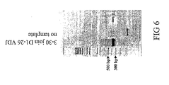
- FIG 6 shows the VDJ recombined product for a VDJ reaction involving a single 3-30 VH sequence as derived from pRF26 and a single non-degenerate joining oligo D1-26.
- Sequencing results in Table 2 show that 2 of 2 clones analyzed are identical to the computer assembled (electronic) copy of 3-30 VDJ: All clones have the same invariant 3-30 VH sequence as defined by plasmid RF26, and single invariant IGHD1-26 and IGHJ4 sequences.
- FIG 7 shows 3-oligo VDJ recombined products using degenerate joining oligos 3 or 4 and the 3-30 VH minigene derived from either plasmid or gDNA in independent recombination reactions.
- Successful recombination is visually shown by the conversion of input template at ⁇ 300 bp, to VDJ recombined template of ⁇ 400 bp.
- Sequence analysis in Table 2 show clones which used 3-30VH from plasmid RF26 have an invariant heavy chain, while clones using 3-30VH from genomic DNA have diverse heavy chains since the 3-30V F & R PCR primers will amplify other heavy chain variable regions in gDNA depending on the stringency of the PCR. Clones from both sets of constructs have diverse D-regions demonstrating successful VDJ recombination with degenerate joining oligos.
- FIG 8 shows another set VDJ recombined products for each joining oligo (Gel 2), using the 3-30VH gene amplified at low stringency (56 °C annealing) from genomic DNA (Gel 1). Conversion of 3-30VH template ( ⁇ 300 bp) to recombined VDJ product can be seen. Recombined VDJ products were cloned into appropriate vectors. Sequence analysis in Table 2 shows that reduced stringency PCR increased the diversity of heavy chains in the sample compared to 3-30VH templates derived by PCR of gDNA at 63 C.
- FIG 9 shows the results of a VDJ recombination experiment using 2 oligos (join4 & JH4 Nhe/Not), instead of 3 oligos (join 4, JH4 Nhe/Not & 3-30R).
- the original PCR of 3-30VH template from plasmid RF26 was used in varying amounts with fixed concentrations of joining oligo 4 and JH4 Nhe/Not.
- Effective conversion of the 3-30VH template ( ⁇ 300 bp) to recombined 3-30H VDJ product ( ⁇ 400 bp) improves with increasing amounts of 3-30VH template.
- the 10x 3-30 VDJ product shown on the gel was cloned directly with out further amplification and sequenced. Results in Table 2 show that 3-30VH is invariant as expected while diverse D-regions were incorporated in the resulting 3-30 VDJ clones.
- FIG. 10 shows an amino acid alignment of the polypeptides encoded by the human germline heavy chain V minigenes.
- the complementarity determining regions are labeled CDR1, CDR2, and CDR3 and the framework regions are labeled FR1, FR2, and FR3.
- the amino acid position is indicated at the top.
- FIG. 11 shows an amino acid alignment of the human germline heavy chain D (top) and J segments (bottom).
- the D regions can be read in multiple reading frames, which are designated RF1, RF2, and RF3.
- RF1, RF2, and RF3 There are seven families ofD regions, labeled D1 through D7 on the left, followed by the designation of each gene name.
- J segments labeled accordingly.
- FIG. 12 shows an amino acid alignment of the polypeptides encoded by the human germline light chain kappa V minigenes.
- the complementarity determining regions are labeled CDR1, CDR2, and CDR3 and the framework regions are labeled FR1, FR2, and FR3.
- the amino acid position is indicated at the top.
- FIG. 13 shows an amino acid alignment of the polypeptides encoded by the human germline light chain lambda V minigenes.
- the complementarity determining regions are labeled CDR1, CDR2, and CDR3 and the framework regions are labeled FR1, FR2, and FR3.
- the amino acid position is indicated at the top.
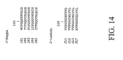
- FIG. 14 shows the polypeptides encoded by the J minigenes for the Kappa locus (top) and Lambda locus (bottom).
- nucleic acid refers to deoxyribonucleic acids (DNA) or ribonucleic acids (RNA) and polymers thereof in either single- or double-stranded form. Unless specifically limited, the term encompasses nucleic acids containing known analogues of natural nucleotides that have similar binding properties as the reference nucleic acid and are metabolized in a manner similar to naturally occurring nucleotides. Unless otherwise indicated, a particular nucleic acid sequence also implicitly encompasses conservatively modified variants thereof (e.g. , degenerate codon substitutions), alleles, orthologs, SNPs, and complementary sequences as well as the sequence explicitly indicated.
- DNA deoxyribonucleic acids
- RNA ribonucleic acids
- degenerate codon substitutions may be achieved by generating sequences in which the third position of one or more selected (or all) codons is substituted with mixed-base and/or deoxyinosine residues ( Batzer et al., Nucleic Acid Res. 19:5081 (1991 ); Ohtsuka et al., J. Biol. Chem. 260:2605-2608 (1985 ); and Rossolini et al., Mol. Cell. Probes 8:91-98 (1994 )).
- the term nucleic acid is used interchangeably with gene, cDNA, and mRNA encoded by a gene.
- the term "gene” means the segment of DNA involved in producing a polypeptide chain; it includes regions preceding and following the coding region (leader and trailer) as well as intervening sequences (introns) between individual coding segments (exons).
- the term "minigene” as applied to antibody genes refers to a polynucleotide sequence corresponding to the V, D, or J genetic element. Each of these genetic elements is incapable of encoding an antibody protein individually unless they are recombined with one another. For instance a functional antibody heavy chain gene comprises a V minigene fused to a D minigene which is fused to a J minigene.
- V minigene (either kappa or lambda) is fused to a J minigene.
- Human V, D, and J minigenes are well known to those in the art. Their sequences can be found online at the National Center for Biotechnology Information (NCBI) or in the literature [ Ruiz, et al. Exp. Clin.Immunogenet. 16: 173-184(1999 ); Pallares, et al. Exp.Clin.Immunogenet. 16: 36-60(1999 )].
- the term "germline” refers to the sequences of the V, D, and J minigenes, prior to the exposure of an antibody to an antigen.
- V-regions describe the genetic element which results from the rearrangement event between V, D, and J (for heavy chains) or V and J minigenes (for light chains).
- An “antibody V-region” refers to the polypeptide region encoded by the V, D, and J element.
- An antibody V-region is encoded by rearranged V,D, and J minigenes.
- V(D)J Recombination refers to any process wherein a V, D, or J minigene is recombined to another V, D, or J minigene.
- a V-region may be part of a full length antibody, an FAb, a scFv, or any other derivative of an antibody (see definition of antibody below).
- a “germline V-region” refers to the sequence of rearranged V, D, and J minigenes prior to significant mutagenic events.
- a germline V-region may have random insertions or deletions at the junctions of the V-D, D-J, or V-J minigenes.
- a non-germline V-region (or a "mature" V-region) will differ from the germline sequences of the minigenes by usually more than 5 residues (not including the junctional deletions or insertions).
- Polypeptide and “peptide” are used interchangeably herein to refer to a polymer of amino acid residues; whereas “protein” may contain one or multiple polypeptide chains. All three terms apply to amino acid polymers in which one or more amino acid residue is an artificial chemical mimetic of a corresponding naturally occurring amino acid, as well as to naturally occurring amino acid polymers and non-naturally occurring amino acid polymers. As used herein, the terms encompass amino acid chains of any length, including full-length proteins, wherein the amino acid residues are linked by covalent peptide bonds.
- an "exogenously rearranged" V-region or antibody gene refers to the location where the V, D, or J minigenes were rearranged to form a functional V-region gene capable of encoding an antibody V-region polypeptide.
- rearrangement typically occurs in cells of the B-lymphoid lineage.
- an exogenously rearranged V-region is one wherein rearrangement occurs outside of a B lymphocyte.
- An exogenously rearranged V-region could have been rearranged in vitro, or in a cell line that does not typically undergo V(D)J rearrangement.
- Such cell lines can be induced to perform V(D)J rearrangement by introduction of the recombination activating genes RAG-1 and RAG-2 and the proper signal sequences adjacent to the V, D, or J minigenes.
- An "endogenously rearranged" V-region or antibody gene means that the gene was rearranged in a B-lymphocyte precursor, in the natural context of an animal.
- amino acid refers to naturally occurring and synthetic amino acids, as well as amino acid analogs and amino acid mimetics that function in a manner similar to the naturally occurring amino acids.
- Naturally occurring amino acids are those encoded by the genetic code, as well as those amino acids that are later modified, e.g. , hydroxyproline, ⁇ -carboxyglutamate, and O-phosphoserine.
- Amino acid analogs refers to compounds that have the same basic chemical structure as a naturally occurring amino acid, i.e., an ⁇ carbon that is bound to a hydrogen, a carboxyl group, an amino group, and an R group, e.g.
- amino acid mimetics refers to chemical compounds that have a structure that is different from the general chemical structure of an amino acid, but that functions in a manner similar to a naturally occurring amino acid.
- Amino acids may be referred to herein by either the commonly known three letter symbols or by the one-letter symbols recommended by the TUPAC-IUB Biochemical Nomenclature Commission. Nucleotides, likewise, may be referred to by their commonly accepted single-letter codes.
- Constantly modified variants applies to both amino acid and nucleic acid sequences. With respect to particular nucleic acid sequences, “conservatively modified variants” refers to those nucleic acids that encode identical or essentially identical amino acid sequences, or where the nucleic acid does not encode an amino acid sequence, to essentially identical sequences. Because of the degeneracy of the genetic code, a large number of functionally identical nucleic acids encode any given protein. For instance, the codons GCA, GCC, GCG and GCU all encode the amino acid alanine. Thus, at every position where an alanine is specified by a codon, the codon can be altered to any of the corresponding codons described without altering the encoded polypeptide.
- nucleic acid variations are "silent variations," which are one species of conservatively modified variations. Every nucleic acid sequence herein that encodes a polypeptide also describes every possible silent variation of the nucleic acid.
- each codon in a nucleic acid except AUG, which is ordinarily the only codon for methionine, and TGG, which is ordinarily the only codon for tryptophan
- TGG which is ordinarily the only codon for tryptophan
- degenerate when applied to nucleotide sequences, describes a nucleotide sequence wherein more than one residue could be located at a given location.
- homologous or “homology” means that one single-stranded nucleic acid sequence may hybridize to a second single-stranded nucleic acid sequence under certain conditions.
- the degree of hybridization may depend on a number of factors including the amount of identity between the sequences and the hybridization conditions such as temperature and salt concentration as discussed later.
- the region of identity is greater than about 5 bp, more preferably the region of identity is greater than 10 bp. If two nucleic acids have "homology,” they can hybridize to one another under appropriate conditions.
- operably linked refers to a linkage of polynucleotide elements in a functional relationship.
- a nucleic acid is “operably linked” when it is placed into a functional relationship with another nucleic acid sequence.
- a promoter or enhancer is operably linked to a coding sequence if it affects the transcription of the coding sequence.
- Nucleic acids encoding proteins that produce multimers, such as heterodimers, heterotrimers, etc. are operably linked when they are expressed together at the same time under conditions where they may interact.
- an “antibody” refers to a protein of the immunoglobulin family or a polypeptide comprising fragments of an immunoglobulin that is capable of noncovalently, reversibly, and in a specific manner binding a corresponding antigen.
- An exemplary antibody structural unit comprises a tetramer. Each tetramer is composed of two identical pairs of polypeptide chains, each pair having one "light” (about 25 kD) and one "heavy" chain (about 50-70 kD), connected through a disulfide bond.
- the recognized immunoglobulin genes include the ⁇ , ⁇ , ⁇ , ⁇ , ⁇ , ⁇ , and ⁇ constant region genes, as well as the myriad immunoglobulin variable region genes.
- Light chains are classified as either ⁇ or ⁇ Heavy chains are classified as ⁇ , ⁇ , ⁇ , ⁇ , or ⁇ , which in turn define the immunoglobulin classes, IgG, IgM, IgA, IgD, and IgE, respectively.
- the N-terminus of each chain defines a variable region of about 100 to 110 or more amino acids primarily responsible for antigen recognition.
- the terms variable light chain (V L ) and variable heavy chain (V H ) refer to these regions of light and heavy chains respectively.
- CDRs Complementarity-determining domains
- the CDRs are the target protein-binding site of the antibody chains that harbors specificity for such target protein. There are three CDRs (CDR1-3, numbered sequentially from the N-terminus) in each human V L or V H , constituting about 15-20% of the variable domains.
- the CDRs are structurally complementary to the epitope of the target protein and are thus directly responsible for the binding specificity.
- the remaining stretches of the V L or V H the so-called framework regions, exhibit less variation in amino acid sequence ( Kuby, Immunology, 4th ed., Chapter 4. W.H. Freeman & Co., New York, 2000 ).
- the positions of the CDRs and framework regions are determined using various well known definitions in the art, e.g. , Kabat, Chothia, international ImMunoGeneTics database (IMGT), and AbM (see, e.g., Johnson et al., Nucleic Acids Res., 29:205-206 (2001 ); Chothia and Lesk, J. Mol. Biol., 196:901-917 (1987 ); Chothia et al., Nature, 342:877-883 (1989 ); Chothia et al., J. Mol.
- an “antibody light chain” or an “antibody heavy chain” refers to a polypeptide comprising the V L or V H , respectively.
- the V L is encoded by the minigenes V (variable) and J (junctional), and the V H by minigenes V, D (diversity), and J.
- Each of V L or V H includes the CDRs as well as the framework regions.
- antibody light chains and/or antibody heavy chains may, from time to time, be collectively referred to as "antibody chains.” These terms encompass antibody chains containing mutations that do not disrupt the basic structure of V L or V H , as one skilled in the art will readily recognize.
- Antibodies exist as intact immunoglobulins or as a number of well-characterized fragments produced by digestion with various peptidases.
- pepsin digests an antibody below the disulfide linkages in the hinge region to produce F( ab )' 2 , a dimer of Fab ' which itself is a light chain joined to V H -C H 1 by a disulfide bond.
- the F( ab )' 2 may be reduced under mild conditions to break the disulfide linkage in the hinge region, thereby converting the F( ab )' 2 dimer into an F ab ' monomer.
- the F ab ' monomer is essentially F ab with part of the hinge region. Paul, Fundamental Immunology 3d ed. (1993 ).
- antibody fragments are defined in terms of the digestion of an intact antibody, one of skill will appreciate that such fragments may be synthesized de novo either chemically or by using recombinant DNA methodology.
- the term antibody also includes antibody fragments either produced by the modification of whole antibodies, or those synthesized de novo using recombinant DNA methodologies ( e.g. , single chain F v ) or those identified using phage display libraries ( see, e.g., McCafferty et al., Nature, 348:552-554 (1990 )).
- any technique known in the art can be used (see, e.g., Kohler & Milstein, Nature, 256:495-497 (1975 ); Kozbor et al., Immunology Today, 4:72 (1983 ); Cole et al., Monoclonal Antibodies and Cancer Therapy, pp. 77-96. Alan R. Liss, Inc. 1985 ).
- Techniques for the production of single chain antibodies U.S. Patent No. 4,946,778
- transgenic mice, or other organisms such as other mammals may be used to express humanized antibodies.
- phage display technology can be used to identify antibodies, and heteromeric F ab fragments, or scFv fragments that specifically bind to selected antigens (see, e.g., McCafferty et al., supra ; Marks et al., Biotechnology, 10:779-783, (1992 )).
- endopeptidase activity refers to the ability of an enzyme to catalyze the hydrolysis of at least one non-terminal peptide bond between two amino acid residues within a polypeptide of any length.
- serine protease activity is supported by a highly conserved tertiary structure, which comprises a serine-histidine-aspartate triad. Studies have shown that the aspartate residue is not always essential for catalytic activity.
- the "serine protease dyad” as used herein is the minimal structure of the catalytic site for a protease to maintain at least a portion of its proteolytic activity.
- This structure comprises a histidine residue and a serine residue located within CDR3 and CDR2, respectively, of an antibody light chain, where the residues are in a spatial relation to each other similar to their spatial alignment in a serine protease triad, such that the histidine can abstract the proton from the serine hydroxyl group, allowing the serine to act as a nucleophile and attack the carbonyl group of the amide bond within the protein substrate.
- “Mutating” or “mutation” refers to the deletion, insertion, or substitution of any nucleotide, by chemical, enzymatic, or any other means, in a nucleic acid encoding an antibody germline gene such that the amino acid sequence of the resulting polypeptide is altered at one or more amino acid residues.
- a "library" of germline antibody members refers to a repertoire of recombinant polypeptides comprising at least two different germline V-region genes or proteins.
- array refers to an ordered spatial arrangement, particularly an arrangement of immobilized biomolecules or polymeric anchoring structures.
- addressable array refers to an array wherein the individual elements have precisely defined x and y coordinates, so that a given element can be pinpointed.
- Primer extension refers to the process whereby: a homologous polynucleotide hybridizes to a second homologous polynucleotide, wherein at least one of the ends of the hybridized molecule contains a single-stranded region and under conditions wherein a polymerase converts at least a portion of the single stranded region to a double-stranded polynucleotide.
- the present invention provides a method to generate full length antibody germline V-region genes, and the proteins which they encode.
- the method first produces a V minigene, by a method such as gene amplification or chemical synthesis, and then uses a hybrid primer capable of hybridizing to a V mini gene and either a D or J minigene.
- a hybrid primer facilitates recombination of a V minigene to a D or J minigene to produce a full length V-region gene.
- a full length V-region gene may be produced from a similar process comprising first obtaining the sequence of a D or J minigene and subsequent recombination using a hybrid primer capable of hybridizing to a V minigene and a D or J minigene.
- V-regions that include but are not limited to: a) degenerate codons in germline antibody CDRs, b) germline V-regions of different lengths, c) germline V-region genes encoding unique functional activities such as protease function, d) protein molecules encoded by said germline V-region genes, e) cells transfected with said germline V-region genes, including hybridoma cells or hybridoma fusion partners, f) transgenic mice comprising the said rearranged germline V-region genes, g) germline V-region genes used in display technologies like phage display, ribosome display, RNA display, or plasmid display, and h) germline V-region genes as part of an addressable array.
- Kb kilobases
- bp base pairs
- Kb kilobases
- bp base pairs
- kD kilo-Daltons
- Proteins sizes are estimated from gel electrophoresis, from sequenced proteins, from derived amino acid sequences, or from published protein sequences.
- Oligonucleotides that are not commercially available can be chemically synthesized according to the solid phase phosphoramidite triester method first described by Beaucage and Caruthers, Tetrahedron Letters, 22:1859-1862 (1981 ), using an automated synthesizer, as described in Van Devanter et al., Nucleic Acids Res., 12:6159-6168 (1984 ). Purification of oligonucleotides is by either native polyacrylamide gel electrophoresis or by anon-exchange chromatography as described in Pearson & Reanier, J. Chrom., 255:137-149 (1983 ).
- sequence of the cloned genes and synthetic oligonucleotides can be verified after cloning using, e.g., the chain termination method for sequencing double-stranded templates of Wallace et al., Gene, 16:21-26 (1981 ).
- V minigenes are typically between 250 and 350 nucleotides in length, and could be produced by standard gene synthesis, or by amplification from a template nucleic acid that comprises unrearranged V minigenes.
- the D or J minigenes can be produced in a similar manner.
- a V minigene is amplified directly from germline DNA.
- An example of germline DNA is genomic DNA prepared from a cell line or tissue. Such a cell line or tissue is preferably derived from a mammal. Germline DNA is preferably not from a mature B-cell that produces an antibody molecule.
- germline DNA is genomic DNA from a fibroblast.
- Production of a polynucleotide sequence encoding a germline V region begins with obtaining the sequence of a V minigene or a J minigene, which may then be joined with at least a J minigene (optionally with a D minigene in between) or a V minigene (optionally with a D mini gene in between), respectively.
- Both chemical methods and enzymatic methods are useful for obtaining the V or J minigene.
- the initial V or J minigene may be directly synthesized or may be obtained from a genomic DNA library using amplification methods such as polymerase chain reaction (PCR). Amplification of the V minigene requires primers for an amplification process.
- Such an amplification process includes the polymerase chain reaction or other isothermal amplification processes (see e.g. Kurn,U.S. Pat. 6,251,639 ).
- Primers for amplification may comprise nucleic acids or a derivative thereof, and may be at least 10 nucleotides in length. Preferably the primers are between 15 and 100 nucleotides long.
- the primers can be designed to amplify the full V minigene, and may or may not include the 5' sequence encoding the leader and intron or the 3' recombination signal sequence. Restriction sites may be included in the primer to facilitate cloning of the V mingene.
- the primers should be designed as forward and reverse primers such that the minigene is amplified after several rounds of thermocycling. Requirements for design of primers for PCR are well known to those of skill in the art, and are described in several references both generally and specifically for antibody genes.
- Table 3 shows a set of oligonucleotide primers for amplifying the repertoire of germline heavy chain V minigenes from human genomic DNA
- Table 3 Multiple family V-region heavy chain primers Forward Primers: VH Family: Primer Name: V-Regions Amplified: Sequence: VH1 VH1FA VH1-45 CAGATGCAGCTGGTGCAGTCTGGG VH1FB VH1-58 CAAATGCAGCTGGTGCAGTCTGGG VH1FC VH1-2, VH1-46, VH1-69, VH1-8 CAGGTGCAGCTGGTGCAGTCTG VHIFD VH1-3, VH1-18 CAGGTTCAGCTGGTGCAGTCTGG VH1FE VH1-24 CAGGTCCAGCTGGTACAGTCTGG VH2 VH2FA VH2-26 CAGGTCACCTTGAAGGAGTCTGG VH2FB VH2-70 CAGGTCACCTTGAAGGAGTCTGG VH2FC VH2-5 CAGATCACCTTGAAGGAGTCTGG VH
- D and J minigenes are typically less than 100 nucleotides long. Thus, these minigenes can be synthesized chemically, by standard oligonucleotide synthesis.
- the D and J minigenes may be single-stranded or double stranded. Sequences of human D and J minigenes are publicly available at the National Center for Biotechnology Information (NCBI), or in the literature [ Ruiz, et al. Exp. Clin. Immunogenet. 16: 173-184(1999 ); Pallares, et al.
- FIGS XX-XX Amino acid sequences ancoded by the minigenes are shown in FIGS XX-XX, thus any nucleotide sequence encoding the amino acid sequences of FIGS XX-XX can be considered V, D, or J minigenes, respectively. Efficient rearrangement of a V minigene to a D minigene can utilize a primer that can hybridize with both a V minigene and either a D or J minigene.
- the primer may hybridize at its 5' end with a V minigene and at its 3' end with a D or J minigene, or the primer may hybridize at its 3' end with a V minigene and at its 5' end with a D or J minigene.
- the primer may also be capable of hybridizing with both a D and J minigene. In fact, since several D minigenes are less than 30 nucleotides in length, a primer may include an entire D minigene between the sequences that can hybridize to a V and J minigene.
- such a hybrid primer is utilized in an amplification reaction along with a back primer that can hybridize with the 5' end of the V minigene, and a forward primer capable of hybridizing to the 3' end of the J minigene as well as the J minigene sequence present in the hybrid primer.
- An amplification reaction can then occur, where the V minigene is ultimately fused to the D and/or J minigene in the final product.
- the success of the recombination can be determined using agarose gel electrophoresis as in FIG. 3 or FIG. 4 , and comparing the size of the final product to the size of the original V minigene.
- the rearranged V(D)J V-region can be directly sequenced using standard techniques, or cloned into a plasmid vector for DNA sequencing or restriction enzyme analysis.
- the method generally described above to generate rearranged antibody genes can be specifically applied to generate germline light chain antibody genes.
- Antibody light chains utilize only V minigenes and J minigenes.
- the hybrid primer described above should hybridize to both a germline V minigene and a germline J minigene.
- the V and J minigenes may be from either the Kappa or Lambda families, and should preferably be derived from a mammal.
- a V minigene and a J minigene should be produced.
- One method to produce a light chain V minigene is to amplify the V minigene by a method such as PCR using genomic DNA as a template.
- a J minigene is typically less than 100 nucleotides and can be produced by standard oligonucleotide synthesis.
- the V or J minigenes can be single or double-stranded at this stage.
- a hybrid nucleic acid primer can be used which hybridizes to the V minigene as well as the J minigene. Preferably this primer is greater than 10 nucleotides.
- a reverse primer hybridizing to the 5' end of the V minigene and a forward primer hybridizing to the 3' end of the J minigene can also be included in the recombination reaction.
- the reaction can use PCR and standard amplification conditions.
- the success of the recombination can be determined using agarose gel electrophoresis as in FIG. 3 or FIG.
- the rearranged VJ light chain V-region can be directly sequenced using standard techniques, or cloned into a plasmid vector for DNA sequencing or restriction enzyme analysis.
- the method generally described above to generate rearranged antibody germline genes can be specifically applied to generate germline heavy chain antibody genes.
- Antibody heavy chains typically utilize V, D, and J minigenes, however they could utilize only V and J minigenes. Additionally, more than one D-region may be used between V and J regions.
- the hybrid primer described above should hybridize to both a germline V minigene and a germline J minigene, but may additionally contain a region capable of hybridizing to a D minigene.
- the V, D, and J minigenes may be from should preferably be derived from a mammal. In order to produce a full length heavy chain V-region, a V, D, and J minigene should be produced.
- One method to produce a heavy chain V minigene is to amplify the V minigene by a method such as PCR using genomic DNA as a template.
- D and J minigenes are typically less than 100 nucleotides and can be produced by standard oligonucleotide synthesis.
- the D and J minigenes need not be produced as separate molecules. They could be produced as a single hybrid primer comprised of regions of homology to V, D, and J minigenes.
- the V, D, and J minigenes can be single or double-stranded at this stage.
- a hybrid nucleic acid primer can be used which hybridizes to the V minigene as well as the J minigene, and optionally including a region capable of hybridizing to a D minigene located between the V and J regions. Preferably this primer is greater than 10 nucleotides.
- a recombination reaction can then be performed containing at least two primers, but optionally containing three.
- a forward primer hybridizing to the 5' end of the V minigene and a reverse primer hybridizing to the 3' end of the J minigene can be included in the recombination reaction.
- the reaction can use PCR and standard amplification conditions. The success of the recombination can be determined using agarose gel electrophoresis as in FIG. 3 or FIG.
- the rearranged VJ light chain V-region can be directly sequenced using standard techniques, or cloned into a plasmid vector for DNA sequencing or restriction enzyme analysis.
- degeneracy may be present in the primer componenets such that diversity is generated in the final rearranged V-region.
- Codon based degeneracy is well known to those in the art and can be accomplished by standard techniques.
- degenerate oligonucleotides can be designed as primer sets and PCR can be performed under suitable conditions (see, e.g., White et al., PCR Protocols: Current Methods and Applications, 1993 ; Griffin and Griffin, PCR Technology, CRC Press Inc. 1994 ) to amplify a segment of nucleotide sequence from a human cDNA or genomic library.
- mutagenesis could be accomplished in order to enhance its binding affinity or another useful activity such as a catalytic function.
- a library can be created consisting of mutagenized versions of the parental germline V-region.
- Current methods in widespread use for creating mutant proteins in a library format are error-prone polymerase chain reaction [Caldwell and Joyce (1992); Gram, et al. Proc Natl Acad Sci 89: 3576-80(1992 )] and cassette mutagenesis [ Stemmer and Morris Biotechniques 13: 214-20(1992 ); Arkin and Youvan Proc Natl Acad Sci 89: 7811-5(1992 ); Oliphant, et al.
- Error-prone PCR uses low-fidelity polymerization conditions to introduce a low level of point mutations randomly over a long sequence. Error prone PCR can also be used to mutagenize a mixture of fragments of unknown sequence. Error-prone PCR can randomly mutate genes by altering the concentrations of respective dNTP's in the presence of dITP [Caldwell and Joyce (1992); Leung and Miyamoto Nucleic Acids Res 17: 1177-95(1989 ); Spee, et al. Nucleic Acids Res 21: 777-8(1993 )]. Methods of saturation mutagenesis utilizing random or partially degenerate primers that incorporate restriction sites have also been described [ Oliphant, et al.
- Cassette mutagenesis is another method for creating libraries of mutant proteins [ Hill, et al. Methods Enzymol 155: 558-68(1987 ); Shiraishi and Shimura Gene 64: 313-9(1988 ); Bock, et al. U S 5,830,720 (1995 ); Stemmer and Crameri U S 5,830,721 (1998 ); Miller, et al. U S 5,830,740 (1998 ); Christou and McCabe 5,830,728 [1998 )].
- Cassette mutagenesis typically replaces a sequence block length of a template with a partially randomized sequence. The maximum information content that can be obtained is thus limited statistically to the number of random sequences in the randomized portion of the cassette.
- Zaccolo and Gherardi (1999) describe a method of random mutagenesis utilizing pyrimidine and purine nucleoside analogs [ Zaccolo and Gherardi J Mol Biol 285: 775-83(1999 )]. This method was successful in achieving substitution mutations which rendered ⁇ -lactamase with an increased catalytic rate against the cephalosporin cefotaxime. Crea describes a "walk through” method, wherein a predetermined amino acid is introduced into a targeted sequence at pre-selected positions [ Crea U S 5,798,208 [1998 )].
- DNA Shuffling The technique most often used to evolve proteins in vitro is known as "DNA Shuffling".
- a library of gene modifications is created by fragmenting homologous sequences of a gene, allowing the fragments to randomly anneal to one another, and filling in the overhangs with polymerase.
- a full length gene library is then reconstructed with polymerase chain reaction (PCR).
- PCR polymerase chain reaction
- the utility of this method occurs at the step of annealing, whereby homologous sequences may anneal to one another, producing sequences with attributes of both starting sequences. In effect, the method affects recombination between two or more genes that are homologous, but that contain significant differences at several positions.
- fragmentation of the initial DNA can be accomplished by premature termination of the polymerase in an extension reaction by inducing adduct formation in the target gene [ Short U S 5, 965, 408 (1999 )].
- a library is created by inducing incremental truncations in each of two homologs to produce a library of fusion genes, each of which contains domains donated from each homolog [ Ostermeier, et al. Nat. Biotechnol. 17: 1205-1209(1999 )].
- the advantage of this approach is that significant homology amongst the starting sequences is not required since the annealing step of previous methods is omitted. It is unclear, however, whether this modified technique actually will lead to generation of improved gene function after selection techniques are applied to the library.
- the nucleic acids encoding recombinant polypeptides of the present invention are typically cloned into an intermediate vector before transformation into prokaryotic or eukaryotic cells for replication and/or expression.
- the intermediate vector is typically a prokaryote vector such as a plasmid or shuttle vector.
- V-region To obtain high level expression of a cloned V-region one typically subclones the DNA into an expression vector that contains a strong promoter to direct transcription, a transcription/translation terminator, and a ribosome binding site for translational initiation. Additionally, the V-region may optionally be fused to a C-region to produce an antibody comprising constant regions. Suitable bacterial promoters are well known in the art and fully described in scientific literature such as Sambrook and Russell, supra, and Ausubel et al, supra. Bacterial expression systems for expressing antibody chains of the recombinant catalytic polypeptide are available in, e.g., E.
- Kits for such expression systems are commercially available.
- Eukaryotic expression systems for mammalian cells, yeast, and insect cells are well known in the art and are also commercially available.
- the promoter used to direct expression of a heterologous nucleic acid depends on the particular application.
- the promoter is preferably positioned about the same distance from the heterologous transcription start site as it is from the transcription start site in its natural setting. As is known in the art, however, some variation in this distance can be accommodated without loss of promoter function.
- the expression vector typically contains a transcription unit or expression cassette that contains all the additional elements required for the expression of the antibody chain in host cells.
- a typical expression cassette thus contains a promoter operably linked to the nucleic acid sequence encoding the germline antibody chain and signals required for efficient polyadenylation of the transcript, ribosome binding sites, and translation termination. Additional elements of the cassette may include enhancers and, if genomic DNA is used as the structural gene, introns with functional splice donor and acceptor sites.
- the expression cassette should also contain a transcription termination region downstream of the structural gene to provide for efficient termination.
- the termination region may be obtained from the same gene as the promoter sequence or may be obtained from different genes.
- the particular expression vector used to transport the genetic information into the cell is not particularly critical. Any of the conventional vectors used for expression in eukaryotic or prokaryotic cells may be used. Standard bacterial expression vectors include plasmids such as pBR322 based plasmids, pSKF, pET23D, and fusion expression systems such as MBP, GST, and LacZ. Epitope tags can also be added to recombinant proteins to provide convenient methods of isolation, e.g., c-myc or histidine tags.
- Expression vectors containing regulatory elements from eukaryotic viruses are typically used in eukaryotic expression vectors, e.g., SV40 vectors, papilloma virus vectors, and vectors derived from Epstein-Barr virus.
- eukaryotic vectors include pMSG, pAV009/A + , pMT010/A + , pMAMneo-5, baculovirus pDSVE, and any other vector allowing expression of proteins under the direction of the CMV promoter, SV40 early promoter, SV40 later promoter, metallothionein promoter, murine mammary tumor virus promoter, Rous sarcoma virus promoter, polyhedrin promoter, or other promoters shown effective for expression in eukaryotic cells.
- Some expression systems have markers that provide gene amplification such as thymidine kinase and dihydrofolate reductase.
- markers that provide gene amplification such as thymidine kinase and dihydrofolate reductase.
- high yield expression systems not involving gene amplification are also suitable, such as using a baculovirus vector in insect cells, with a nucleic acid sequence encoding a germline antibody chain under the direction of the polyhedrin promoter or other strong baculovirus promoters.
- the elements that are typically included in expression vectors also include a replicon that functions in E. coli, a gene encoding antibiotic resistance to permit selection of bacteria that harbor recombinant plasmids, and unique restriction sites in nonessential regions of the plasmid to allow insertion of eukaryotic sequences.
- the particular antibiotic resistance gene chosen is not critical, any of the many resistance genes known in the art are suitable.
- the prokaryotic sequences are preferably chosen such that they do not interfere with the replication of the DNA in eukaryotic cells, if necessary.
- Standard transfection methods are used to produce bacterial, mammalian, yeast, or insect cell lines that express large quantity of antibody chains, which is then purified using standard techniques (see, e.g., Colley et al., J. Biol. Chem., 264:17619-17622 (1989 ); Guide to Protein Purification, in Methods in Enzymology, vol. 182 (Deutscher, ed.), 1990 ). Transformation of eucaryotic and prokaryotic cells are performed according to standard techniques ( see, e.g., Morrison, J. Bact., 132:349-351 (1977 ); Clark-Curtiss and Curtiss, Methods in Enzymology, 101:347-362 (Wu et al., eds), (1983 )).
- Any of the well-known procedures for introducing foreign nucleotide sequences into host cells may be used. These include the use of calcium phosphate transfection, polybrene, protoplast fusion, electroporation, biolistics, liposomes, microinjection, plasma vectors, viral vectors and any of the other well known methods for introducing cloned genomic DNA, cDNA, synthetic DNA, or other foreign genetic material into a host cell (see, e.g., Sambrook and Russell, supra). It is only necessary that the particular genetic engineering procedure used be capable of successfully introducing at least both genes into the host cell capable of expressing germline antibody polypeptide.
- the transfected cells are cultured under conditions favoring expression of the germline antibody chain, which is recovered from the culture using standard techniques identified below.
- a current focus of interest in molecular biology and biotechnology is in the display of large libraries of proteins and peptides and in means of searching them by affinity selection.
- the key to genetic exploitation of a selection method is a physical link between individual molecules of the library (phenotype) and the genetic information encoding them (genotype).
- the libraries of the present invention can be prepared in a number of formats, including those described below.
- phage display in which proteins or peptides are expressed individually on the surface of phage as fusions to a coat protein, while the same phage particle carries the DNA encoding the protein or peptide. Selection of the phage is achieved through a specific binding reaction involving recognition of the protein or peptide, enabling the particular phage to be isolated and cloned and the DNA for the protein or peptide to be recovered and propagated or expressed.
- a particularly desirable application of display technology is the selection of antibody combining sites from combinatorial libraries. Screening for high affinity antibodies to specific antigens has been widely carried out by phage display of antibody fragments ( Winter, G.et.al.(1994) Annu. Rev. Immunol. 12, 433-455 ). Combinations of the variable (V) regions of heavy (H) and light (L) chains are displayed on the phage surface and recombinant phage are selected by binding to immobilized antigen.
- V H /K Another type of single chain antibody fragment is termed V H /K, in which the V H domain is linked to the complete light chain, i.e. V H -linlcer-V L -C L ( He, M. et.al. (1995) Immunology 84, 662-668 .).
- V H -linlcer-V L -C L He, M. et.al. (1995) Immunology 84, 662-668 .
- This has a number of advantages, including stability of expression in E. coli and the use of the C L domain as a spacer and as a tag in detection systems such as ELISA and Western blotting.
- Antibody V H and V L region genes are readily obtained by the methods of the current invention.
- Single chain antibody libraries are potentially of a size of >10 10 members.
- Libraries can also be generated by mutagenesis of cloned DNA fragments encoding specific V H /V L combinations and screened for mutants having improved properties of affinity or specificity. Mutagenesis is carried out preferably on the CDR regions, and particularly on the highly variable H-CDR3, where the potential number of variants which could be constructed from a region of 10 amino acids is 20 10 or 10 13 .
- One such method is the display of proteins or peptides in nascent form on the surface of ribosomes, such that a stable complex with the encoding mRNA is also formed; the complexes are selected with a ligand for the protein or peptide and the genetic information obtained by reverse transcription of the isolated mRNA.
- This is known as ribosome or polysome display.
- a description of such a method is to be found in two U.S. patents, granted to G. Kawasaki/Optein Inc. ( Kawasaki, G. U.S. Pat. Nos. 5,643,768 Cell free synthesis and isolation of novel genes and polypeptides (Jul. 1, 1997) and 5,658,754 (Aug. 19, 1997 )).
- RNA display A further recent display method was described by Robert and Szostak ( Roberts R. W. and Szostak J. W. (1997) Proc. Nat. Acad. Sci USA 94, 12297-12302 ), in which the nascent protein is caused to bind covalently to its mRNA through a puromycin link (termed RNA display).
- Robert and Szostak Roberts R. W. and Szostak J. W. (1997) Proc. Nat. Acad. Sci USA 94, 12297-12302
- selection is carried out on these protein-mRNA fusions after dissociation of the ribosome.
- gene expression can be detected at nucleic acid level.
- a variety of methods of specific DNA and RNA measurement using nucleic acid hybridization techniques are commonly used (e.g. , Sambrook and Russell, supra ). Some methods involve an electrophoretic separation (e.g. , Southern blot for detecting DNA and Northern blot for detecting RNA), but detection of DNA or RNA can be carried out without electrophoresis as well (such as by dot blot).
- the presence of nucleic acid encoding recombinant germline antibodies in transfected cells can also be detected by PCR or RT-PCR using sequence-specific primers.
- gene expression can be detected at the polypeptide level.
- Various immunological assays are routinely used by those skilled in the art to measure the level of a gene product, particularly using polyclonal or monoclonal antibodies that react specifically with a recombinant polypeptide of the present invention, such as an antibody light chain or heavy chain (e.g. , Harlow and Lane, Antibodies, A Labor-crtory Manual, Chapter 14, Cold Spring Harbor, 1988 ; Kohler and Milstein, Nature, 256:495-497 (1975 )).
- antibody light chain or heavy chain e.g. , Harlow and Lane, Antibodies, A Labor-crtory Manual, Chapter 14, Cold Spring Harbor, 1988 ; Kohler and Milstein, Nature, 256:495-497 (1975 )
- Such techniques require antibody preparation by selecting antibodies with high specificity against the recombinant polypeptide or an antigenic portion thereof.
- Antibody chains of the present invention can be purified for use in functional assays.
- the recombinant germline antibodies of the invention may be purified to substantial purity by standard techniques, including selective precipitation with such substances as ammonium sulfate; column chromatography, gel filtration, immunopurification methods, and others (see, e.g., U.S. Patent No. 4,673,641 ; Scopes, Protein Purification: Principles and Practice, 1982 ; Sambrook and Russell, supra ; and Ausubel et al., supra ).
- proteins having established molecular adhesion properties can be reversibly fused to polypeptides of the invention.
- the polypeptides can be selectively adsorbed to a purification column and then freed from the column in a relatively pure form. The fused protein is then removed by enzymatic cleavage. Finally the polypeptide can be purified using affinity columns.
- polypeptides When recombinant polypeptides are expressed by the transformed bacteria in large amounts, typically after promoter induction, although expression can be constitutive, the polypeptides may form insoluble aggregates.
- recombinant polypeptides When recombinant polypeptides are expressed by the transformed bacteria in large amounts, typically after promoter induction, although expression can be constitutive, the polypeptides may form insoluble aggregates.
- protocols that are suitable for purification of polypeptide inclusion bodies and are described in detail in numerous scientific publications (such as Sambrook and Russell, supra, and Ausubel et al., supra ). Numerous variations will be apparent to those of skill in the art.
- the cell suspension is generally centrifuged and the pellet containing the inclusion bodies resuspended in buffer which does not dissolve but washes the inclusion bodies, e.g., 20 mM Tris-HCl (pH 7.2), 1 mM EDTA, 150 mM NaCl and 2% Triton-X 100, a non-ionic detergent. It may be necessary to repeat the wash step to remove as much cellular debris as possible.
- the remaining pellet of inclusion bodies may be resuspended in an appropriate buffer (e.g. , 20 mM sodium phosphate, pH 6.8, 150 mM NaCl).
- an appropriate buffer e.g. , 20 mM sodium phosphate, pH 6.8, 150 mM NaCl.
- Other appropriate buffers will be apparent to those of skill in the art.
- the periplasmic fraction of the bacteria can be isolated by cold osmotic shock in addition to other methods known to those of skill in the art (e.g. , Ausubel et al., supra).
- the bacterial cells are centrifuged to form a pellet. The pellet is resuspended in a buffer containing 20% sucrose.
- the bacteria are centrifuged and the pellet is resuspended in ice-cold 5 mM MgSO 4 and kept in an ice bath for approximately 10 minutes.
- the cell suspension is centrifuged and the supernatant decanted and saved.
- the recombinant polypeptides present in the supernatant can be separated from the host proteins by standard separation techniques well known to those of skill in the art. These methods include, but are not limited to, the following steps: solubility fractionation, size differential filtration, and column chromatography.
- a germline antibody polypeptide to a second antibody polypeptide, wherein the second polypeptide is either a germline antibody or a non-germline antibody polypeptide.
- the second polypeptide is either a germline antibody or a non-germline antibody polypeptide.
- An antibody light chain and an antibody heavy chain can be joined by recombinant DNA technology prior to their expression (see, e.g., Chaudhary et al, Nature, 339:394-397 (1989 ); Pantoliano et al., Biochemistry, 30:10117-10125 (1991 ); Kim et al., Mol. Immunol., 34:891-906 (1997 )).
- a polynucleotide sequence can be introduced to connect the coding sequences for the antibody light and heavy chains (e.g. to construct a scFv) by employing various tools and techniques such as enzymatic digestion/ligation and /or PCR.
- the precise length of the insertion is essential in that the open reading frame of the coding sequence down stream from the insertion should not be disrupted.
- one single polypeptide is generated, which contains both the antibody light and heavy chains and a peptide linker of appropriate length joining them.
- the two antibody chains may also be joined by chemical means following their expression and purification.
- Chemical modifications include, for example, derivitization for the purpose of linking the antibody chains to each other, either directly or through a linking compound, by methods that are well known in the art of protein chemistry.
- Both covalent and noncovalent attachment means may be used with the recombinant germline antibodies of the present invention.
- the procedure for linking the two antibody chains will vary according to the chemical structure of the moieties where the chains are joined.
- one antibody chain typically contain a variety of functional groups such as carboxylic acid (-COOH), free amine (-NH 2 ), or sulfhydryl (-SH) groups, which are available for reaction with a suitable functional group on the other antibody chain to result in a linkage.
- one antibody chain can be derivatized to expose or to attach additional reactive functional groups.
- the derivatization may involve attachment of any of a number of linker molecules such as those available from Pierce Chemical Company, Rockford Illinois.
- the linker is capable of foaming covalent bonds to both antibody chains.
- Suitable linkers are well known to those of skill in the art and include, but are not limited to, straight or branched-chain carbon linkers, heterocyclic carbon linkers, or peptide linkers. Since the antibody chains are polypeptides, the lingers may be joined to the constituent amino acids through their side groups (for example, through a disulfide linkage to cysteine).
- the linkers may also be joined to the alpha carbon amino and carboxyl groups of the terminal amino acids.
- Hybridoma cells can be generated by fusing B cells producing a desired antibody with an immortalized cell line, usually a myeloma cell line, so that the resulting fusion cells will be an immortalized cell line that secrets a particular antibody.
- myeloma cells can be first transfected with a nucleic acid encoding a germline antibody V-region and can be screened for the expression of the germline V-region. Those myeloma cells with highest level of proteolytic light chain expression can be subsequently fused with B cells that produce an antibody with desired target protein specificity.
- the fusion cells will produce two types of antibodies: one is a heterologous antibody containing an endogenous antibody chain (either heavy or light) operably joined to the recombinant germline V-region (either heavy or light), and the other is the same antibody that the parental B cells would secrete ( e.g. both endogenous heavy and light chains).
- the operably joined heterologous heavy and light chains can be isolated by conventional methods such as chromatography and identification can be confirmed by target protein binding assays, assays identifying a unique tag of the germline polypeptide, or endopeptidase activity assays described in other sections of this disclosure. In some cases, where the heterologous antibody is the predominant type in quantity among the two types of antibodies, such isolation may not be needed.
- an assay is available to determine whether an antibody polypeptide contains endopeptidase activity.
- any assay that can detect hydrolysis of a secondary amide bond may be used to determine endopeptidase activity.
- Commonly used assays utilize peptide analogs conjugated to reporter molecules that can be detected when released from the peptide.
- a commonly used assay involves a peptide-methylcoumarinamide (MCA) derivative, such that hydrolysis of the peptide-MCA bond produces the leaving group aminomethylcoumarin whose fluorescence is measured at an excitation of 370 nm and an emmission of 460 nm.
- MCA peptide-methylcoumarinamide
- Such an assay has been practiced to detect proteolytic activity of murine light chains ( Gao, et al, J. Biol.
- any method that allows detection of a cleaved peptide bond in a target protein is suitable for use in the present invention. Since hydrolysis of a peptide bond necessarily produces more that one polypeptide product, several standard size or mass analysis techniques well known in the art can be used to identify peptide bond hydrolysis. These techniques include electrophoretic mobility techniques such as SDS polyacrylamide gel electrophoresis, high performance liquid chromatography (HPLC), and mass spectrometry methods such as MALDI-TOF. Alternatively, a protein labeled with a radioisotope can be precipitated in TCA, wherein hydrolysis of a peptide bond will be indicated by the amount of TCA soluble radioactivity ( Gao, et al, J. Biol. Chem.
- a nucleic acid sequence encoding a germline antibody polypeptide of the present invention can be introduced into a non-human mammal to generate a transgenic animal that expresses the germline antibody polypeptide.
- the transgene expressed by the transgenic mammals of the present invention need not replace at least one allele of the endogenous coding sequence responsible for the variable regions of antibody chains following somatic recombination. Due to allelic exclusion, the presence of an exogenous, post-somatic rearrangement version of the germline V-region DNA will inhibit the endogenous alleles of pre-somatic rearrangement V minigenes from undergoing somatic rearrangement and contributing to the makeup of antibody chains this mammal may produce.
- heterologous antibodies comprising one endogenously rearranged antibody chain, and one transgenic gene which was rearranged a priori.
- heterologous antibodies are invaluable in research and in treating certain conditions in live subjects.
- a method that directs the integration of the transgene to the locus of an endogenous allele will fully serve the purpose of practicing the present invention as well.
- transgenic animal of the present invention is mouse, whereas a number of other transgenic animals can also be produced using the same general method.
- These animals include, but are not limited to: rabbits, sheep, cattle, and pigs ( Jaenisch Science 240:1468-1474 (1988 ); Hammer et al., J. Animal. Sci. 63:269 (1986 ); Hammer et al. Nature 315:680 (1985 ); Wagner et al., Theriogenology 21:29 (1984 )).
- spatially addressable arrays i.e., gene chips, microtiter plates, etc.
- oligonucleotides and polynucleotides or corresponding oligopeptides and polypeptides
- at least one of the biopolymers present on the spatially addressable array comprises an oligonucleotide or polynucleotide sequence first disclosed in at least one germline antibody V-region, or an amino acid sequence encoded thereby.
- Addressable arrays comprising germline antibody V-regions can be used to identify and characterize the temporal and tissue specific expression of an antibody as well as analyze its affinity for a given antigen.
- These addressable arrays incorporate oligonucleotide or peptide sequences of sufficient length to confer the required specificity, yet be within the limitations of the production technology.
- the length of these probes is within a range of between about 8 to about 2000 nucleotides.
- the probes consist of 60 nucleotides and more preferably 25 nucleotides from the germline antibody V-regions.
- a series of the described oligonucleotide sequences, or the complements thereof, can be used in chip format to represent all or a portion of the germline antibody repertoire.
- the oligonucleotides typically between about 16 to about 40 (or any whole number within the stated range) nucleotides in length can partially overlap each other and/or the sequence may be represented using oligonucleotides that do not overlap.
- the described polynucleotide sequences shall typically comprise at least about two or three distinct oligonucleotide sequences of at least about 8 nucleotides in length that encode an antibody germline V-region.
- Such oligonucleotide sequences can begin at any nucleotide present within a germline V-region and proceed in either a sense (5'-to-3') orientation vis-a-vis the described sequence or in an antisense orientation.
- Microanay-based analysis allows the discovery of broad patterns of genetic activity, providing new understanding of gene functions and generating novel and unexpected insight into transcriptional processes and biological mechanisms.
- V minigenes for human A18b or A2c were amplified from genomic DNA using primers hybridizing to the 5' and 3' ends of the minigenes. Primers were annealed to 100 ng of human genomic fibroblast DNA at 56°C for 30 seconds, followed by extension at 70°C for 30 seconds and denaturation at 94°C for 30 seconds. Thirty thermocycles following this pattern were completed. In a subsequent "joining" reaction, a primer comprising the 3' sequence of A18b or A2c and the 5' region of human JK1 was included in a PCR reaction along with a back primer specific for the 5' end of either A18b or A2c, as well as a forward primer specific for the 3' end of JK1.
- the 3' forward primer included a BsiWI restriction site that allowed fusion to the human CK gene.
- the 5' back primer included an Sfi I site that allowed cloning into a bacterial periplasmic expression vector containing the CK constant region fused to six histidines at the C-terminus. Amplification conditions were the same as described above. The rearranged antibody genes were cloned into the vector and expressed as described below.
- the A18b and A2c genes in a pCANTAB derived vector were electroporated into E.coli strain TOP10F', single colonies were isolated and grown at 30°C for 12 hrs in Luria-Bertani broth, and expression induced with 100 mM IPTG for 8 hours.
- Periplasmic extracts were prepared by osmotic lysis, and subjected to two rounds of immobilized metal chelate chromatography to purify the antibody light chains.
- Protease activity was determined by incubating 200 ng of recombinant germline antibody with PFR-MCA protease substrate for 14 hours.
- Peptide hydrolysis was determined by measuring fluorescence of the methylcoumarinamide (MCA) leaving group at ex370/em465. Activity was quantitated using known MCA concentrations to produce a standard curve.
- V minigenes for human 3-30 V were amplified from genomic DNA or plasmid containing 3-30 V, using primers hybridizing to the 5' and 3' ends of the minigenes. PCR was performed on 100 ng of gDNA or 10 ng of plasmid RF26 at 63°C primer annealing for 35 cycles.
- Figure 7 shows a standard 3-oligo joining reaction comprised of the 3-30 V minigene as template (1/10 volume of original 3-30 V PCR), the 3-30 R primer from the 5' end of 3-30 V, joining oligos 3 or 4 which are partially complementary to the 3' end of the 3-30 V minigene and to the 5' end of the J-region primer, and the J-region primer JH4 Nhe-I/Not-I with cloning sites and which is the reverse PCR primer for assembled recombined VDJ products.
- Figure 9 shows a 2-oligo joining reaction which is identical to the above except that the 3-30 R primer for the 5'end of the 3-30 V minigene was left out.
- V-regions are highly homologous to each other. Changing the stringency of a primer pair annealing to genomic DNA in PCR will result in different populations of V minigenes for each PCR condition.
- VDJ recombination on 2 different populations of 3-30 VH minigenes-one derived from PCR at 56°C and the other from PCR at 63°C ( Figure 7 & 8 ). Sequencing results for the 2 different populations of VDJ rearranged clones are shown within Table 2.
- Reduced stringency PCR 56°C
Abstract
Description
- This application claims priority to
U.S. Provisional Application No. 60/501,073, filed September 9, 2003 - The immune system of a mammal is one of the most versatile biological systems as probably greater than 107 antibody specificities can be produced. Indeed, much of contemporary biological and medical research is directed toward tapping this repertoire. Recently there has been a dramatic increase in the ability to harness the output of the vast immunological repertoire. The development of the hybridoma methodology by Kohler and Milstein has made it possible to produce monoclonal antibodies, i.e., a composition of antibody molecules of a single specificity, from the repertoire of antibodies induced during an immune response.
- Unfortunately, current methods for generating monoclonal antibodies are not capable of efficiently surveying the entire antibody response induced by a particular immunogen. In an individual animal there are at least 5-10,000 different B-cell clones capable of generating unique antibodies to a small relatively rigid immunogens, such as, for example dinitrophenol. Further, because of the process of somatic mutation during the generation of antibody diversity, essentially an unlimited number of unique antibody molecules may be generated. In contrast to this vast potential for different antibodies, current hybridoma methodologies typically yield only a few hundred different monoclonal antibodies per fusion.
- Approaches to mimicking the first stage randomisation process which have been described in the literature include those based on the construction of 'naïve' or 'germline' combinatorial antibody libraries prepared by isolating panels of immunoglobulin heavy chain variable (VH) domains and recombining these with panels of light variable chains (VL) domains (see, for example, Gram et al, Proc. Natl. Acad. Sa, USA, 89, 3576-3580, 1992). Naive libraries of antibody fragments have been constructed, for example, by cloning the rearranged V-genes from the IgM RNA of B cells of un-immunised donors isolated from peripheral blood lymphocytes, bone marrow or spleen cells (see, for example, Griffiths et al, EMBO Journal, 12(2), 725-734, 1993, Marks et al, J. Mol. Biol., 222, 581-597, 1991). Such libraries can be screened for antibodies against a range of different antigens.
- Germline antibody genes form precursors to the high affinity antibodies characterized by the secondary immune response. Germline antibody diversity can reach nearly 2x10⁷ different antibodies that derive from the combinatorial use of different V, D, or J minigenes (see
FIG. 1 ). Further diversity is generated by insertional or deletional events occurring at the V-D, D-J, or V-J junctions. Antibody proteins encoded by germline genes are important for several reasons: (i) they form the basis from which higher affinity and more specific antibodies can be derived by further mutation, (ii) all germline heavy or light chain proteins can efficiently pair with other one another, (iii) germline antibody proteins have a flexible structure, allowing polyspecificity and antigen induced complimentarity, and (iv) germline encoded proteins can confer unique activities such as protease function to antibodies. - Thus, germline antibody genes have a commercial use as precursors to more high affinity antibodies, can be useful in the generation of efficiently pairing libraries of heavy and light chains, and could uniquely confer properties like protease activity to antibodies that contain them. Prior to the present invention, no known methods existed to recombine human antibody germline minigenes in vitro in order to produce functional antibody genes. The aforementioned techniques to produce antibodies suffer from limitations which are overcome by the present invention. For example, all of the above techniques rely on the in vivo recombination of antibody genes. In an animal, negative and positive selection events act upon antibody producing B-cells to limit the antibody repertoire. Thus, antibodies or antibody libraries from an animal can be "skewed" towards those antibody sequences compatible with a particular organism or biological environment. In the pharmaceutical industry, drug targets for antibody therapeutics are often "self" antigens. Antibodies to "self" antigens (or antigens endogenously produced by the animal) would be negatively selected, and removed from the animal, in order to avoid autoimmune disease. Although fully synthetic methods to produce antibody genes in vitro have been described, such methods produce significant changes in the antibody genes which could render them immunogenic as a therapeutic. The present invention allows a completely in vitro approach to produce germline antibody genes, which mimicks the natural process of V(D)J recombination that occurs in vivo. Such antibody genes are completely human and native in their sequence, and libraries of such antibody genes can be constructed which represent an unselected population representing the entire antibody repertoire. This invention addresses these and other related needs.
- The present invention provides a method to generate full length antibody germline V-region genes, and the proteins which they encode. The method utilizes gene amplification to produce a V minigene, and a hybrid primer capable of hybridizing to a V minigene and either a D or J minigene. Such a hybrid primer facilitates recombination of a V minigene to a D or J minigene to produce a full length V-region gene. The method described herein allows production of V-regions comprising: a) degenerate codons in germline antibody CDRs, b) germline V-regions of different lengths, c) germline V-region genes encoding unique functional activities such as protease function, d) protein molecules encoded by said germline V-region genes, e) cells transfected with said germline V-region genes, including hybridoma cells or hybridoma fusion partners, f) transgenic mice comprising the rearranged germline V-region genes, g) germline V-region genes used in display technologies like phage display, ribosome display, RNA display, or plasmid display, h) germline V-region genes as part of an addressable array.
- The present invention additionally provides for libraries of exogenously rearranged germline antibody genes. Such libraries can comprise antibody genes of human origin, and may include light chain V-regions, heavy chain V-regions, or light chain V-regions operably linked to heavy chain V-regions. Protein molecules produced from such libraries can be monomers, heterodimers, or homodimers. The library format can be phage display, ribosome display, RNA display, plasmid display, or any other display technology compatible with antibody expression. Additionally, the libraries could form part of an addressable array.
Also described herein is: - (1) A method for producing a polynucleotide encoding a human germline antibody V-region, comprising the steps of:
- (a) obtaining a V minigene or a J minigene; and
- (b) joining the V minigene with at least one J minigene, or joining the J minigene with a V minigene, wherein the J minigene is located at the 3'end of the V minigene.
- (2) The method of (1), wherein a D minigene is further joined to the 3' end of the V minigene and the 5'end of the J minigene.
- (3) The method of (1), wherein the V minigene or the J minigene in step (a) is obtained by chemical synthesis.
- (4) The method of (1), wherein the V minigene or the J minigene in step (a) is obtained by amplification from a germline DNA library.
- (5) The method of (1), wherein step (b) is performed by primer extension using at least two oligonucleotide primers.
- (6) The method of (2), wherein step (b) is performed by primer extension using at least three oligonucleotide primers.
- (7) The method of (5), wherein one of the primers comprises horology to both the V minigene and the J minigene.
- (8) The method of (6), wherein one of the primers comprises homology to both the V minigene and the D minigene.
- (9) The method of (6), wherein one of the primers comprises homology to both the D minigene and the J minigene.
- (10) The method of (6), wherein at least one of the oligonucleotide primers comprises degeneracy at one nucleotide position.
- (11) The method of (1), wherein the V minigene is derived from human immunoglobulin kappa locus.
- (12) The method of (1), wherein the V minigene is derived from human immunoglobulin lambda locus.
- (13) The method of (1), wherein the V minigene is derived from human immunoglobulin heavy chain locus.
- (14) The method of (1), wherein the V-region comprises a serine protease triad.
- (15) A library comprising member polynucleotides encoding exogenously rearranged human germline antibody V-regions.
- (16) The library of (15), wherein the germline V-regions are light chain V-regions.
- (17) The library of (16), wherein each of the light chain V-regions is operably linked to an endogenously rearranged heavy chain V-region.
- (18) The library of (15), wherein the germline V-regions are heavy chain V-regions.
- (19) The library of (18), wherein each of the heavy chain V-regions is operably linked to an endogenously rearranged light chain V-region.
- (20) The library of (15), wherein the germline V-regions comprise operably linked heavy chain and light chain V-regions.
- (21) The library of (15), which is a phage library.
- (22) The library of (15), which resides in a eukaryotic cell.
- (23) The library of (15), which is a ribosome display library.
- (24) The library of (15), which is an RNA display library.
- (25) The library of (15), which is a plasmid display library.
-
FIG. 1 shows a schematic diagram of the antibody heavy chain germline locus. V, D, and J minigenes are arranged on the chromosome and are recombined to produce a functional antibody V-region. Combinatorial and junctional diversity generate a diverse array of antibody germline genes. -
FIG. 2 shows a schematic diagram of the V(D)J recombination method for generating germline V-region antibody genes described herein. -
FIG. 3 shows an agarose gel of the A17 V minigene as amplified by polymerase chain reaction, and the result of recombination of the A17 V minigene and the JK1 minigene. -
FIG. 4 shows an agarose gel of several V minigenes recombined with the JK1 minigene. -
FIG 5 shows the 3-30 VH minigene PCR amplified with primers annealing at 63° C to human genomic DNA and plasmid RF26 that contains the 3-30 VH gene. The derived PCR products served as template in VDJ reactions forFigures 6 ,7 , and9 . -
FIG 6 shows the VDJ recombined product for a VDJ reaction involving a single 3-30 VH sequence as derived from pRF26 and a single non-degenerate joining oligo D1-26. Sequencing results in Table 2 show that 2 of 2 clones analyzed are identical to the computer assembled (electronic) copy of 3-30 VDJ: All clones have the same invariant 3-30 VH sequence as defined by plasmid RF26, and single invariant IGHD1-26 and IGHJ4 sequences. -
FIG 7 shows 3-oligo VDJ recombined products usingdegenerate joining oligos -
FIG 8 shows another set VDJ recombined products for each joining oligo (Gel 2), using the 3-30VH gene amplified at low stringency (56 °C annealing) from genomic DNA (Gel 1). Conversion of 3-30VH template (∼300 bp) to recombined VDJ product can be seen. Recombined VDJ products were cloned into appropriate vectors. Sequence analysis in Table 2 shows that reduced stringency PCR increased the diversity of heavy chains in the sample compared to 3-30VH templates derived by PCR of gDNA at 63 C. -
FIG 9 shows the results of a VDJ recombination experiment using 2 oligos (join4 & JH4 Nhe/Not), instead of 3 oligos (join 4, JH4 Nhe/Not & 3-30R). The original PCR of 3-30VH template from plasmid RF26 was used in varying amounts with fixed concentrations of joiningoligo 4 and JH4 Nhe/Not. Effective conversion of the 3-30VH template (∼300 bp) to recombined 3-30H VDJ product (∼400 bp) improves with increasing amounts of 3-30VH template. The 10x 3-30 VDJ product shown on the gel was cloned directly with out further amplification and sequenced. Results in Table 2 show that 3-30VH is invariant as expected while diverse D-regions were incorporated in the resulting 3-30 VDJ clones. -
FIG. 10 shows an amino acid alignment of the polypeptides encoded by the human germline heavy chain V minigenes. The complementarity determining regions are labeled CDR1, CDR2, and CDR3 and the framework regions are labeled FR1, FR2, and FR3. There are seven families of sequences, which are labeled VH1 through VH7 on the left, followed by the designation of each minigene. The amino acid position is indicated at the top. -
FIG. 11 shows an amino acid alignment of the human germline heavy chain D (top) and J segments (bottom). The D regions can be read in multiple reading frames, which are designated RF1, RF2, and RF3. There are seven families ofD regions, labeled D1 through D7 on the left, followed by the designation of each gene name. There are six J segments, labeled accordingly. -
FIG. 12 shows an amino acid alignment of the polypeptides encoded by the human germline light chain kappa V minigenes. The complementarity determining regions are labeled CDR1, CDR2, and CDR3 and the framework regions are labeled FR1, FR2, and FR3. There are six families of sequences, which are labeled VKI through VKVI on the left, followed by the designation of each minigene. The amino acid position is indicated at the top. -
FIG. 13 shows an amino acid alignment of the polypeptides encoded by the human germline light chain lambda V minigenes. The complementarity determining regions are labeled CDR1, CDR2, and CDR3 and the framework regions are labeled FR1, FR2, and FR3. There are ten families of sequences, which are labeled VL1 through VL10 on the left, followed by the designation of each minigene. The amino acid position is indicated at the top. -
FIG. 14 shows the polypeptides encoded by the J minigenes for the Kappa locus (top) and Lambda locus (bottom). - The term "nucleic acid" or "polynucleotide" refers to deoxyribonucleic acids (DNA) or ribonucleic acids (RNA) and polymers thereof in either single- or double-stranded form. Unless specifically limited, the term encompasses nucleic acids containing known analogues of natural nucleotides that have similar binding properties as the reference nucleic acid and are metabolized in a manner similar to naturally occurring nucleotides. Unless otherwise indicated, a particular nucleic acid sequence also implicitly encompasses conservatively modified variants thereof (e.g., degenerate codon substitutions), alleles, orthologs, SNPs, and complementary sequences as well as the sequence explicitly indicated. Specifically, degenerate codon substitutions may be achieved by generating sequences in which the third position of one or more selected (or all) codons is substituted with mixed-base and/or deoxyinosine residues (Batzer et al., Nucleic Acid Res. 19:5081 (1991); Ohtsuka et al., J. Biol. Chem. 260:2605-2608 (1985); and Rossolini et al., Mol. Cell. Probes 8:91-98 (1994)). The term nucleic acid is used interchangeably with gene, cDNA, and mRNA encoded by a gene.
- The term "gene" means the segment of DNA involved in producing a polypeptide chain; it includes regions preceding and following the coding region (leader and trailer) as well as intervening sequences (introns) between individual coding segments (exons). The term "minigene" as applied to antibody genes refers to a polynucleotide sequence corresponding to the V, D, or J genetic element. Each of these genetic elements is incapable of encoding an antibody protein individually unless they are recombined with one another. For instance a functional antibody heavy chain gene comprises a V minigene fused to a D minigene which is fused to a J minigene. For functional light chain genes, a V minigene (either kappa or lambda) is fused to a J minigene. Human V, D, and J minigenes are well known to those in the art. Their sequences can be found online at the National Center for Biotechnology Information (NCBI) or in the literature [Ruiz, et al. Exp. Clin.Immunogenet. 16: 173-184(1999); Pallares, et al. Exp.Clin.Immunogenet. 16: 36-60(1999)]. The term "germline" refers to the sequences of the V, D, and J minigenes, prior to the exposure of an antibody to an antigen.
- Rearranged "V-regions" describe the genetic element which results from the rearrangement event between V, D, and J (for heavy chains) or V and J minigenes (for light chains). An "antibody V-region" refers to the polypeptide region encoded by the V, D, and J element. An antibody V-region is encoded by rearranged V,D, and J minigenes. The term "V(D)J Recombination" refers to any process wherein a V, D, or J minigene is recombined to another V, D, or J minigene. A V-region may be part of a full length antibody, an FAb, a scFv, or any other derivative of an antibody (see definition of antibody below). A "germline V-region" refers to the sequence of rearranged V, D, and J minigenes prior to significant mutagenic events. A germline V-region may have random insertions or deletions at the junctions of the V-D, D-J, or V-J minigenes. A non-germline V-region (or a "mature" V-region) will differ from the germline sequences of the minigenes by usually more than 5 residues (not including the junctional deletions or insertions).
- "Polypeptide" and "peptide" are used interchangeably herein to refer to a polymer of amino acid residues; whereas "protein" may contain one or multiple polypeptide chains. All three terms apply to amino acid polymers in which one or more amino acid residue is an artificial chemical mimetic of a corresponding naturally occurring amino acid, as well as to naturally occurring amino acid polymers and non-naturally occurring amino acid polymers. As used herein, the terms encompass amino acid chains of any length, including full-length proteins, wherein the amino acid residues are linked by covalent peptide bonds.
- An "exogenously rearranged" V-region or antibody gene refers to the location where the V, D, or J minigenes were rearranged to form a functional V-region gene capable of encoding an antibody V-region polypeptide. In an animal, rearrangement typically occurs in cells of the B-lymphoid lineage. Thus, an exogenously rearranged V-region is one wherein rearrangement occurs outside of a B lymphocyte. An exogenously rearranged V-region could have been rearranged in vitro, or in a cell line that does not typically undergo V(D)J rearrangement. Such cell lines can be induced to perform V(D)J rearrangement by introduction of the recombination activating genes RAG-1 and RAG-2 and the proper signal sequences adjacent to the V, D, or J minigenes. An "endogenously rearranged" V-region or antibody gene means that the gene was rearranged in a B-lymphocyte precursor, in the natural context of an animal.
- The term "amino acid" refers to naturally occurring and synthetic amino acids, as well as amino acid analogs and amino acid mimetics that function in a manner similar to the naturally occurring amino acids. Naturally occurring amino acids are those encoded by the genetic code, as well as those amino acids that are later modified, e.g., hydroxyproline, γ-carboxyglutamate, and O-phosphoserine. Amino acid analogs refers to compounds that have the same basic chemical structure as a naturally occurring amino acid, i.e., an α carbon that is bound to a hydrogen, a carboxyl group, an amino group, and an R group, e.g., homoserine, norleucine, methionine sulfoxide, methionine methyl sulfonium. Such analogs have modified R groups (e.g., norleucine) or modified peptide backbones, but retain the same basic chemical structure as a naturally occurring amino acid. "Amino acid mimetics" refers to chemical compounds that have a structure that is different from the general chemical structure of an amino acid, but that functions in a manner similar to a naturally occurring amino acid.
- Amino acids may be referred to herein by either the commonly known three letter symbols or by the one-letter symbols recommended by the TUPAC-IUB Biochemical Nomenclature Commission. Nucleotides, likewise, may be referred to by their commonly accepted single-letter codes.
- "Conservatively modified variants" applies to both amino acid and nucleic acid sequences. With respect to particular nucleic acid sequences, "conservatively modified variants" refers to those nucleic acids that encode identical or essentially identical amino acid sequences, or where the nucleic acid does not encode an amino acid sequence, to essentially identical sequences. Because of the degeneracy of the genetic code, a large number of functionally identical nucleic acids encode any given protein. For instance, the codons GCA, GCC, GCG and GCU all encode the amino acid alanine. Thus, at every position where an alanine is specified by a codon, the codon can be altered to any of the corresponding codons described without altering the encoded polypeptide. Such nucleic acid variations are "silent variations," which are one species of conservatively modified variations. Every nucleic acid sequence herein that encodes a polypeptide also describes every possible silent variation of the nucleic acid. One of skill will recognize that each codon in a nucleic acid (except AUG, which is ordinarily the only codon for methionine, and TGG, which is ordinarily the only codon for tryptophan) can be modified to yield a functionally identical molecule. Accordingly, each silent variation of a nucleic acid that encodes a polypeptide is implicit in each described sequence.
- The term "degenerate", when applied to nucleotide sequences, describes a nucleotide sequence wherein more than one residue could be located at a given location. Degenerate nucleotides are given the following notation: R=A/G, Y=C/T, M=A/C, K=G/T, S=C/G, W=A/T, B=C/G/T, D=A/G/T, H=A/C/T, V=A/C/G, and N=A/C/G/T.
- The term "homologous" or "homology" means that one single-stranded nucleic acid sequence may hybridize to a second single-stranded nucleic acid sequence under certain conditions. The degree of hybridization may depend on a number of factors including the amount of identity between the sequences and the hybridization conditions such as temperature and salt concentration as discussed later. Preferably the region of identity is greater than about 5 bp, more preferably the region of identity is greater than 10 bp. If two nucleic acids have "homology," they can hybridize to one another under appropriate conditions.
- As used herein, the term "operably linked" refers to a linkage of polynucleotide elements in a functional relationship. A nucleic acid is "operably linked" when it is placed into a functional relationship with another nucleic acid sequence. For instance, a promoter or enhancer is operably linked to a coding sequence if it affects the transcription of the coding sequence. Nucleic acids encoding proteins that produce multimers, such as heterodimers, heterotrimers, etc., are operably linked when they are expressed together at the same time under conditions where they may interact.
- An "antibody" refers to a protein of the immunoglobulin family or a polypeptide comprising fragments of an immunoglobulin that is capable of noncovalently, reversibly, and in a specific manner binding a corresponding antigen. An exemplary antibody structural unit comprises a tetramer. Each tetramer is composed of two identical pairs of polypeptide chains, each pair having one "light" (about 25 kD) and one "heavy" chain (about 50-70 kD), connected through a disulfide bond. The recognized immunoglobulin genes include the κ, λ, α, γ, δ, ε, and µ constant region genes, as well as the myriad immunoglobulin variable region genes. Light chains are classified as either κ or λ Heavy chains are classified as γ, µ, α, δ, or ε, which in turn define the immunoglobulin classes, IgG, IgM, IgA, IgD, and IgE, respectively. The N-terminus of each chain defines a variable region of about 100 to 110 or more amino acids primarily responsible for antigen recognition. The terms variable light chain (VL) and variable heavy chain (VH) refer to these regions of light and heavy chains respectively.
- "Complementarity-determining domains" or "CDRs" refers to the hypervariable regions of VL and VH. The CDRs are the target protein-binding site of the antibody chains that harbors specificity for such target protein. There are three CDRs (CDR1-3, numbered sequentially from the N-terminus) in each human VL or VH, constituting about 15-20% of the variable domains. The CDRs are structurally complementary to the epitope of the target protein and are thus directly responsible for the binding specificity. The remaining stretches of the VL or VH the so-called framework regions, exhibit less variation in amino acid sequence (Kuby, Immunology, 4th ed., ).
- The positions of the CDRs and framework regions are determined using various well known definitions in the art, e.g., Kabat, Chothia, international ImMunoGeneTics database (IMGT), and AbM (see, e.g., Johnson et al., Nucleic Acids Res., 29:205-206 (2001); Chothia and Lesk, J. Mol. Biol., 196:901-917 (1987); Chothia et al., Nature, 342:877-883 (1989); Chothia et al., J. Mol. Biol., 227:799-817 (1992); Al-Lazikani et al., J.Mol.Biol., 273:927-748 (1997)). Definitions of antigen combining sites are also described in the following: Ruiz et al., Nucleic Acids Res., 28:219-221 (2000); and Lefranc, M.P., Nucleic Acids Res., 29:207-209 (2001); MacCallum et al., J. Mol. Biol., 262:732-745 (1996); and Martin et al, Proc. Natl. Acad. Sci. USA, 86:9268-9272 (1989); Martin et al., Methods Enzymol., 203:121-153 (1991); and Rees et al., In Sternberg M.J.E. (ed.), Protein Structure Prediction, Oxford University Press, Oxford, 141-172 (1996).
- An "antibody light chain" or an "antibody heavy chain" as used herein refers to a polypeptide comprising the VL or VH, respectively. The VL is encoded by the minigenes V (variable) and J (junctional), and the VH by minigenes V, D (diversity), and J. Each of VL or VH includes the CDRs as well as the framework regions. In this application, antibody light chains and/or antibody heavy chains may, from time to time, be collectively referred to as "antibody chains." These terms encompass antibody chains containing mutations that do not disrupt the basic structure of VL or VH, as one skilled in the art will readily recognize.
- Antibodies exist as intact immunoglobulins or as a number of well-characterized fragments produced by digestion with various peptidases. Thus, for example, pepsin digests an antibody below the disulfide linkages in the hinge region to produce F(ab)'2, a dimer of Fab' which itself is a light chain joined to VH-
C H1 by a disulfide bond. The F(ab)'2 may be reduced under mild conditions to break the disulfide linkage in the hinge region, thereby converting the F(ab)'2 dimer into an Fab' monomer. The Fab' monomer is essentially Fab with part of the hinge region. Paul, Fundamental Immunology 3d ed. (1993). While various antibody fragments are defined in terms of the digestion of an intact antibody, one of skill will appreciate that such fragments may be synthesized de novo either chemically or by using recombinant DNA methodology. Thus, the term antibody, as used herein, also includes antibody fragments either produced by the modification of whole antibodies, or those synthesized de novo using recombinant DNA methodologies (e.g., single chain Fv) or those identified using phage display libraries (see, e.g., McCafferty et al., Nature, 348:552-554 (1990)). - For preparation of monoclonal or polyclonal antibodies, any technique known in the art can be used (see, e.g., Kohler & Milstein, Nature, 256:495-497 (1975); Kozbor et al., Immunology Today, 4:72 (1983); Cole et al., Monoclonal Antibodies and Cancer Therapy, pp. 77-96. Alan R. Liss, Inc. 1985). Techniques for the production of single chain antibodies (
U.S. Patent No. 4,946,778 ) can be adapted to produce antibodies to polypeptides of this invention. Also, transgenic mice, or other organisms such as other mammals, may be used to express humanized antibodies. Alternatively, phage display technology can be used to identify antibodies, and heteromeric Fab fragments, or scFv fragments that specifically bind to selected antigens (see, e.g., McCafferty et al., supra; Marks et al., Biotechnology, 10:779-783, (1992)). - The term "endopeptidase activity" as used herein refers to the ability of an enzyme to catalyze the hydrolysis of at least one non-terminal peptide bond between two amino acid residues within a polypeptide of any length.
- Despite the diversity in primary amino acid sequence among individual members of the family, serine protease activity is supported by a highly conserved tertiary structure, which comprises a serine-histidine-aspartate triad. Studies have shown that the aspartate residue is not always essential for catalytic activity. The "serine protease dyad" as used herein is the minimal structure of the catalytic site for a protease to maintain at least a portion of its proteolytic activity. This structure comprises a histidine residue and a serine residue located within CDR3 and CDR2, respectively, of an antibody light chain, where the residues are in a spatial relation to each other similar to their spatial alignment in a serine protease triad, such that the histidine can abstract the proton from the serine hydroxyl group, allowing the serine to act as a nucleophile and attack the carbonyl group of the amide bond within the protein substrate.
- "Mutating" or "mutation" refers to the deletion, insertion, or substitution of any nucleotide, by chemical, enzymatic, or any other means, in a nucleic acid encoding an antibody germline gene such that the amino acid sequence of the resulting polypeptide is altered at one or more amino acid residues.
- A "library" of germline antibody members refers to a repertoire of recombinant polypeptides comprising at least two different germline V-region genes or proteins.
- As used herein, the term "array" refers to an ordered spatial arrangement, particularly an arrangement of immobilized biomolecules or polymeric anchoring structures.
- As used herein, the term "addressable array" refers to an array wherein the individual elements have precisely defined x and y coordinates, so that a given element can be pinpointed.
- "Primer extension" refers to the process whereby: a homologous polynucleotide hybridizes to a second homologous polynucleotide, wherein at least one of the ends of the hybridized molecule contains a single-stranded region and under conditions wherein a polymerase converts at least a portion of the single stranded region to a double-stranded polynucleotide.
- The present invention provides a method to generate full length antibody germline V-region genes, and the proteins which they encode. For example, the method first produces a V minigene, by a method such as gene amplification or chemical synthesis, and then uses a hybrid primer capable of hybridizing to a V mini gene and either a D or J minigene. Such a hybrid primer facilitates recombination of a V minigene to a D or J minigene to produce a full length V-region gene. Likewise, a full length V-region gene may be produced from a similar process comprising first obtaining the sequence of a D or J minigene and subsequent recombination using a hybrid primer capable of hybridizing to a V minigene and a D or J minigene. The method described herein allows production of V-regions that include but are not limited to: a) degenerate codons in germline antibody CDRs, b) germline V-regions of different lengths, c) germline V-region genes encoding unique functional activities such as protease function, d) protein molecules encoded by said germline V-region genes, e) cells transfected with said germline V-region genes, including hybridoma cells or hybridoma fusion partners, f) transgenic mice comprising the said rearranged germline V-region genes, g) germline V-region genes used in display technologies like phage display, ribosome display, RNA display, or plasmid display, and h) germline V-region genes as part of an addressable array.
- This invention relies on routine techniques in the field of recombinant genetics. Basic texts disclosing the general methods of use in this invention include Sambrook and Russell, Molecular Cloning: A Laboratory Manual 3d ed. (2001); Kriegler, Gene Transfer and Expression: A Laboratory Manual (1990); and Ausubel et al., Current Protocols in Molecular Biology (1994).
- For nucleic acids, sizes are given in either kilobases (Kb) or base pairs (bp). These are estimates derived from agarose or polyacrylamide gel electrophoresis, from sequenced nucleic acids, or from published DNA sequences. For proteins, sizes are given in kilo-Daltons (kD) or amino acid residue numbers. Proteins sizes are estimated from gel electrophoresis, from sequenced proteins, from derived amino acid sequences, or from published protein sequences.
- Oligonucleotides that are not commercially available can be chemically synthesized according to the solid phase phosphoramidite triester method first described by Beaucage and Caruthers, Tetrahedron Letters, 22:1859-1862 (1981), using an automated synthesizer, as described in Van Devanter et al., Nucleic Acids Res., 12:6159-6168 (1984). Purification of oligonucleotides is by either native polyacrylamide gel electrophoresis or by anon-exchange chromatography as described in Pearson & Reanier, J. Chrom., 255:137-149 (1983). The sequence of the cloned genes and synthetic oligonucleotides can be verified after cloning using, e.g., the chain termination method for sequencing double-stranded templates of Wallace et al., Gene, 16:21-26 (1981).
- The production of a full length antibody V-region requires the V, D, and J minigenes. V minigenes are typically between 250 and 350 nucleotides in length, and could be produced by standard gene synthesis, or by amplification from a template nucleic acid that comprises unrearranged V minigenes. The D or J minigenes can be produced in a similar manner. In one embodiment, a V minigene is amplified directly from germline DNA. An example of germline DNA is genomic DNA prepared from a cell line or tissue. Such a cell line or tissue is preferably derived from a mammal. Germline DNA is preferably not from a mature B-cell that produces an antibody molecule. One example of germline DNA is genomic DNA from a fibroblast.
- Production of a polynucleotide sequence encoding a germline V region begins with obtaining the sequence of a V minigene or a J minigene, which may then be joined with at least a J minigene (optionally with a D minigene in between) or a V minigene (optionally with a D mini gene in between), respectively. Both chemical methods and enzymatic methods are useful for obtaining the V or J minigene. For instance, the initial V or J minigene may be directly synthesized or may be obtained from a genomic DNA library using amplification methods such as polymerase chain reaction (PCR). Amplification of the V minigene requires primers for an amplification process. Such an amplification process includes the polymerase chain reaction or other isothermal amplification processes (see e.g.
Kurn,U.S. Pat. 6,251,639 ). Primers for amplification may comprise nucleic acids or a derivative thereof, and may be at least 10 nucleotides in length. Preferably the primers are between 15 and 100 nucleotides long. The primers can be designed to amplify the full V minigene, and may or may not include the 5' sequence encoding the leader and intron or the 3' recombination signal sequence. Restriction sites may be included in the primer to facilitate cloning of the V mingene. If polymerase chain reaction is used to amplify the V minigene, the primers should be designed as forward and reverse primers such that the minigene is amplified after several rounds of thermocycling. Requirements for design of primers for PCR are well known to those of skill in the art, and are described in several references both generally and specifically for antibody genes. - Table 3 shows a set of oligonucleotide primers for amplifying the repertoire of germline heavy chain V minigenes from human genomic DNA
Table 3: Multiple family V-region heavy chain primers Forward Primers: VH Family: Primer Name: V-Regions Amplified: Sequence: VH1 VH1FA VH1-45 CAGATGCAGCTGGTGCAGTCTGGG VH1FB VH1-58 CAAATGCAGCTGGTGCAGTCTGGG VH1FC VH1-2, VH1-46, VH1-69, VH1-8 CAGGTGCAGCTGGTGCAGTCTG VHIFD VH1-3, VH1-18 CAGGTTCAGCTGGTGCAGTCTGG VH1FE VH1-24 CAGGTCCAGCTGGTACAGTCTGG VH2 VH2FA VH2-26 CAGGTCACCTTGAAGGAGTCTGG VH2FB VH2-70 CAGGTCACCTTGAGGGAGTCTGG VH2FC VH2-5 CAGATCACCTTGAAGGAGTCTGG VH3 VH3FA* VH3-53, VH3-13, VH3-35, VH3-38, VH3-48, VH3-49 VH3-64, VH3-72, VH3-7, VH3-66, VH3-21, VH3-20, VH3-16, VH3-15 GAGGTGCAGCTGGTGGAGTCTGG VH3FC* VH3-30, VH3-33, VH3-11 CAGGTGCAGCTGGTGGAGTCTGG VH3FD* VH3-74, VH3-73 GAGGTGCAGCTGGTGGAGTCCG VH3FE VH3-43, VH3-9 GAAGTGCAGCTGGTGGAGTCTGGG VH3FG VH3-23 GAGGTGCAGCTGTTGGAGTCTGG VH4 VH4FA VH4-39 CAGCTGCAGCTGCAGGAGTCGGG VH4FB VH4-4, VH4-59, VH4-61, VH4-3 1, VH4-28 CAGGTGCAGCTGCAGGAGTCGG VH4FC VH4-34 CAGGTGCAGCTACAGCAGTGG VH5 VH5F VH5-51 GAGGTGCAGCTGGTGCAGTCTG VH6 VH6F VH6-1 CAGGTACAGCTGCAGCAGTCAG VH7 VH7F VH7-81 CAGGTGCAGCTGGTGCAGTCTGG Reverse Primers Family: Name: V regions: Sequence: VH1 VH1RA VH1-45 TATCTTGCACAGTAATACATGG VH1RB VH1-58 TCTGCCGCACAGTAATACACGGC VH1C*,3A,4B VH1-46, VH1-2, VH1-69, VH1-18, VH3-53, VH3-11, VH4-4, VH4-59, VH4-61, VH4-31 TCTCTCGCACAGTAATACACOG VHR1D,3C VH1-3, VH3-30, VH3-33, VH3-7, VH3-66, VH3-21, VH3-64, VH3-48 TCTCTCGCACAGTAATACACAGC VH1RE VH1-24 TCTGTTGCACAGTAATACACGGC VH1RF* VH1-8 CCTCTCGCACAGTAATACACGGC VH2 VH2RA VH2-26 GTATCCGTGCACAGTAATATGTGG VH2RB VH2-70 GTATCCGTGCACAATAATACG VH2RC VH2-5 GTCTGTGTGCACAGTAATATGTGG VH3 VH3RB VH3-16, VH3-35 TTTCTCACACAGTAATACACAGC VH3RD VH3-74, VH3-13 TCTCTTGCACAGTAATACACAGCC VH3RE VH3-43,VH3-9 TATCTTTTGCACAGTAATACAAGG VH3RG VH3-23 TCTTTCGCACAGTAATATACGGC VH3RH VH3-15 TCTGTGGTACAGTAATACACGG VH3RI VH3-49 TCTCTAGTACAGTAATACACGG VH3RJ VH3-73 TGTCTAGTACAGTAATACACGG VH3RK VH3-72 TCTCTAGCACAGTAATACACGG VH3RL VH3-38 TATCTGGCACAGTAATACACGGC VH4 VH4RA VH4-39 TGTCTCGCACAGTAATACACAGCC VH4RD VH4-34 CCTCTCGCACAGTAATACACAGC VH4RC VH4-28 TTTCTCGCACAGTAATACACGG VH5 VH5R VH5-51 TGTCTCGCACAGTAATACATGG VH6 VH6R VH6-1 TCTCTTGCACAGTAATACACAG VH7 VH7R VH7-81 TATCTCGCACAGTAATACATGG - The rearrangement of a V minigene to either a D minigene or a J minigene requires polynucleotides encoding the D and/or J minigenes. D and J minigenes are typically less than 100 nucleotides long. Thus, these minigenes can be synthesized chemically, by standard oligonucleotide synthesis. The D and J minigenes may be single-stranded or double stranded. Sequences of human D and J minigenes are publicly available at the National Center for Biotechnology Information (NCBI), or in the literature [Ruiz, et al. Exp. Clin. Immunogenet. 16: 173-184(1999); Pallares, et al. Exp .Clin. Immunogenet. 16: 36-60(1999)]. Amino acid sequences ancoded by the minigenes are shown in FIGS XX-XX, thus any nucleotide sequence encoding the amino acid sequences of FIGS XX-XX can be considered V, D, or J minigenes, respectively. Efficient rearrangement of a V minigene to a D minigene can utilize a primer that can hybridize with both a V minigene and either a D or J minigene. The primer may hybridize at its 5' end with a V minigene and at its 3' end with a D or J minigene, or the primer may hybridize at its 3' end with a V minigene and at its 5' end with a D or J minigene. The primer may also be capable of hybridizing with both a D and J minigene. In fact, since several D minigenes are less than 30 nucleotides in length, a primer may include an entire D minigene between the sequences that can hybridize to a V and J minigene. In one embodiment, such a hybrid primer is utilized in an amplification reaction along with a back primer that can hybridize with the 5' end of the V minigene, and a forward primer capable of hybridizing to the 3' end of the J minigene as well as the J minigene sequence present in the hybrid primer. An amplification reaction can then occur, where the V minigene is ultimately fused to the D and/or J minigene in the final product. The success of the recombination can be determined using agarose gel electrophoresis as in
FIG. 3 orFIG. 4 , and comparing the size of the final product to the size of the original V minigene. Furthermore, the rearranged V(D)J V-region can be directly sequenced using standard techniques, or cloned into a plasmid vector for DNA sequencing or restriction enzyme analysis. - Given the description of the general principle and methodology used in producing polynucleotide sequences encoding germline V-regions, one of skill in the art would recognize that various modifications can be made to the methods specifically described herein and achieve essentially the same results.
- The method generally described above to generate rearranged antibody genes can be specifically applied to generate germline light chain antibody genes. Antibody light chains utilize only V minigenes and J minigenes. Thus, the hybrid primer described above should hybridize to both a germline V minigene and a germline J minigene. The V and J minigenes may be from either the Kappa or Lambda families, and should preferably be derived from a mammal. In order to produce a full length light chain V-region, a V minigene and a J minigene should be produced. One method to produce a light chain V minigene is to amplify the V minigene by a method such as PCR using genomic DNA as a template. A J minigene is typically less than 100 nucleotides and can be produced by standard oligonucleotide synthesis. The V or J minigenes can be single or double-stranded at this stage. A hybrid nucleic acid primer can be used which hybridizes to the V minigene as well as the J minigene. Preferably this primer is greater than 10 nucleotides. A reverse primer hybridizing to the 5' end of the V minigene and a forward primer hybridizing to the 3' end of the J minigene can also be included in the recombination reaction. The reaction can use PCR and standard amplification conditions. The success of the recombination can be determined using agarose gel electrophoresis as in
FIG. 3 orFIG. 4 , and comparing the size of the final product to the size of the original V minigene. Furthermore, the rearranged VJ light chain V-region can be directly sequenced using standard techniques, or cloned into a plasmid vector for DNA sequencing or restriction enzyme analysis. - The method generally described above to generate rearranged antibody germline genes can be specifically applied to generate germline heavy chain antibody genes. Antibody heavy chains typically utilize V, D, and J minigenes, however they could utilize only V and J minigenes. Additionally, more than one D-region may be used between V and J regions. Thus, the hybrid primer described above should hybridize to both a germline V minigene and a germline J minigene, but may additionally contain a region capable of hybridizing to a D minigene. The V, D, and J minigenes may be from should preferably be derived from a mammal. In order to produce a full length heavy chain V-region, a V, D, and J minigene should be produced. One method to produce a heavy chain V minigene is to amplify the V minigene by a method such as PCR using genomic DNA as a template. Both D and J minigenes are typically less than 100 nucleotides and can be produced by standard oligonucleotide synthesis. The D and J minigenes need not be produced as separate molecules. They could be produced as a single hybrid primer comprised of regions of homology to V, D, and J minigenes. The V, D, and J minigenes can be single or double-stranded at this stage. A hybrid nucleic acid primer can be used which hybridizes to the V minigene as well as the J minigene, and optionally including a region capable of hybridizing to a D minigene located between the V and J regions. Preferably this primer is greater than 10 nucleotides. A recombination reaction can then be performed containing at least two primers, but optionally containing three. A forward primer hybridizing to the 5' end of the V minigene and a reverse primer hybridizing to the 3' end of the J minigene can be included in the recombination reaction. The reaction can use PCR and standard amplification conditions. The success of the recombination can be determined using agarose gel electrophoresis as in
FIG. 3 orFIG. 4 , and comparing the size of the final product to the size of the original V minigene. Furthermore, the rearranged VJ light chain V-region can be directly sequenced using standard techniques, or cloned into a plasmid vector for DNA sequencing or restriction enzyme analysis. - In performing the rearrangement reaction to produce a rearranged germline V-region, degeneracy may be present in the primer componenets such that diversity is generated in the final rearranged V-region. Codon based degeneracy is well known to those in the art and can be accomplished by standard techniques. Based on sequence homology, degenerate oligonucleotides can be designed as primer sets and PCR can be performed under suitable conditions (see, e.g., White et al., PCR Protocols: Current Methods and Applications, 1993; Griffin and Griffin, PCR Technology, CRC Press Inc. 1994) to amplify a segment of nucleotide sequence from a human cDNA or genomic library.
- Following the generation of the germline V-region, further mutagenesis could be accomplished in order to enhance its binding affinity or another useful activity such as a catalytic function. Furthermore, a library can be created consisting of mutagenized versions of the parental germline V-region. Current methods in widespread use for creating mutant proteins in a library format are error-prone polymerase chain reaction [Caldwell and Joyce (1992); Gram, et al. Proc Natl Acad Sci 89: 3576-80(1992)] and cassette mutagenesis [Stemmer and Morris Biotechniques 13: 214-20(1992); Arkin and Youvan Proc Natl Acad Sci 89: 7811-5(1992); Oliphant, et al. Gene 44: 177-83(1986); Hermes, et al. Proc Natl Acad. Sci 87: 696-700(1990)], in which the specific region to be optimized is replaced with a synthetically mutagenized oligonucleotide. Alternatively, mutator strains of host cells have been employed to add mutational frequency [Greener, et al. Mol Biotechnol 7: 189-95(1997)]. In each case, a 'mutant cloud' [Kauffman New York (1993)] is generated around certain sites in the original sequence.
- Error-prone PCR uses low-fidelity polymerization conditions to introduce a low level of point mutations randomly over a long sequence. Error prone PCR can also be used to mutagenize a mixture of fragments of unknown sequence. Error-prone PCR can randomly mutate genes by altering the concentrations of respective dNTP's in the presence of dITP [Caldwell and Joyce (1992);Leung and Miyamoto Nucleic Acids Res 17: 1177-95(1989); Spee, et al. Nucleic Acids Res 21: 777-8(1993)]. Methods of saturation mutagenesis utilizing random or partially degenerate primers that incorporate restriction sites have also been described [Oliphant, et al. Gene 44: 177-83(1986); Hill, et al. Methods Enzymol 155: 558-68(1987); Reidhaar-Olson, et al. Methods Enzymol 208: 564-86(1991)].
- "Cassette" mutagenesis is another method for creating libraries of mutant proteins [Hill, et al. Methods Enzymol 155: 558-68(1987); Shiraishi and Shimura Gene 64: 313-9(1988);
Bock, et al. U S 5,830,720 (1995 );Stemmer and Crameri U S 5,830,721 (1998 );Miller, et al. U S 5,830,740 (1998 );Christou and McCabe 5,830,728 [1998 )]. Cassette mutagenesis typically replaces a sequence block length of a template with a partially randomized sequence. The maximum information content that can be obtained is thus limited statistically to the number of random sequences in the randomized portion of the cassette. - A protocol has also been developed by which synthesis of an oligonucleotide is "doped" with non-native phosphoramidites, resulting in randomization of the gene section targeted for random mutagenesis [Wang and Hoover J Bacteriol 179: 5812-9(1997)]. This method allows control of position selection, while retaining a random substitution rate.
- Zaccolo and Gherardi (1999) describe a method of random mutagenesis utilizing pyrimidine and purine nucleoside analogs [Zaccolo and Gherardi J Mol Biol 285: 775-83(1999)]. This method was successful in achieving substitution mutations which rendered β-lactamase with an increased catalytic rate against the cephalosporin cefotaxime. Crea describes a "walk through" method, wherein a predetermined amino acid is introduced into a targeted sequence at pre-selected positions [
Crea U S 5,798,208 [1998 )]. - The technique most often used to evolve proteins in vitro is known as "DNA Shuffling". In this method, a library of gene modifications is created by fragmenting homologous sequences of a gene, allowing the fragments to randomly anneal to one another, and filling in the overhangs with polymerase. A full length gene library is then reconstructed with polymerase chain reaction (PCR). The utility of this method occurs at the step of annealing, whereby homologous sequences may anneal to one another, producing sequences with attributes of both starting sequences. In effect, the method affects recombination between two or more genes that are homologous, but that contain significant differences at several positions. It has been shown that creation of the library using several homologous sequences allows a sampling of more sequence space than using a randomly mutated single starting sequence [Crameri, et al. Nature 391: 288-291(1998)]. This effect is likely due to the fact that years of evolution have already selected for different, advantageous or neutral mutations amongst the homologs of the different species. Starting with homologs, then, appreciably limits the number of deleterious mutations in the creation of the library which is to be screened. Combinatorially rearranging the advantageous positions of the homologs can apparently allow for an optimized secondary protein structure for catalyzing a biochemical reaction. The resulting evolved protein appears to contain positive features contributed from each of the starting sequences, which results in drastically improved function following selection.
- Alterations to the DNA shuffling technique have been devised. One process is termed the 'staggered extension' process, or StEP. Instead of reassembling the pool of fragments created by the extended primers, full-length genes are assembled directly in the presence of the template(s). The StEP consists of repeated cycles of denaturation followed by extremely abbreviated annealing/extension steps. In each cycle the extended fragments can anneal to different templates based on complementarity and extend a little further to create "recombinant cassettes." Due to this template switching, most of the polynucleotides contain sequences from different parental genes (i.e. are novel recombinants). This process is repeated until full-length genes form. It can be followed by an optional gene amplification step [
Arnold, et al. U S 6,177,263 (2001 )]. - In another technique, fragmentation of the initial DNA can be accomplished by premature termination of the polymerase in an extension reaction by inducing adduct formation in the target gene [
Short U S 5, 965, 408 (1999 - The nucleic acids encoding recombinant polypeptides of the present invention are typically cloned into an intermediate vector before transformation into prokaryotic or eukaryotic cells for replication and/or expression. The intermediate vector is typically a prokaryote vector such as a plasmid or shuttle vector.
- To obtain high level expression of a cloned V-region one typically subclones the DNA into an expression vector that contains a strong promoter to direct transcription, a transcription/translation terminator, and a ribosome binding site for translational initiation. Additionally, the V-region may optionally be fused to a C-region to produce an antibody comprising constant regions. Suitable bacterial promoters are well known in the art and fully described in scientific literature such as Sambrook and Russell, supra, and Ausubel et al, supra. Bacterial expression systems for expressing antibody chains of the recombinant catalytic polypeptide are available in, e.g., E. coli, Bacillus sp., and Salmonella (Palva et al., Gene, 22:229-235 (1983); Mosbach et al., Nature, 302:543-545 (1983)). Kits for such expression systems are commercially available. Eukaryotic expression systems for mammalian cells, yeast, and insect cells are well known in the art and are also commercially available.
- Selection of the promoter used to direct expression of a heterologous nucleic acid depends on the particular application. The promoter is preferably positioned about the same distance from the heterologous transcription start site as it is from the transcription start site in its natural setting. As is known in the art, however, some variation in this distance can be accommodated without loss of promoter function.
- In addition to the promoter, the expression vector typically contains a transcription unit or expression cassette that contains all the additional elements required for the expression of the antibody chain in host cells. A typical expression cassette thus contains a promoter operably linked to the nucleic acid sequence encoding the germline antibody chain and signals required for efficient polyadenylation of the transcript, ribosome binding sites, and translation termination. Additional elements of the cassette may include enhancers and, if genomic DNA is used as the structural gene, introns with functional splice donor and acceptor sites.
- In addition to a promoter sequence, the expression cassette should also contain a transcription termination region downstream of the structural gene to provide for efficient termination. The termination region may be obtained from the same gene as the promoter sequence or may be obtained from different genes.
- The particular expression vector used to transport the genetic information into the cell is not particularly critical. Any of the conventional vectors used for expression in eukaryotic or prokaryotic cells may be used. Standard bacterial expression vectors include plasmids such as pBR322 based plasmids, pSKF, pET23D, and fusion expression systems such as MBP, GST, and LacZ. Epitope tags can also be added to recombinant proteins to provide convenient methods of isolation, e.g., c-myc or histidine tags.
- Expression vectors containing regulatory elements from eukaryotic viruses are typically used in eukaryotic expression vectors, e.g., SV40 vectors, papilloma virus vectors, and vectors derived from Epstein-Barr virus. Other exemplary eukaryotic vectors include pMSG, pAV009/A+, pMT010/A+, pMAMneo-5, baculovirus pDSVE, and any other vector allowing expression of proteins under the direction of the CMV promoter, SV40 early promoter, SV40 later promoter, metallothionein promoter, murine mammary tumor virus promoter, Rous sarcoma virus promoter, polyhedrin promoter, or other promoters shown effective for expression in eukaryotic cells.
- Some expression systems have markers that provide gene amplification such as thymidine kinase and dihydrofolate reductase. Alternatively, high yield expression systems not involving gene amplification are also suitable, such as using a baculovirus vector in insect cells, with a nucleic acid sequence encoding a germline antibody chain under the direction of the polyhedrin promoter or other strong baculovirus promoters.
- The elements that are typically included in expression vectors also include a replicon that functions in E. coli, a gene encoding antibiotic resistance to permit selection of bacteria that harbor recombinant plasmids, and unique restriction sites in nonessential regions of the plasmid to allow insertion of eukaryotic sequences. The particular antibiotic resistance gene chosen is not critical, any of the many resistance genes known in the art are suitable. The prokaryotic sequences are preferably chosen such that they do not interfere with the replication of the DNA in eukaryotic cells, if necessary.
- Standard transfection methods are used to produce bacterial, mammalian, yeast, or insect cell lines that express large quantity of antibody chains, which is then purified using standard techniques (see, e.g., Colley et al., J. Biol. Chem., 264:17619-17622 (1989); Guide to Protein Purification, in Methods in Enzymology, vol. 182 (Deutscher, ed.), 1990). Transformation of eucaryotic and prokaryotic cells are performed according to standard techniques (see, e.g., Morrison, J. Bact., 132:349-351 (1977); Clark-Curtiss and Curtiss, Methods in Enzymology, 101:347-362 (Wu et al., eds), (1983)).
- Any of the well-known procedures for introducing foreign nucleotide sequences into host cells may be used. These include the use of calcium phosphate transfection, polybrene, protoplast fusion, electroporation, biolistics, liposomes, microinjection, plasma vectors, viral vectors and any of the other well known methods for introducing cloned genomic DNA, cDNA, synthetic DNA, or other foreign genetic material into a host cell (see, e.g., Sambrook and Russell, supra). It is only necessary that the particular genetic engineering procedure used be capable of successfully introducing at least both genes into the host cell capable of expressing germline antibody polypeptide.
- After the expression vector is introduced into the cells, the transfected cells are cultured under conditions favoring expression of the germline antibody chain, which is recovered from the culture using standard techniques identified below.
- A current focus of interest in molecular biology and biotechnology is in the display of large libraries of proteins and peptides and in means of searching them by affinity selection. The key to genetic exploitation of a selection method is a physical link between individual molecules of the library (phenotype) and the genetic information encoding them (genotype). The libraries of the present invention can be prepared in a number of formats, including those described below.
- A number of cell-based methods are available, such as on the surfaces of phages (Smith, G. P. (1985) Science 228 1315-1317), bacteria (Georgiou, G., et.al. (1993) TIBTECH 11 6-10.) and animal viruses (Kasahara, N et.al. (1994) Science 266, 1373-1376). Of these, the most widely used is phage display, in which proteins or peptides are expressed individually on the surface of phage as fusions to a coat protein, while the same phage particle carries the DNA encoding the protein or peptide. Selection of the phage is achieved through a specific binding reaction involving recognition of the protein or peptide, enabling the particular phage to be isolated and cloned and the DNA for the protein or peptide to be recovered and propagated or expressed.
- A particularly desirable application of display technology is the selection of antibody combining sites from combinatorial libraries. Screening for high affinity antibodies to specific antigens has been widely carried out by phage display of antibody fragments (Winter, G.et.al.(1994) Annu. Rev. Immunol. 12, 433-455). Combinations of the variable (V) regions of heavy (H) and light (L) chains are displayed on the phage surface and recombinant phage are selected by binding to immobilized antigen. Single-chain (sc) Fv fragments, in which the VH and VL domains are linked by a flexible linker peptide, have been widely used to construct such libraries. Another type of single chain antibody fragment is termed VH /K, in which the VH domain is linked to the complete light chain, i.e. VH -linlcer-VL -CL (He, M. et.al. (1995) Immunology 84, 662-668.). This has a number of advantages, including stability of expression in E. coli and the use of the CL domain as a spacer and as a tag in detection systems such as ELISA and Western blotting. Antibody VH and VL region genes are readily obtained by the methods of the current invention. Single chain antibody libraries are potentially of a size of >1010 members. Libraries can also be generated by mutagenesis of cloned DNA fragments encoding specific VH /VL combinations and screened for mutants having improved properties of affinity or specificity. Mutagenesis is carried out preferably on the CDR regions, and particularly on the highly variable H-CDR3, where the potential number of variants which could be constructed from a region of 10 amino acids is 2010 or 1013 .
- One such method is the display of proteins or peptides in nascent form on the surface of ribosomes, such that a stable complex with the encoding mRNA is also formed; the complexes are selected with a ligand for the protein or peptide and the genetic information obtained by reverse transcription of the isolated mRNA. This is known as ribosome or polysome display. A description of such a method is to be found in two U.S. patents, granted to G. Kawasaki/Optein Inc. (
Kawasaki, G. U.S. Pat. Nos. 5,643,768 Cell free synthesis and isolation of novel genes and polypeptides (Jul. 1, 1997) and5,658,754 (Aug. 19, 1997 )).
A further recent display method was described by Robert and Szostak (Roberts R. W. and Szostak J. W. (1997) Proc. Nat. Acad. Sci USA 94, 12297-12302), in which the nascent protein is caused to bind covalently to its mRNA through a puromycin link (termed RNA display). In this system, selection is carried out on these protein-mRNA fusions after dissociation of the ribosome. - Following the transfection procedure, cells are screened for the expression of antibody chains of the recombinant germline antibody polypeptides.
- Several general methods for screening gene expression are well known among those skilled in the art. First, gene expression can be detected at nucleic acid level. A variety of methods of specific DNA and RNA measurement using nucleic acid hybridization techniques are commonly used (e.g., Sambrook and Russell, supra). Some methods involve an electrophoretic separation (e.g., Southern blot for detecting DNA and Northern blot for detecting RNA), but detection of DNA or RNA can be carried out without electrophoresis as well (such as by dot blot). The presence of nucleic acid encoding recombinant germline antibodies in transfected cells can also be detected by PCR or RT-PCR using sequence-specific primers.
- Second, gene expression can be detected at the polypeptide level. Various immunological assays are routinely used by those skilled in the art to measure the level of a gene product, particularly using polyclonal or monoclonal antibodies that react specifically with a recombinant polypeptide of the present invention, such as an antibody light chain or heavy chain (e.g., Harlow and Lane, Antibodies, A Labor-crtory Manual, Chapter 14, Cold Spring Harbor, 1988; Kohler and Milstein, Nature, 256:495-497 (1975)). Such techniques require antibody preparation by selecting antibodies with high specificity against the recombinant polypeptide or an antigenic portion thereof. The methods of raising polyclonal and monoclonal antibodies are well established and their descriptions can be found in the literature, see, e.g., Harlow and Lane, supra; Kohler and Milstein, Eur. J. Immunol., 6:511-519 (1976).
- Antibody chains of the present invention can be purified for use in functional assays. The recombinant germline antibodies of the invention may be purified to substantial purity by standard techniques, including selective precipitation with such substances as ammonium sulfate; column chromatography, gel filtration, immunopurification methods, and others (see, e.g.,
U.S. Patent No. 4,673,641 ; Scopes, Protein Purification: Principles and Practice, 1982; Sambrook and Russell, supra; and Ausubel et al., supra). - A number of procedures can be employed when recombinant germline antibodies are purified. For example, proteins having established molecular adhesion properties can be reversibly fused to polypeptides of the invention. With the appropriate ligand, the polypeptides can be selectively adsorbed to a purification column and then freed from the column in a relatively pure form. The fused protein is then removed by enzymatic cleavage. Finally the polypeptide can be purified using affinity columns.
- When recombinant polypeptides are expressed by the transformed bacteria in large amounts, typically after promoter induction, although expression can be constitutive, the polypeptides may form insoluble aggregates. There are several protocols that are suitable for purification of polypeptide inclusion bodies and are described in detail in numerous scientific publications (such as Sambrook and Russell, supra, and Ausubel et al., supra). Numerous variations will be apparent to those of skill in the art.
- The cell suspension is generally centrifuged and the pellet containing the inclusion bodies resuspended in buffer which does not dissolve but washes the inclusion bodies, e.g., 20 mM Tris-HCl (pH 7.2), 1 mM EDTA, 150 mM NaCl and 2% Triton-
X 100, a non-ionic detergent. It may be necessary to repeat the wash step to remove as much cellular debris as possible. The remaining pellet of inclusion bodies may be resuspended in an appropriate buffer (e.g., 20 mM sodium phosphate, pH 6.8, 150 mM NaCl). Other appropriate buffers will be apparent to those of skill in the art. - Alternatively, it is possible to purify recombinant germline antibody polypeptides from bacteria periplasm. Where the polypeptide is exported into the periplasm of the bacteria, the periplasmic fraction of the bacteria can be isolated by cold osmotic shock in addition to other methods known to those of skill in the art (e.g., Ausubel et al., supra). To isolate recombinant polypeptides from the periplasm, the bacterial cells are centrifuged to form a pellet. The pellet is resuspended in a buffer containing 20% sucrose. To lyse the cells, the bacteria are centrifuged and the pellet is resuspended in ice-cold 5 mM MgSO4 and kept in an ice bath for approximately 10 minutes. The cell suspension is centrifuged and the supernatant decanted and saved. The recombinant polypeptides present in the supernatant can be separated from the host proteins by standard separation techniques well known to those of skill in the art. These methods include, but are not limited to, the following steps: solubility fractionation, size differential filtration, and column chromatography.
- There is particular value in joining a germline antibody polypeptide to a second antibody polypeptide, wherein the second polypeptide is either a germline antibody or a non-germline antibody polypeptide. There are several methods to join the antibody light chain and heavy chain of the recombinant germline antibodies. For example, one skilled in the art will recognize that when genes encoding two antibody chains are expressed in transfected cells simultaneously, they will be joined during the process. The two antibody chains may also be joined at nucleic acid level or at polypeptide level, before or after their expression.
- An antibody light chain and an antibody heavy chain can be joined by recombinant DNA technology prior to their expression (see, e.g., Chaudhary et al, Nature, 339:394-397 (1989); Pantoliano et al., Biochemistry, 30:10117-10125 (1991); Kim et al., Mol. Immunol., 34:891-906 (1997)). As a person of ordinary skill in the art will know, a polynucleotide sequence can be introduced to connect the coding sequences for the antibody light and heavy chains (e.g. to construct a scFv) by employing various tools and techniques such as enzymatic digestion/ligation and /or PCR. The precise length of the insertion is essential in that the open reading frame of the coding sequence down stream from the insertion should not be disrupted. Upon transfection and expression, one single polypeptide is generated, which contains both the antibody light and heavy chains and a peptide linker of appropriate length joining them.
- The two antibody chains may also be joined by chemical means following their expression and purification. Chemical modifications include, for example, derivitization for the purpose of linking the antibody chains to each other, either directly or through a linking compound, by methods that are well known in the art of protein chemistry. Both covalent and noncovalent attachment means may be used with the recombinant germline antibodies of the present invention.
- The procedure for linking the two antibody chains will vary according to the chemical structure of the moieties where the chains are joined. As a polypeptide one antibody chain typically contain a variety of functional groups such as carboxylic acid (-COOH), free amine (-NH2), or sulfhydryl (-SH) groups, which are available for reaction with a suitable functional group on the other antibody chain to result in a linkage.
- Alternatively, one antibody chain can be derivatized to expose or to attach additional reactive functional groups. The derivatization may involve attachment of any of a number of linker molecules such as those available from Pierce Chemical Company, Rockford Illinois. The linker is capable of foaming covalent bonds to both antibody chains. Suitable linkers are well known to those of skill in the art and include, but are not limited to, straight or branched-chain carbon linkers, heterocyclic carbon linkers, or peptide linkers. Since the antibody chains are polypeptides, the lingers may be joined to the constituent amino acids through their side groups (for example, through a disulfide linkage to cysteine). The linkers may also be joined to the alpha carbon amino and carboxyl groups of the terminal amino acids.
- Hybridoma cells can be generated by fusing B cells producing a desired antibody with an immortalized cell line, usually a myeloma cell line, so that the resulting fusion cells will be an immortalized cell line that secrets a particular antibody. By the same principle, myeloma cells can be first transfected with a nucleic acid encoding a germline antibody V-region and can be screened for the expression of the germline V-region. Those myeloma cells with highest level of proteolytic light chain expression can be subsequently fused with B cells that produce an antibody with desired target protein specificity. The fusion cells will produce two types of antibodies: one is a heterologous antibody containing an endogenous antibody chain (either heavy or light) operably joined to the recombinant germline V-region (either heavy or light), and the other is the same antibody that the parental B cells would secrete (e.g. both endogenous heavy and light chains). The operably joined heterologous heavy and light chains can be isolated by conventional methods such as chromatography and identification can be confirmed by target protein binding assays, assays identifying a unique tag of the germline polypeptide, or endopeptidase activity assays described in other sections of this disclosure. In some cases, where the heterologous antibody is the predominant type in quantity among the two types of antibodies, such isolation may not be needed.
- Several assays are available to determine whether an antibody polypeptide contains endopeptidase activity. Generally, any assay that can detect hydrolysis of a secondary amide bond may be used to determine endopeptidase activity. Commonly used assays utilize peptide analogs conjugated to reporter molecules that can be detected when released from the peptide. A commonly used assay involves a peptide-methylcoumarinamide (MCA) derivative, such that hydrolysis of the peptide-MCA bond produces the leaving group aminomethylcoumarin whose fluorescence is measured at an excitation of 370 nm and an emmission of 460 nm. Such an assay has been practiced to detect proteolytic activity of murine light chains (Gao, et al, J. Biol. Chem. 269:32389-32393 (1994); Sun et al, J. Mol. Biol. 271:374-385 (1997)). Other similar methods are known in the art to conjugate peptides to molecules that have altered spectral properties when they are cleaved (e.g., nitroaniline conjugates).
- Any method that allows detection of a cleaved peptide bond in a target protein is suitable for use in the present invention. Since hydrolysis of a peptide bond necessarily produces more that one polypeptide product, several standard size or mass analysis techniques well known in the art can be used to identify peptide bond hydrolysis. These techniques include electrophoretic mobility techniques such as SDS polyacrylamide gel electrophoresis, high performance liquid chromatography (HPLC), and mass spectrometry methods such as MALDI-TOF. Alternatively, a protein labeled with a radioisotope can be precipitated in TCA, wherein hydrolysis of a peptide bond will be indicated by the amount of TCA soluble radioactivity (Gao, et al, J. Biol. Chem. 269: 32389-32393 (1994)). Other methods for detecting target protein hydrolysis include coupling a labeled target protein to a solid support, and measuring release of the labeled protein following exposure to the catalytic polypeptide. Furthermore, Smith and Kohorn (PNAS 88: 5159-5162 (1991)), Lawler and Snyder (Anal. Biochem. 269: 133-138 (1999)), Dasmahaptra, et al (PNAS 89: 4159-4162(1992)), Murray, et al (Gene 134: 123-128 (1993)), and Kim, et al (Biochem. Biophys. Res. Commun. 296: 419 (2002)) describe genetic mechanisms for detecting proteolytic activity using variants of the yeast two-hybrid system. This system could be modified to accommodate recombinant germline antibodies of the present invention.
- A nucleic acid sequence encoding a germline antibody polypeptide of the present invention can be introduced into a non-human mammal to generate a transgenic animal that expresses the germline antibody polypeptide. Unlike the transgenic animal models more commonly seen, the transgene expressed by the transgenic mammals of the present invention need not replace at least one allele of the endogenous coding sequence responsible for the variable regions of antibody chains following somatic recombination. Due to allelic exclusion, the presence of an exogenous, post-somatic rearrangement version of the germline V-region DNA will inhibit the endogenous alleles of pre-somatic rearrangement V minigenes from undergoing somatic rearrangement and contributing to the makeup of antibody chains this mammal may produce. Thus, when exposed to a particular antigen, the mammal will generate heterologous antibodies comprising one endogenously rearranged antibody chain, and one transgenic gene which was rearranged a priori. Such heterologous antibodies are invaluable in research and in treating certain conditions in live subjects. On the other hand, a method that directs the integration of the transgene to the locus of an endogenous allele will fully serve the purpose of practicing the present invention as well.
- The general methods of generating transgenic animals have been well established and frequently practiced. For reviews and protocols for generating transgenic animals and related methods for genetic manipulations, see, e.g., Mansour et al., Nature 336:348-352 (1988); Capecchi et al., Trends Genet. 5:70-76 (1989); Capecchi, Science 244:1288-1292 (1989); Capecchi et al., Current Communications in Molecular Biology, pp45-52, Capecchi, M.R. (ed.), Cold Spring Harbor Press, Cold Spring Harbor, N.Y. (1989); Frohman et al., Cell 56: 145-147 (1989); Brinster et al., Proc. Natl. Acad. Sci. USA 82:4438-4442 (1985); Evans et. al., Nature 292:154-156 (1981); Bradley et al., Nature 309:255-258 (1984); Gossler et al., Proc. Natl. Acad. Sci. USA 83:9065-9069 (1986); Robertson et al., Nature 322:445-448 (1986); Jaenisch Science 240:1468-1474 (1988); and Siedel, G. E., Jr., "Critical review of embryo transfer procedures with cattle" in Fertilization and Embryonic Development in Vitro, page 323, L. Mastroianni, Jr. and J. D. Biggers, ed., Plenum Press, New York, N.Y. (1981).
- An exemplary transgenic animal of the present invention is mouse, whereas a number of other transgenic animals can also be produced using the same general method. These animals include, but are not limited to: rabbits, sheep, cattle, and pigs (Jaenisch Science 240:1468-1474 (1988); Hammer et al., J. Animal. Sci. 63:269 (1986); Hammer et al. Nature 315:680 (1985); Wagner et al., Theriogenology 21:29 (1984)).
- Of particular note are spatially addressable arrays (i.e., gene chips, microtiter plates, etc.) of oligonucleotides and polynucleotides, or corresponding oligopeptides and polypeptides, wherein at least one of the biopolymers present on the spatially addressable array comprises an oligonucleotide or polynucleotide sequence first disclosed in at least one germline antibody V-region, or an amino acid sequence encoded thereby. Methods for attaching biopolymers to, or synthesizing biopolymers on, solid support matrices, and conducting binding studies thereon are disclosed in, inter alia,
U.S. Pat. Nos. 5,700,637 ;5,556,752 ,5,744,305 ,4,631,211 ,5,445,934 ,5,252,743 ,4,713,326 ,5,424,186 , and4,689,405 the-disclosures of which are herein incorporated by reference in their entirety. - Addressable arrays comprising germline antibody V-regions can be used to identify and characterize the temporal and tissue specific expression of an antibody as well as analyze its affinity for a given antigen. These addressable arrays incorporate oligonucleotide or peptide sequences of sufficient length to confer the required specificity, yet be within the limitations of the production technology. The length of these probes is within a range of between about 8 to about 2000 nucleotides. Preferably the probes consist of 60 nucleotides and more preferably 25 nucleotides from the germline antibody V-regions.
- For example, a series of the described oligonucleotide sequences, or the complements thereof, can be used in chip format to represent all or a portion of the germline antibody repertoire. The oligonucleotides, typically between about 16 to about 40 (or any whole number within the stated range) nucleotides in length can partially overlap each other and/or the sequence may be represented using oligonucleotides that do not overlap. Accordingly, the described polynucleotide sequences shall typically comprise at least about two or three distinct oligonucleotide sequences of at least about 8 nucleotides in length that encode an antibody germline V-region. Such oligonucleotide sequences can begin at any nucleotide present within a germline V-region and proceed in either a sense (5'-to-3') orientation vis-a-vis the described sequence or in an antisense orientation.
- Microanay-based analysis allows the discovery of broad patterns of genetic activity, providing new understanding of gene functions and generating novel and unexpected insight into transcriptional processes and biological mechanisms.
- The following examples are provided by way of illustration only and not by way of limitation. Those of skill in the art will readily recognize a variety of non-critical parameters that could be changed or modified to yield essentially similar results.
- The V minigenes for human A18b or A2c were amplified from genomic DNA using primers hybridizing to the 5' and 3' ends of the minigenes. Primers were annealed to 100 ng of human genomic fibroblast DNA at 56°C for 30 seconds, followed by extension at 70°C for 30 seconds and denaturation at 94°C for 30 seconds. Thirty thermocycles following this pattern were completed. In a subsequent "joining" reaction, a primer comprising the 3' sequence of A18b or A2c and the 5' region of human JK1 was included in a PCR reaction along with a back primer specific for the 5' end of either A18b or A2c, as well as a forward primer specific for the 3' end of JK1. The 3' forward primer included a BsiWI restriction site that allowed fusion to the human CK gene. The 5' back primer included an Sfi I site that allowed cloning into a bacterial periplasmic expression vector containing the CK constant region fused to six histidines at the C-terminus. Amplification conditions were the same as described above. The rearranged antibody genes were cloned into the vector and expressed as described below.
- The A18b and A2c genes in a pCANTAB derived vector (Amersham) were electroporated into E.coli strain TOP10F', single colonies were isolated and grown at 30°C for 12 hrs in Luria-Bertani broth, and expression induced with 100 mM IPTG for 8 hours. Periplasmic extracts were prepared by osmotic lysis, and subjected to two rounds of immobilized metal chelate chromatography to purify the antibody light chains.
- Protease activity was determined by incubating 200 ng of recombinant germline antibody with PFR-MCA protease substrate for 14 hours. Peptide hydrolysis was determined by measuring fluorescence of the methylcoumarinamide (MCA) leaving group at ex370/em465. Activity was quantitated using known MCA concentrations to produce a standard curve.
- The V minigenes for human 3-30 V were amplified from genomic DNA or plasmid containing 3-30 V, using primers hybridizing to the 5' and 3' ends of the minigenes. PCR was performed on 100 ng of gDNA or 10 ng of plasmid RF26 at 63°C primer annealing for 35 cycles.
Figure 7 shows a standard 3-oligo joining reaction comprised of the 3-30 V minigene as template (1/10 volume of original 3-30 V PCR), the 3-30 R primer from the 5' end of 3-30 V, joiningoligos Figure 9 shows a 2-oligo joining reaction which is identical to the above except that the 3-30 R primer for the 5'end of the 3-30 V minigene was left out. Conversion of the 3-30 V template to recombined 3-30 VDJ product occurred with increasing input amounts of the 3-30 V template. The oligonucleotide primers used in VDJ recombinations described in this example are listed in Table 1.Table 1: 3-30 VDJ oligos Oligo Sequence 5' Comment 3-30F 
3-30 VH gene primer / forward primer of assembled VDJ products / cloning sites 3-30R 
3-30 VH gene primer 3-30joinD1-26 
no degeneracy 3-30join2 
degeneracy at 2 amino acids 3-30join3 
degeneracy at 3 amino acids; length increased by 1 amino acid 3-30join4 
degeneracy at 6 amino acids JH4-Nhe/Not 
IGHJ4 gene primer / reverse primer for assembled VDJ products /cloning sites Note: "N" = any of 4 nucleotides (A, C, G, T)
Note: "K" = any of 2 nucleotides (G, T) - Many V-regions are highly homologous to each other. Changing the stringency of a primer pair annealing to genomic DNA in PCR will result in different populations of V minigenes for each PCR condition. We performed VDJ recombination on 2 different populations of 3-30 VH minigenes-one derived from PCR at 56°C and the other from PCR at 63°C (
Figure 7 &8 ). Sequencing results for the 2 different populations of VDJ rearranged clones are shown within Table 2. Reduced stringency PCR (56°C) resulted in 5 of 5 clones (J4A-J4E) having unique V-regions from 2 different heavy chain families (1 & 3). In contrast, 6 clones (L1A-L1F) from higher stringency PCR (63°C), were limited to 3 V-regions from a single heavy chain family (3) which has 22 V-regions.Table 2: Sequence Analysis of 3-30 VDJ clones Clone heavy chain ID D name Diversity sequence Diversity sequence translation Joining Oligo V template: plasmid RF26, join primer: D1-26, Figure 6 D126electronic IGHV3-30*18 IGHD1-26*01 
CAKGYSGSYYYFDYW D126A IGHV3-30*18 IGHD1-26*01 
CAKGYSGSYYYFDYW 3-30 joinD1-26 D126B IGHV3-30*1 IGHD1-26*01 
CAKGYSGSYYYFDYW 330 joinD1-26 V template: plasmid RF26, join primer: join 3&4, Figure 7 L2A IGHV3-30*18 IGHD1-26*01 
CAKVYSGSYVEYFDW 3-30join3 L2B IGHV3-30*18 IGHD1-26*01 
CAKDSGSYGDYFDW* 3-30join4 L2C IGHV3-30*18 IGHD3-16*01 
CAKITAEEVYFDYW 3-30join4 L2D IGHV3-30*18 IGHD3-3 *01 
CAKRQRMFVXYFDYW 3-30join3 L2E IGHV3-30*18 IGHD1-26*01 
CAKAYSGSYVGYFDYW 3-30join3 L2F IGHV3-30* 18 IGHD1-26*01 
CAKDYSGSYX*YFDYW 3-30join3 V template: human gDNA @ 63°C, join primer:join 3&4, Figure 7 L1A IGHV3-30*14 IGHD2-8*01 
CAKMVSARLYFDYW 3-30join4 L1B IGHV3-33*01 IGHD2-2*01 
CAKGLTCXMNYFDYW 3-30join4 L1C IGHV3-11*01 IGHD3-10*01 
CAKYGVGREYFDYW 3-30join4 L1D IGHV3-33*01 IGHD1-26*01 
CAKGYSGSYXYYFDYW 3-30join3 L1E IGHV3-30*18 IGHD1-26*01 
CAKDYSGSYGMYFDYW 3-30join3 LF TGHV3-30*19 IGHD1-1*01 
CAKXKGTTGYFDYW 3-30join4 V template: human gDNA @ 56°C, join primer: join 4, Figure 8 J4A IGHV3-66*02 IGHD2-21*01 
CAKIGHRCSYFDYW 3-30join4 J4B IGHV1-46*03 IGHD3-9*01 
CAKYWDRLAYFDYW 3-30join4 J4C IGHV1-2*02 IGHD2-21*01 
CAKWGG*RRYFDYW 3-30join4 J4D IGHV3-35*01 IGHD2-15*01 
CAKTVPVAAYFDYW 3-30join4 J4E IGHV1-69*01 IGHD2-8*02 
CAKQRRVPAYFDYW 3-30join4 V template: plasmid RF26, join promer: join 4, Figure 9 L3A IGHV3-30*18 IGHD6-19*01 
CAKVLRLGTYFDYW 3-30join4 L3B IGHV3-30*18 truncated sequence truncated sequence truncated sequence L3C IGHV3-30*18 IGHD1-26*01 
CAKDSGSYSPGYW 3-30join4 L3D IGHV3-30*18 IGHD2-8*01 
CAKEGRMXTYFDYW 3-30join4 L3E IGHV3-30*18 IGHD2-8*01 
CAKXXMG?GYFDYW 3-30join4 - All patents, patent applications, and other publications cited in this application, including published amino acid or polynucleotide sequences, are incorporated by reference in the entirety for all purposes.


Claims (13)
- An in vitro method for producing a polynucleotide encoding a human germline antibody heavy chain V-region, comprising the steps of:(a) obtaining a V minigene, a D minigene, and a J minigene; and(b) joining the D minigene to the 3' end of the V minigene and the 5' end of the J minigene, wherein the joining is performed by primer extension using at least three oligonucleotide primers.
- The method of claim 1, wherein the V minigene, the D minigene, or the J minigene in step (a) is obtained by amplification from germline DNA.
- The method of claim 1, wherein one of the primers comprises homology to both the V minigene and the D minigene.
- The method of claim 1, wherein one of the primers comprises homology to both the D minigene and the J minigene.
- The method of claim 1, wherein at least one of the oligonucleotide primers comprises degeneracy at one nucleotide position.
- A library comprising member polynucleodites encoding exogenously rearranged human germline antibody heavy chain V-regions, which are produced by the method of claim 1.
- The library of claim 6, wherein each of the heavy chain V-regions is operably linked to a rearranged light chain V-region.
- The library of claim 6, which is a phage library.
- The library of claim 6, which resides in a eukaryotic cell.
- The library of claim 6, which is a ribosome display library.
- The library of claim 6, which is an RNA display library.
- The library of claim 6, which is a plasmid display library.
- A library comprising member polynucleotides encoding exogenously rearranged human germline antibody V-regions.
Applications Claiming Priority (2)
| Application Number | Priority Date | Filing Date | Title |
|---|---|---|---|
| US50107303P | 2003-09-09 | 2003-09-09 | |
| EP04788690.8A EP1664343B1 (en) | 2003-09-09 | 2004-09-09 | Methods and compositions for generation of germline human antibody genes |
Related Parent Applications (2)
| Application Number | Title | Priority Date | Filing Date |
|---|---|---|---|
| EP04788690.8A Division-Into EP1664343B1 (en) | 2003-09-09 | 2004-09-09 | Methods and compositions for generation of germline human antibody genes |
| EP04788690.8 Division | 2004-09-09 |
Publications (2)
| Publication Number | Publication Date |
|---|---|
| EP2325339A2 true EP2325339A2 (en) | 2011-05-25 |
| EP2325339A3 EP2325339A3 (en) | 2011-11-02 |
Family
ID=34273014
Family Applications (3)
| Application Number | Title | Priority Date | Filing Date |
|---|---|---|---|
| EP10185743A Withdrawn EP2325339A3 (en) | 2003-09-09 | 2004-09-09 | Methods and compositions for generation of germline human antibody genes |
| EP04788690.8A Not-in-force EP1664343B1 (en) | 2003-09-09 | 2004-09-09 | Methods and compositions for generation of germline human antibody genes |
| EP14166974.7A Withdrawn EP2824190A1 (en) | 2003-09-09 | 2004-09-09 | Methods and compositions for generation of germline human antibody genes |
Family Applications After (2)
| Application Number | Title | Priority Date | Filing Date |
|---|---|---|---|
| EP04788690.8A Not-in-force EP1664343B1 (en) | 2003-09-09 | 2004-09-09 | Methods and compositions for generation of germline human antibody genes |
| EP14166974.7A Withdrawn EP2824190A1 (en) | 2003-09-09 | 2004-09-09 | Methods and compositions for generation of germline human antibody genes |
Country Status (4)
| Country | Link |
|---|---|
| US (2) | US20120202710A1 (en) |
| EP (3) | EP2325339A3 (en) |
| DK (1) | DK1664343T3 (en) |
| WO (1) | WO2005023993A2 (en) |
Families Citing this family (37)
| Publication number | Priority date | Publication date | Assignee | Title |
|---|---|---|---|---|
| US20040067532A1 (en) | 2002-08-12 | 2004-04-08 | Genetastix Corporation | High throughput generation and affinity maturation of humanized antibody |
| NL1029605C2 (en) * | 2005-07-25 | 2007-01-26 | Li-Te Chin | New human antibody capable of human CD152 (CTLA-4) binding or an antibody fragment, useful for diagnosing or treating human diseases caused by over- and/or under-expression of CD152 |
| BRPI0617549A2 (en) | 2005-09-26 | 2011-07-26 | Medarex Inc | isolated human monoclonal antibody, isolated human monoclonal antibody or antigen-binding portion thereof, composition, immunoconjugate, isolated nucleic acid molecule, expression vector, host cell, method of preparing an anti-cd70 antibody, method of treating or preventing a Disease, method for treating an autoimmune disease in an individual, Method for treating an inflammation in an individual, Method for treating a viral infection in an individual, antibody or antigen-binding portion thereof and use of an antibody or a portion thereof antigen thereof |
| GB0614825D0 (en) * | 2006-07-26 | 2006-09-06 | Medical Res Council | Method for amplification of ligation reactions |
| US8877688B2 (en) | 2007-09-14 | 2014-11-04 | Adimab, Llc | Rationally designed, synthetic antibody libraries and uses therefor |
| BRPI0816785A2 (en) | 2007-09-14 | 2017-05-02 | Adimab Inc | rationally designed synthetic antibody libraries, and uses thereof |
| US8628927B2 (en) | 2008-11-07 | 2014-01-14 | Sequenta, Inc. | Monitoring health and disease status using clonotype profiles |
| US8748103B2 (en) | 2008-11-07 | 2014-06-10 | Sequenta, Inc. | Monitoring health and disease status using clonotype profiles |
| US9528160B2 (en) | 2008-11-07 | 2016-12-27 | Adaptive Biotechnolgies Corp. | Rare clonotypes and uses thereof |
| AU2009313551B2 (en) | 2008-11-07 | 2015-12-17 | Fabrus Llc | Anti-DLL4 antibodies and uses thereof |
| US8685896B2 (en) | 2009-05-29 | 2014-04-01 | Morphosys Ag | Collection and methods for its use |
| AU2010315101B2 (en) | 2009-11-04 | 2016-01-28 | Fabrus Llc | Methods for affinity maturation-based antibody optimization |
| EP4219805A1 (en) | 2010-07-16 | 2023-08-02 | Adimab, LLC | Antibody libraries |
| RU2603080C2 (en) | 2010-11-19 | 2016-11-20 | МорфоСис АГ | Collection and methods for its use |
| EP3904536A1 (en) * | 2011-12-09 | 2021-11-03 | Adaptive Biotechnologies Corporation | Diagnosis of lymphoid malignancies and minimal residual disease detection |
| KR102423377B1 (en) | 2013-08-05 | 2022-07-25 | 트위스트 바이오사이언스 코포레이션 | De novo synthesized gene libraries |
| US10066265B2 (en) | 2014-04-01 | 2018-09-04 | Adaptive Biotechnologies Corp. | Determining antigen-specific t-cells |
| CA2975852A1 (en) | 2015-02-04 | 2016-08-11 | Twist Bioscience Corporation | Methods and devices for de novo oligonucleic acid assembly |
| CA2975855A1 (en) | 2015-02-04 | 2016-08-11 | Twist Bioscience Corporation | Compositions and methods for synthetic gene assembly |
| WO2016172377A1 (en) | 2015-04-21 | 2016-10-27 | Twist Bioscience Corporation | Devices and methods for oligonucleic acid library synthesis |
| CN108368482A (en) | 2015-09-18 | 2018-08-03 | 特韦斯特生物科学公司 | Oligonucleotide Mutant libraries and its synthesis |
| WO2017053450A1 (en) | 2015-09-22 | 2017-03-30 | Twist Bioscience Corporation | Flexible substrates for nucleic acid synthesis |
| CA3006867A1 (en) | 2015-12-01 | 2017-06-08 | Twist Bioscience Corporation | Functionalized surfaces and preparation thereof |
| EP3500672A4 (en) | 2016-08-22 | 2020-05-20 | Twist Bioscience Corporation | De novo synthesized nucleic acid libraries |
| US10417457B2 (en) | 2016-09-21 | 2019-09-17 | Twist Bioscience Corporation | Nucleic acid based data storage |
| US10907274B2 (en) | 2016-12-16 | 2021-02-02 | Twist Bioscience Corporation | Variant libraries of the immunological synapse and synthesis thereof |
| CA3054303A1 (en) | 2017-02-22 | 2018-08-30 | Twist Bioscience Corporation | Nucleic acid based data storage |
| CN110913865A (en) | 2017-03-15 | 2020-03-24 | 特韦斯特生物科学公司 | Library of variants of immune synapses and synthesis thereof |
| KR102628876B1 (en) | 2017-06-12 | 2024-01-23 | 트위스트 바이오사이언스 코포레이션 | Methods for seamless nucleic acid assembly |
| WO2018231864A1 (en) | 2017-06-12 | 2018-12-20 | Twist Bioscience Corporation | Methods for seamless nucleic acid assembly |
| AU2018328847A1 (en) | 2017-09-11 | 2020-04-23 | Twist Bioscience Corporation | GPCR binding proteins and synthesis thereof |
| KR102637566B1 (en) | 2017-10-20 | 2024-02-16 | 트위스트 바이오사이언스 코포레이션 | Heated nanowells for polynucleotide synthesis |
| WO2019136175A1 (en) | 2018-01-04 | 2019-07-11 | Twist Bioscience Corporation | Dna-based digital information storage |
| EP3814497A4 (en) | 2018-05-18 | 2022-03-02 | Twist Bioscience Corporation | Polynucleotides, reagents, and methods for nucleic acid hybridization |
| SG11202109283UA (en) | 2019-02-26 | 2021-09-29 | Twist Bioscience Corp | Variant nucleic acid libraries for antibody optimization |
| EP3930753A4 (en) | 2019-02-26 | 2023-03-29 | Twist Bioscience Corporation | Variant nucleic acid libraries for glp1 receptor |
| JP2022550497A (en) | 2019-06-21 | 2022-12-02 | ツイスト バイオサイエンス コーポレーション | Barcode-based nucleic acid sequence assembly |
Citations (20)
| Publication number | Priority date | Publication date | Assignee | Title |
|---|---|---|---|---|
| US4631211A (en) | 1985-03-25 | 1986-12-23 | Scripps Clinic & Research Foundation | Means for sequential solid phase organic synthesis and methods using the same |
| US4673641A (en) | 1982-12-16 | 1987-06-16 | Molecular Genetics Research And Development Limited Partnership | Co-aggregate purification of proteins |
| US4689405A (en) | 1983-01-20 | 1987-08-25 | Gesellschaft Fur Biotechnologische Forschung Mbh (Gbf) | Process for the simultaneous synthesis of several oligonucleotides on a solid phase |
| US4713326A (en) | 1983-07-05 | 1987-12-15 | Molecular Diagnostics, Inc. | Coupling of nucleic acids to solid support by photochemical methods |
| US4946778A (en) | 1987-09-21 | 1990-08-07 | Genex Corporation | Single polypeptide chain binding molecules |
| US5252743A (en) | 1989-11-13 | 1993-10-12 | Affymax Technologies N.V. | Spatially-addressable immobilization of anti-ligands on surfaces |
| US5424186A (en) | 1989-06-07 | 1995-06-13 | Affymax Technologies N.V. | Very large scale immobilized polymer synthesis |
| US5445934A (en) | 1989-06-07 | 1995-08-29 | Affymax Technologies N.V. | Array of oligonucleotides on a solid substrate |
| US5556752A (en) | 1994-10-24 | 1996-09-17 | Affymetrix, Inc. | Surface-bound, unimolecular, double-stranded DNA |
| US5643768A (en) | 1989-10-05 | 1997-07-01 | Optein, Inc. | Cell-free synthesis and isolation of novel genes and polypeptides |
| US5700637A (en) | 1988-05-03 | 1997-12-23 | Isis Innovation Limited | Apparatus and method for analyzing polynucleotide sequences and method of generating oligonucleotide arrays |
| US5744305A (en) | 1989-06-07 | 1998-04-28 | Affymetrix, Inc. | Arrays of materials attached to a substrate |
| US5798208A (en) | 1990-04-05 | 1998-08-25 | Roberto Crea | Walk-through mutagenesis |
| US5830728A (en) | 1990-02-26 | 1998-11-03 | Agracetus, Inc. | Plant transformation process with early identification of germ line transformation events |
| US5830721A (en) | 1994-02-17 | 1998-11-03 | Affymax Technologies N.V. | DNA mutagenesis by random fragmentation and reassembly |
| US5830720A (en) | 1989-04-21 | 1998-11-03 | Boehringer Mannheim Gmbh | Recombinant DNA and expression vector for the repressible and inducible expression of foreign genes |
| US5830740A (en) | 1994-07-20 | 1998-11-03 | Research Corporation Technologies, Inc. | Serine protease operative between 75°C. and 103°C. |
| US5965408A (en) | 1996-07-09 | 1999-10-12 | Diversa Corporation | Method of DNA reassembly by interrupting synthesis |
| US6177263B1 (en) | 1997-03-25 | 2001-01-23 | California Institute Of Technology | Recombination of polynucleotide sequences using random or defined primers |
| US6251639B1 (en) | 1999-09-13 | 2001-06-26 | Nugen Technologies, Inc. | Methods and compositions for linear isothermal amplification of polynucleotide sequences, using a RNA-DNA composite primer |
Family Cites Families (6)
| Publication number | Priority date | Publication date | Assignee | Title |
|---|---|---|---|---|
| JP2919890B2 (en) * | 1988-11-11 | 1999-07-19 | メディカル リサーチ カウンスル | Single domain ligand, receptor consisting of the ligand, method for producing the same, and use of the ligand and the receptor |
| ATE463573T1 (en) * | 1991-12-02 | 2010-04-15 | Medimmune Ltd | PRODUCTION OF AUTOANTIBODIES ON PHAGE SURFACES BASED ON ANTIBODIES SEGMENT LIBRARIES |
| DK0672142T3 (en) * | 1992-12-04 | 2001-06-18 | Medical Res Council | Multivalent and multi-specific binding proteins as well as their preparation and use |
| CA2402452A1 (en) * | 2000-03-31 | 2001-10-11 | Cambridge Antibody Technology Limited | Improvements to ribosome display |
| WO2003040342A2 (en) * | 2001-11-07 | 2003-05-15 | Integrigen, Inc. | Method for generation of modular polynucleotides using solid supports |
| JP2006501856A (en) * | 2002-10-09 | 2006-01-19 | インテグリジェン, インコーポレイテッド | Recombinant catalytic polypeptide and uses thereof |
-
2004
- 2004-09-09 EP EP10185743A patent/EP2325339A3/en not_active Withdrawn
- 2004-09-09 WO PCT/US2004/029617 patent/WO2005023993A2/en active Application Filing
- 2004-09-09 DK DK04788690.8T patent/DK1664343T3/en active
- 2004-09-09 EP EP04788690.8A patent/EP1664343B1/en not_active Not-in-force
- 2004-09-09 EP EP14166974.7A patent/EP2824190A1/en not_active Withdrawn
- 2004-09-09 US US10/571,574 patent/US20120202710A1/en not_active Abandoned
-
2014
- 2014-07-09 US US14/327,486 patent/US20150183853A1/en not_active Abandoned
Patent Citations (21)
| Publication number | Priority date | Publication date | Assignee | Title |
|---|---|---|---|---|
| US4673641A (en) | 1982-12-16 | 1987-06-16 | Molecular Genetics Research And Development Limited Partnership | Co-aggregate purification of proteins |
| US4689405A (en) | 1983-01-20 | 1987-08-25 | Gesellschaft Fur Biotechnologische Forschung Mbh (Gbf) | Process for the simultaneous synthesis of several oligonucleotides on a solid phase |
| US4713326A (en) | 1983-07-05 | 1987-12-15 | Molecular Diagnostics, Inc. | Coupling of nucleic acids to solid support by photochemical methods |
| US4631211A (en) | 1985-03-25 | 1986-12-23 | Scripps Clinic & Research Foundation | Means for sequential solid phase organic synthesis and methods using the same |
| US4946778A (en) | 1987-09-21 | 1990-08-07 | Genex Corporation | Single polypeptide chain binding molecules |
| US5700637A (en) | 1988-05-03 | 1997-12-23 | Isis Innovation Limited | Apparatus and method for analyzing polynucleotide sequences and method of generating oligonucleotide arrays |
| US5830720A (en) | 1989-04-21 | 1998-11-03 | Boehringer Mannheim Gmbh | Recombinant DNA and expression vector for the repressible and inducible expression of foreign genes |
| US5744305A (en) | 1989-06-07 | 1998-04-28 | Affymetrix, Inc. | Arrays of materials attached to a substrate |
| US5424186A (en) | 1989-06-07 | 1995-06-13 | Affymax Technologies N.V. | Very large scale immobilized polymer synthesis |
| US5445934A (en) | 1989-06-07 | 1995-08-29 | Affymax Technologies N.V. | Array of oligonucleotides on a solid substrate |
| US5643768A (en) | 1989-10-05 | 1997-07-01 | Optein, Inc. | Cell-free synthesis and isolation of novel genes and polypeptides |
| US5658754A (en) | 1989-10-05 | 1997-08-19 | Optein, Inc. | Cell-free synthesis and isolation of novel genes and polypeptides |
| US5252743A (en) | 1989-11-13 | 1993-10-12 | Affymax Technologies N.V. | Spatially-addressable immobilization of anti-ligands on surfaces |
| US5830728A (en) | 1990-02-26 | 1998-11-03 | Agracetus, Inc. | Plant transformation process with early identification of germ line transformation events |
| US5798208A (en) | 1990-04-05 | 1998-08-25 | Roberto Crea | Walk-through mutagenesis |
| US5830721A (en) | 1994-02-17 | 1998-11-03 | Affymax Technologies N.V. | DNA mutagenesis by random fragmentation and reassembly |
| US5830740A (en) | 1994-07-20 | 1998-11-03 | Research Corporation Technologies, Inc. | Serine protease operative between 75°C. and 103°C. |
| US5556752A (en) | 1994-10-24 | 1996-09-17 | Affymetrix, Inc. | Surface-bound, unimolecular, double-stranded DNA |
| US5965408A (en) | 1996-07-09 | 1999-10-12 | Diversa Corporation | Method of DNA reassembly by interrupting synthesis |
| US6177263B1 (en) | 1997-03-25 | 2001-01-23 | California Institute Of Technology | Recombination of polynucleotide sequences using random or defined primers |
| US6251639B1 (en) | 1999-09-13 | 2001-06-26 | Nugen Technologies, Inc. | Methods and compositions for linear isothermal amplification of polynucleotide sequences, using a RNA-DNA composite primer |
Non-Patent Citations (93)
| Title |
|---|
| "Guide to Protein Purification, in Methods in Enzymology", vol. 182, 1990 |
| AL-LAZILCANI ET AL., J:MOL.BIOL., vol. 273, 1997, pages 927 - 748 |
| ARLDN; YOUVAN, PROC NATL ACAD SCI, vol. 89, 1992, pages 7811 - 5 |
| AUSUBEL ET AL.: "Current Protocols in Molecular Biology", 1994 |
| BATZER ET AL., NUCLEIC ACID RES., vol. 19, 1991, pages 5081 |
| BEAUCAGE; CARUTHERS, TETRAHEDRON LETTERS, vol. 22, 1981, pages 1859 - 1862 |
| BRADLEY ET AL., NATURE, vol. 309, 1984, pages 255 - 258 |
| BRINSTER ET AL., PROC. NATL. ACAD. SCI. USA, vol. 82, 1985, pages 4438 - 4442 |
| CAPECCHI ET AL., TRENDS GENET., vol. 5, 1989, pages 70 - 76 |
| CAPECCHI, SCIENCE, vol. 244, 1989, pages 1288 - 1292 |
| CAPECCLII ET AL.: "Current Communications in Molecular Biology", 1989, COLD SPRING HARBOR PRESS, pages: 45 - 52 |
| CHAUDHARY ET AL., NATURE, vol. 339, 1989, pages 394 - 397 |
| CHOTHIA ET AL., J. MOL. BIOL., vol. 227, 1992, pages 799 - 817 |
| CHOTHIA ET AL., NATURE, vol. 342, 1989, pages 877 - 883 |
| CHOTHIA; LESK, J.MOL. BIOL., vol. 196, 1987, pages 901 - 917 |
| CLARK-CURTISS; CURTISS ET AL.: "Methods in Enzymology", vol. 101, 1983, pages: 347 - 362 |
| COLE ET AL.: "Monoclonal Antibodies and Cancer Therapy", 1985, ALAN R. LISS, INC., pages: 77 - 96 |
| COLLEY ET AL., J BIOL. CHEM., vol. 264, 1989, pages 17619 - 17622 |
| CRAMERI ET AL., NATURE, vol. 391, 1998, pages 288 - 291 |
| DASMAHAPTRA ET AL., PNAS, vol. 89, 1992, pages 4159 - 4162 |
| EVANS, NATURE, vol. 292, 1981, pages 154 - 156 |
| FROHMAN ET AL., CELL, vol. 56, 1989, pages 145 - 147 |
| GAO ET AL., J BIOL. CHEM., vol. 269, 1994, pages 32389 - 32393 |
| GAO ET AL., J. BIOL. CHEM., vol. 269, 1994, pages 32389 - 32393 |
| GEORGIOU, G., TIBTECH, vol. 11, 1993, pages 6 - 10 |
| GOSSLER ET AL., PROC. NATL. ACAD. SCI. USA, vol. 83, 1986, pages 9065 - 9069 |
| GRAM ET AL., PROC NATL ACAD SCI, vol. 89, 1992, pages 3576 - 80 |
| GRAM ET AL., PROC. NATL. ACAD. SA, USA, vol. 89, 1992, pages 3576 - 3580 |
| GREENER ET AL., MOL BIOTECHNOL, vol. 7, 1997, pages 189 - 95 |
| GRIFFIN; GRIFFIN: "PCR Technology", 1994, CRC PRESS INC. |
| GRIFFITHS ET AL., EMBO JOURNAL, vol. 12, no. 2, 1993, pages 725 - 734 |
| HAMMER ET AL., J. ANIMAL. SCI., vol. 63, 1986, pages 269 |
| HAMMER ET AL., NATURE, vol. 315, 1985, pages 680 |
| HARLOW; LANE: "Antibodies, A Laboratory Manual", 1988, COLD SPRING HARBOR |
| HE, M., IMMUNOLOGY, vol. 84, 1995, pages 662 - 668 |
| HERMES ET AL., PROC NATL ACAD SCI, vol. 87, 1990, pages 696 - 700 |
| HILL ET AL., METHODS ENZYMOL, vol. 155, 1987, pages 558 - 68 |
| JAENISCH, SCIENCE, vol. 240, 1988, pages 1468 - 1474 |
| JOLMSON ET AL., NUCLEIC ACIDS RES., vol. 29, 2001, pages 205 - 206 |
| KASAHARA, N, SCIENCE, vol. 266, 1994, pages 1373 - 1376 |
| KIM ET AL., BIOCHEM. BIOPHYS. RES. COMMUN., vol. 296, 2002, pages 419 |
| KIM ET AL., MOL. IMMUNOL., vol. 34, 1997, pages 891 - 906 |
| KOHLER; MILSTEIN, EUR. J IMMUNOL., vol. 6, 1976, pages 511 - 519 |
| KOHLER; MILSTEIN, NATURE, vol. 256, 1975, pages 495 - 497 |
| KOZBOR ET AL., IMMUNOLOGY TODAY, vol. 4, 1983, pages 72 |
| KRIEGLER: "Gene Transfer and Expression: A Laboratory Manual", 1990 |
| KUBY: "Immunology, 4th ed.", 2000, W.H. FREEMAN & CO. |
| LAWLER; SNYDER, ANAL. BIOCHEM., vol. 269, 1999, pages 133 - 138 |
| LEFRANC, M.P., NUCLEIC ACIDS RES., vol. 29, 2001, pages 207 - 209 |
| LEUNG; MIYAMOTO, NUCLEIC ACIDS RES, vol. 17, 1989, pages 1177 - 95 |
| MACCALLUM ET AL., J. MOL. BIOL., vol. 262, 1996, pages 732 - 745 |
| MANSOUR ET AL., NATURE, vol. 336, 1988, pages 348 - 352 |
| MARKS ET AL., BIOTECHNOLOGY, vol. 10, 1992, pages 779 - 783 |
| MARKS ET AL., J. MOL. BIOL., vol. 222, 1991, pages 581 - 597 |
| MARTIN ET AL., METHODS ENZYMOL., vol. 203, 1991, pages 121 - 153 |
| MARTIN ET AL., PROC. NATL. ACAD. SCI. USA, vol. 86, 1989, pages 9268 - 9272 |
| MCCAFFERTY ET AL., NATURE, vol. 348, 1990, pages 552 - 554 |
| MORRISON, J., BACT., vol. 132, 1977, pages 349 - 351 |
| MOSBACH ET AL., NATURE, vol. 302, 1983, pages 543 - 545 |
| MURRAY ET AL., GENE, vol. 134, 1993, pages 123 - 128 |
| OHTSUKA ET AL., J BIOL. CHEM., vol. 260, 1985, pages 2605 - 2608 |
| OLIPHANT ET AL., GENE, vol. 44, 1986, pages 177 - 83 |
| OSTERMEIER ET AL., NAT. BIOTECHNOL., vol. 17, 1999, pages 1205 - 1209 |
| PALLARES ET AL., EXP .CLIN. IMMUNOGENET., vol. 16, 1999, pages 36 - 60 |
| PALLARES ET AL., EXP.CLIN.IMMUNOGENET., vol. 16, 1999, pages 36 - 60 |
| PALVA ET AL., GENE, vol. 22, 1983, pages 229 - 235 |
| PANTOLIANO ET AL., BIOCHEMISTRY, vol. 30, 1991, pages 10117 - 10125 |
| PAUL: "Fundamental Immunology 3d ed.", 1993 |
| PEARSON; REANIER, J. CHROM., vol. 255, 1983, pages 137 - 149 |
| REES ET AL.: "Protein Structure Prediction", 1996, OXFORD UNIVERSITY PRESS, pages: 141 - 172 |
| REIDHAAR-OLSON ET AL., METHODS ENZYMOL, vol. 208, 1991, pages 564 - 86 |
| ROBERTS R. W.; SZOSTAK J. W., PROC. NAT. ACAD. SCI USA, vol. 94, 1997, pages 12297 - 12302 |
| ROBERTSON ET AL., NATURE, vol. 322, 1986, pages 445 - 448 |
| ROSSOLINI ET AL., MOL. CELL. PROBES, vol. 8, 1994, pages 91 - 98 |
| RUIZ ET AL., EXP. CLIN. IMMUNOGENET, vol. 16, 1999, pages 173 - 184 |
| RUIZ ET AL., EXP. CLIN.IMMUNOGENET., vol. 16, 1999, pages 173 - 184 |
| RUIZ ET AL., NUCLEIC ACIDS RES., vol. 28, 2000, pages 219 - 221 |
| SAMBROOK; RUSSELL: "Molecular Cloning: A Laboratoiy Manual 3d ed.", 2001 |
| SCOPES, PROTEIN PURIFICATION: PRINCIPLES AND PRACTICE, 1982 |
| SHIRAISHI; SHIMURA, GENE, vol. 64, 1988, pages 313 - 9 |
| SIEDEL, G. E., JR.: "Fertilization and Embryonic Development in Vitro", 1981, PLENUM PRESS, article "Critical review of embryo transfer procedures with cattle", pages: 323 |
| SMITH, G. P., SCIENCE, vol. 228, 1985, pages 1315 - 1317 |
| SMITH; KOHORN, PNAS, vol. 88, 1991, pages 5159 - 5162 |
| SPEE ET AL., NUCLEIC ACIDS RES, vol. 21, 1993, pages 777 - 8 |
| STEMMER; MORRIS, BIOTECHNIQUES, vol. 13, 1992, pages 214 - 20 |
| SUN ET AL., J MOL. BIOL., vol. 271, 1997, pages 374 - 385 |
| VAN DEVANTER ET AL., NUCLEIC ACIDS RES., vol. 12, 1984, pages 6159 - 6168 |
| WAGNER ET AL., THERIOGENOLOGY, vol. 21, 1984, pages 29 |
| WALLACE ET AL., GENE, vol. 16, 1981, pages 21 - 26 |
| WANG; HOOVER, JBACTERIOL, vol. 179, 1997, pages 5812 - 9 |
| WHITE ET AL., PCR PROTOCOLS: CURRENT METHODS AND APPLICATIONS, 1993 |
| WINTER, G., ANNU. REV. IMMUNOL., vol. 12, 1994, pages 433 - 455 |
| ZACCOLO; GHERARDI, JAM BIOL, vol. 285, 1999, pages 775 - 83 |
Also Published As
| Publication number | Publication date |
|---|---|
| WO2005023993A3 (en) | 2005-12-15 |
| EP1664343B1 (en) | 2014-05-07 |
| US20150183853A1 (en) | 2015-07-02 |
| EP1664343A4 (en) | 2007-07-18 |
| DK1664343T3 (en) | 2014-08-11 |
| EP2325339A3 (en) | 2011-11-02 |
| US20120202710A1 (en) | 2012-08-09 |
| EP2824190A1 (en) | 2015-01-14 |
| EP1664343A2 (en) | 2006-06-07 |
| WO2005023993A2 (en) | 2005-03-17 |
Similar Documents
| Publication | Publication Date | Title |
|---|---|---|
| EP1664343B1 (en) | Methods and compositions for generation of germline human antibody genes | |
| AU2020202185B2 (en) | Humanized non-human animals with restricted immunoglobulin heavy chain loci | |
| EP2432878B2 (en) | Synthetic polypeptide libraries and methods for generating naturally diversified polypeptide variants | |
| Dall’Acqua et al. | Antibody humanization by framework shuffling | |
| KR102290949B1 (en) | Novel method for generating a antibody library and the generated library therefrom | |
| EP2513312B1 (en) | Synthetic polypeptide libraries and methods for generating naturally diversified polypeptide variants | |
| TW201028432A (en) | Improved antibody libraries | |
| JPH06510671A (en) | Production of chimeric antibodies - combinatorial approach | |
| CN113234142A (en) | Screening and engineering methods for hyperstable immunoglobulin variable domains and uses thereof | |
| JP6731702B2 (en) | Identification of affinity matured human antibodies | |
| EP2825652B1 (en) | Composition and method for diversification of target sequences | |
| US20150005201A1 (en) | Synthetic polypeptide libraries and methods for generating naturally diversified polypeptide variants | |
| CN111566262A (en) | Dynamic human antibody light chain library | |
| US20230295271A1 (en) | Methods and compositions for in vitro affinity maturation of monoclonal antibodies | |
| KR102507515B1 (en) | A novel method for generating an antibody library and the generated library therefrom | |
| MC et al. | Novato, CA 94949 (US) | |
| US20220177870A1 (en) | Variable heavy chain only libraries, methods of preparation thereof, and uses thereof | |
| US20140206579A1 (en) | Dna libraries encoding frameworks with synthetic cdr regions | |
| Leow et al. | Monoclonal IgY Antibodies 13 |
Legal Events
| Date | Code | Title | Description |
|---|---|---|---|
| PUAI | Public reference made under article 153(3) epc to a published international application that has entered the european phase |
Free format text: ORIGINAL CODE: 0009012 |
|
| AC | Divisional application: reference to earlier application |
Ref document number: 1664343 Country of ref document: EP Kind code of ref document: P |
|
| AK | Designated contracting states |
Kind code of ref document: A2 Designated state(s): AT BE BG CH CY CZ DE DK EE ES FI FR GB GR HU IE IT LI LU MC NL PL PT RO SE SI SK TR |
|
| PUAL | Search report despatched |
Free format text: ORIGINAL CODE: 0009013 |
|
| AK | Designated contracting states |
Kind code of ref document: A3 Designated state(s): AT BE BG CH CY CZ DE DK EE ES FI FR GB GR HU IE IT LI LU MC NL PL PT RO SE SI SK TR |
|
| RIC1 | Information provided on ipc code assigned before grant |
Ipc: C12N 15/13 20060101ALI20110923BHEP Ipc: C12Q 1/68 20060101AFI20110923BHEP Ipc: C12N 9/50 20060101ALN20110923BHEP Ipc: C07H 21/04 20060101ALI20110923BHEP Ipc: C12P 19/34 20060101ALI20110923BHEP Ipc: C12N 9/00 20060101ALI20110923BHEP Ipc: C07K 16/00 20060101ALI20110923BHEP |
|
| STAA | Information on the status of an ep patent application or granted ep patent |
Free format text: STATUS: THE APPLICATION IS DEEMED TO BE WITHDRAWN |
|
| 18D | Application deemed to be withdrawn |
Effective date: 20120503 |