EP1632238A1 - Encapsulated cells adapted for implantation into the aqueous and vitreous humor of the eye - Google Patents
Encapsulated cells adapted for implantation into the aqueous and vitreous humor of the eye Download PDFInfo
- Publication number
- EP1632238A1 EP1632238A1 EP05023484A EP05023484A EP1632238A1 EP 1632238 A1 EP1632238 A1 EP 1632238A1 EP 05023484 A EP05023484 A EP 05023484A EP 05023484 A EP05023484 A EP 05023484A EP 1632238 A1 EP1632238 A1 EP 1632238A1
- Authority
- EP
- European Patent Office
- Prior art keywords
- cells
- implantation
- capsule
- encapsulated
- capsules
- Prior art date
- Legal status (The legal status is an assumption and is not a legal conclusion. Google has not performed a legal analysis and makes no representation as to the accuracy of the status listed.)
- Withdrawn
Links
Images


Classifications
-
- A—HUMAN NECESSITIES
- A61—MEDICAL OR VETERINARY SCIENCE; HYGIENE
- A61K—PREPARATIONS FOR MEDICAL, DENTAL OR TOILETRY PURPOSES
- A61K9/00—Medicinal preparations characterised by special physical form
- A61K9/0012—Galenical forms characterised by the site of application
- A61K9/0019—Injectable compositions; Intramuscular, intravenous, arterial, subcutaneous administration; Compositions to be administered through the skin in an invasive manner
- A61K9/0024—Solid, semi-solid or solidifying implants, which are implanted or injected in body tissue
-
- A—HUMAN NECESSITIES
- A61—MEDICAL OR VETERINARY SCIENCE; HYGIENE
- A61K—PREPARATIONS FOR MEDICAL, DENTAL OR TOILETRY PURPOSES
- A61K35/00—Medicinal preparations containing materials or reaction products thereof with undetermined constitution
- A61K35/12—Materials from mammals; Compositions comprising non-specified tissues or cells; Compositions comprising non-embryonic stem cells; Genetically modified cells
- A61K35/13—Tumour cells, irrespective of tissue of origin
-
- A—HUMAN NECESSITIES
- A61—MEDICAL OR VETERINARY SCIENCE; HYGIENE
- A61K—PREPARATIONS FOR MEDICAL, DENTAL OR TOILETRY PURPOSES
- A61K35/00—Medicinal preparations containing materials or reaction products thereof with undetermined constitution
- A61K35/12—Materials from mammals; Compositions comprising non-specified tissues or cells; Compositions comprising non-embryonic stem cells; Genetically modified cells
- A61K35/22—Urine; Urinary tract, e.g. kidney or bladder; Intraglomerular mesangial cells; Renal mesenchymal cells; Adrenal gland
-
- A—HUMAN NECESSITIES
- A61—MEDICAL OR VETERINARY SCIENCE; HYGIENE
- A61K—PREPARATIONS FOR MEDICAL, DENTAL OR TOILETRY PURPOSES
- A61K9/00—Medicinal preparations characterised by special physical form
- A61K9/0012—Galenical forms characterised by the site of application
- A61K9/0048—Eye, e.g. artificial tears
-
- A—HUMAN NECESSITIES
- A61—MEDICAL OR VETERINARY SCIENCE; HYGIENE
- A61K—PREPARATIONS FOR MEDICAL, DENTAL OR TOILETRY PURPOSES
- A61K9/00—Medicinal preparations characterised by special physical form
- A61K9/0012—Galenical forms characterised by the site of application
- A61K9/0085—Brain, e.g. brain implants; Spinal cord
-
- A—HUMAN NECESSITIES
- A61—MEDICAL OR VETERINARY SCIENCE; HYGIENE
- A61P—SPECIFIC THERAPEUTIC ACTIVITY OF CHEMICAL COMPOUNDS OR MEDICINAL PREPARATIONS
- A61P27/00—Drugs for disorders of the senses
- A61P27/02—Ophthalmic agents
-
- C—CHEMISTRY; METALLURGY
- C07—ORGANIC CHEMISTRY
- C07K—PEPTIDES
- C07K14/00—Peptides having more than 20 amino acids; Gastrins; Somatostatins; Melanotropins; Derivatives thereof
- C07K14/435—Peptides having more than 20 amino acids; Gastrins; Somatostatins; Melanotropins; Derivatives thereof from animals; from humans
- C07K14/475—Growth factors; Growth regulators
- C07K14/48—Nerve growth factor [NGF]
-
- C—CHEMISTRY; METALLURGY
- C12—BIOCHEMISTRY; BEER; SPIRITS; WINE; VINEGAR; MICROBIOLOGY; ENZYMOLOGY; MUTATION OR GENETIC ENGINEERING
- C12N—MICROORGANISMS OR ENZYMES; COMPOSITIONS THEREOF; PROPAGATING, PRESERVING, OR MAINTAINING MICROORGANISMS; MUTATION OR GENETIC ENGINEERING; CULTURE MEDIA
- C12N5/00—Undifferentiated human, animal or plant cells, e.g. cell lines; Tissues; Cultivation or maintenance thereof; Culture media therefor
- C12N5/06—Animal cells or tissues; Human cells or tissues
- C12N5/0602—Vertebrate cells
- C12N5/0613—Cells from endocrine organs
- C12N5/0614—Adrenal gland
-
- C—CHEMISTRY; METALLURGY
- C12—BIOCHEMISTRY; BEER; SPIRITS; WINE; VINEGAR; MICROBIOLOGY; ENZYMOLOGY; MUTATION OR GENETIC ENGINEERING
- C12N—MICROORGANISMS OR ENZYMES; COMPOSITIONS THEREOF; PROPAGATING, PRESERVING, OR MAINTAINING MICROORGANISMS; MUTATION OR GENETIC ENGINEERING; CULTURE MEDIA
- C12N5/00—Undifferentiated human, animal or plant cells, e.g. cell lines; Tissues; Cultivation or maintenance thereof; Culture media therefor
- C12N5/06—Animal cells or tissues; Human cells or tissues
- C12N5/0602—Vertebrate cells
- C12N5/0618—Cells of the nervous system
-
- A—HUMAN NECESSITIES
- A61—MEDICAL OR VETERINARY SCIENCE; HYGIENE
- A61K—PREPARATIONS FOR MEDICAL, DENTAL OR TOILETRY PURPOSES
- A61K38/00—Medicinal preparations containing peptides
-
- A—HUMAN NECESSITIES
- A61—MEDICAL OR VETERINARY SCIENCE; HYGIENE
- A61K—PREPARATIONS FOR MEDICAL, DENTAL OR TOILETRY PURPOSES
- A61K48/00—Medicinal preparations containing genetic material which is inserted into cells of the living body to treat genetic diseases; Gene therapy
-
- A—HUMAN NECESSITIES
- A61—MEDICAL OR VETERINARY SCIENCE; HYGIENE
- A61K—PREPARATIONS FOR MEDICAL, DENTAL OR TOILETRY PURPOSES
- A61K9/00—Medicinal preparations characterised by special physical form
- A61K9/0087—Galenical forms not covered by A61K9/02 - A61K9/7023
- A61K9/0092—Hollow drug-filled fibres, tubes of the core-shell type, coated fibres, coated rods, microtubules or nanotubes
-
- C—CHEMISTRY; METALLURGY
- C12—BIOCHEMISTRY; BEER; SPIRITS; WINE; VINEGAR; MICROBIOLOGY; ENZYMOLOGY; MUTATION OR GENETIC ENGINEERING
- C12N—MICROORGANISMS OR ENZYMES; COMPOSITIONS THEREOF; PROPAGATING, PRESERVING, OR MAINTAINING MICROORGANISMS; MUTATION OR GENETIC ENGINEERING; CULTURE MEDIA
- C12N2500/00—Specific components of cell culture medium
- C12N2500/02—Atmosphere, e.g. low oxygen conditions
-
- C—CHEMISTRY; METALLURGY
- C12—BIOCHEMISTRY; BEER; SPIRITS; WINE; VINEGAR; MICROBIOLOGY; ENZYMOLOGY; MUTATION OR GENETIC ENGINEERING
- C12N—MICROORGANISMS OR ENZYMES; COMPOSITIONS THEREOF; PROPAGATING, PRESERVING, OR MAINTAINING MICROORGANISMS; MUTATION OR GENETIC ENGINEERING; CULTURE MEDIA
- C12N2500/00—Specific components of cell culture medium
- C12N2500/30—Organic components
- C12N2500/34—Sugars
Definitions
- This invention relates to preparing encapsulated cells for implantation in a host by exposing the cells, either in vivo or in vitro , to one or more restrictive conditions that more closely match those at a desired implantation site for a sufficient period of time to establish a desired cell property in response to the restrictive condition.
- Encapsulated cells which produce a biologically active molecule when implanted in a host, may be used to prevent or treat many diseases or disorders or to provide, restore or augment one or more metabolic functions in the host.
- microencapsulation One approach to encapsulating cells is called “microencapsulation", wherein tiny spheres encapsulate a microscopic droplet of a cell-containing solution (Sefton et al., Biotechnology and Bioengineering 29, pp. 1135-1143 (1987); Sugamori et al., Trans. Am. Soc. Artf. Intern. Organs 35, pp. 791-799 (1989)).
- macroencapsulation involves encapsulating a. plurality of cells in a thermoplastic capsule. Typically this is accomplished by loading cells into a hollow fiber and then sealing the extremities.
- macrocapsules are known in the art.
- Dionne et. al. (WO 93/19195) refers to a macrocapsule having cells dispersed in a matrix and a semipermeable surface jacket, and is incorporated herein by reference. See also Aebischer, U.S. patents 5,158,881, 5,283,187 and 5,284,761 which refer to a cell capsule formed by co-extruding a polymer solution and a cell suspension.
- the cells used for encapsulation and implantation are isolated directly from tissue (primary cells), they are disaggregated, washed, and then encapsulated. See, e.g., Aebischer et al., Trans. Am. Soc. Artif. Intern. Organs , 32, pp. 134-7 (1986); Altman et al., Diabetes , 35, pp. G25-33 (1986); Chang et al., US 5,084,350); Darquay and Reach, Diabetologia , 28, pp. 776-80 (1985); Sugamori and Sefton, Trans. Am. Soc.Artif. Intern. Organs , 35, pp. 791-9 (1989).
- immortalized cells or cell lines When immortalized cells or cell lines are to be encapsulated and implanted, they are typically isolated from nutrient-rich cultures. See e.g., Aebischer et al., Biomaterials , 12, pp. 50-55 (1981); Experimental Neurology , 111, pp. 269-75 (1981) (dopamine-secreting PC12 cells), and Ward et al, wo 93/22427 (IgG-secreting MOPC-31C cells).
- Encapsulated cells are usually incubated in vitro and functionally characterized before implantation. Encapsulated cells are often cultured in a defined medium during this pre-implantation stage. Often the medium is a balanced salt solution lacking nutrient additives (e.g. Aebischer, supra ; Altman, supra ; Chang et al., supra ). Alternatively, encapsulated cells are incubated in a nutrient medium such as RPMI 1640, which contains various amino acids, vitamins, inorganic salts and glucose (2 g/L; 11.11 mM) (Animal Cell Culture, Eds. Pollard and Walker, Humana Press Inc., Clifton, New Jersey, pp. 696-700 (1990)), and is typically supplemented with 5%-15% fetal calf or horse serum.
- RPMI 1640 which contains various amino acids, vitamins, inorganic salts and glucose (2 g/L; 11.11 mM)
- Cells that are encapsulated and implanted in a host must undergo at least two severe changes in nutrient conditions as compared to in vitro conditions. The first occurs upon encapsulation.
- cells in an encapsulated environment are nutrient depleted. This depletion is manifested in two ways. There is a nutrient gradient between the external environment and capsule interior which naturally forms across the membrane. This gradient is further accentuated because molecules do not diffuse freely between the outside host tissue and the cells at every position within the capsule. Cells closer to the capsule surface have preferential access to nutrients diffusing across the capsule jacket. In addition, waste products of cells closer to the capsule surface are more readily eliminated.
- nutrients e.g., oxygen and glucose
- changes in the nutrient environment may result in an alteration of one or more cell properties. For example, cell death or reduced long term cell viability can result.
- the change in environmental conditions upon implantation may also affect other properties of remaining viable encapsulated cells, such as cell growth rate or the cells' ability to produce a biologically active molecule. Changes in the growth rate of discrete subpopulations of cells may result in a takeover of the capsule by a faster growing subpopulation of cells, potentially leading to an apparent shift in the capsule output characteristics, or other potentially undesirable effects.
- Changes in oxygen tension and nutrient stress are known to alter the expression of a large number of genes that affect a variety of cellular functions. Such changes can affect the stability and function of certain mRNAs.
- the tyrosine hydroxylase mRNA which encodes the enzyme that catalyzes the rate limiting step in dopamine production, may be affected by nutritional stress.
- various heat shock genes and the expression of metabolic enzymes like those involved in intermediary metabolism (e.g. the glycolytic and gluconeogenic pathways) may be affected by low glucose or amino acid levels.
- hypoxic shock e.g., Wolffe and Tata, FEBS Letters , 176, pp. 8-15 (1984)].
- Functions that are lost or diminished include the synthesis and modification of proteins. This may affect the production and secretion of the very therapeutic factors that cells are intended to supply to the surrounding host tissue.
- hypoxic conditions can in some cases initiate malignant transformation, (see, e.g., H. Goldblatt et at., Biochemical Medicine , 7, pp. 241-52 (1973)).
- This invention relates to a method for implanting encapsulated cells in a host involving exposing the calls, either in vitro or in vivo , to one or more restrictive conditions for a sufficient period of time to establish a desired cell property in response to the restrictive condition prior to implantation at an implantation site in the host.
- the cell property established by the methods of this invention is substantially maintained following implantation.
- the encapsulated cells produce a biologically active molecule which may be useful in preventing or treating a disease or disorder, or in providing, restoring or augmenting a metabolic function in the host. Cells produced according to this invention may also be useful for in vitro applications, for diagnostic or other purposes.
- a “cell property” includes any phenotypic property that can be measured, including cell viability, growth rate, and cellular production of a biologically active molecule.
- a desired cell property can refer to a change in the level of production of one or more biologically active molecules in response to the restrictive condition, or a change in the output ratios of two biologically active molecules.
- the desired cell property can refer to stabilization of output of a biologically active molecule in response to the restrictive condition.
- restrictive condition means that or.e or more conditions in which the cells are normally or optimally cultured in vitro has been altered to more closely match the actual or expected condition in vivo at the desired host implantation site.
- a “biologically active molecule” is one which may (a) function within the cell in which it is made (e.g. bcl-2 to prevent apoptosis), (b) be expressed on the cell surface and affect the cell's interactions with other cells or biologically active molecules (e.g. a neurotransmitter receptor or cell adhesion molecule), or (c) be released or secreted from the cell in which it is made and exert its effect on a separate target cell (e.g. a neurotransmitter, hormone, growth factor, cytokine, or other transducer of intra-or intercellular signalling).
- the biologically active molecules may be useful in treating or preventing a disease or disorder in the host, or may provide, restore or augment one or more metabolic functions in the host.
- host refers to an appropriate animal subject, including mammals and particularly human subjects.
- recipient refers to an animal in which cells are exposed to one or more restrictive conditions such that the cells exhibit a desired cell property.
- host refers to an animal in which the encapsulated cells that exhibit the desired cell property are implanted.
- cells refers to cells in any form, including but not limited to cells retained in tissue, cell clusters, and individually isolated cells.
- the cells in the present invention produce a biologically active molecule.
- the cells may be primary cells or dividing cells that naturally produce the biologically active molecule, or have been genetically engineered to do so.
- a biocompatible capsule means that the capsule, upon implantation in a host mammal, does not elicit a host response sufficient to detrimentally affect the capsule function or to render it inoperable. Such inoperability may occur, for example, by formation of a fibrotic structure around the capsule limiting diffusion of nutrients to the cells therein. Detrimental effects may also include rejection of the capsule or release of toxic or pyrogenic compounds (e.g. synthetic polymer by-products) from the capsule to surrounding host tissue.
- toxic or pyrogenic compounds e.g. synthetic polymer by-products
- An immunoisolatory capsule means that the capsule upon implantation into a mammalian host minimizes the deleterious effects of the host's immune system on the cells within its core, such that the capsule functions for extended periods of time in vivo .
- hydrogel means a three dimensional network of cross-linked hydrophilic polymers.
- the network is in the form of a gel substantially composed of water, preferably but not limited to gels being greater than 90% water.
- Cross-linked hydrogels can also be considered solids because they do not flow or deform without appreciable applied shear stress.
- cells are encapsulated in a biocompatible capsule and are exposed to one or more restrictive conditions in vitro before implantation at an implantation site in a host.
- the cells are exposed to the restrictive condition or conditions for a sufficient period of time to establish a desired cell property in response to the restrictive condition.
- the cell property is substantially maintained following implantation of the encapsulated cells at an implantation site in the host.
- restrictive conditions contemplated include alteration in one or more of the following: the temperature, pH, or the barometric pressure, or in the effective concentration of one or more metabolites or cofactors, including but not limited to a sugar, alcohol, carboxylic acid, amino acid, fatty acid, nucleic acid, gas, metal ion or any other biological actor which contributes to the growth, function or viability of the cells, such that the alteration more closely matches the in vivo conditions at the desired implantation site.
- the restrictive conditions can also alter the effective concentration of one or more biologically active molecules, including but not limited to a hormone, growth factor, neuro-transmitter, cytokine, or other biologically active factor which participates in intra- or intercellular signalling.
- restrictive conditions contemplated by this invention include exposing the encapsulated cells, either in vivo or in vitro , to target cells from a chosen implantation site to produce the desired cell property in the encapsulated cells. See, e.g., Wainer and Heller, "Neuronal Hybrid Cell Lines: Generation, Characterization and Utility", in Neuronal Cell Lines , J. Wood [Ed], IRL Press., p. 20 (1992).
- Any suitable culture media can be used with the methods of this invention.
- One of ordinary skill in the art can modify a defined minimal tissue culture medium to achieve a desired concentration of a nutrient of critical gas or other restrictive conditions that characterize a desired implantation site, When cells are exposed to the restrictive conditions in vitro , the culture conditions may be slowly and continuously altered, or may by altered by one or more step changes to reach the desired restrictive condition.
- cells are exposed to one or more restrictive conditions in vivo by implanting them into a recipient at a chosen implantation site.
- the molecular environment in the vicinity of the chosen implantation site can affect the properties of cells implanted and acclimated at that site.
- the concentration of one or more biologically active factors e.g. hormones, growth factors, neurotransmitters or cytokines
- the cells are retrieved and can be implanted in a host.
- the recipient and the host are both primates, most preferably the host is human.
- the implantation site in the host is the same as in the recipient.
- Cells prepared in vivo according to this embodiment may also be maintained under one or more restrictive conditions and used in vitro for diagnostic or other purposes.
- cells are exposed to multiple restrictive conditions in vitro before encapsulation.
- the cells are then encapsulated and exposed to additional restrictive conditions prior to implantation in the host. Exposure to one or more restrictive conditions prior to encapsulation enables one to choose the surviving cells from the initial population and encapsulate only those cells. If the cells used are post-mitotic or otherwise non-dividing cells, the cells may be chosen from the initial population based on their apparent health, and phenotype, preferably as measured by production of the desired biologically active molecule. If the cells used are actively dividing cells, the surviving cells typically will overgrow the initial population.
- the time sufficient to establish the desired cell property in response to the restrictive condition will vary according to the cell used, as well as the restrictive conditions. Typically cells will undergo a transition period when initially exposed to the restrictive condition or conditions, during which time the cell phenotype continues to change until the cell property becomes established in response to the restrictive condition. The time required for the transition can be determined by routine experimentation.
- BHK cells baby hamster kidney cells
- NGF nerve growth factor
- a collagen/Vitrogen® matrix Preparation of such BHK-hNGF cells has been described, e.g., in PCT/US94/09299, incorporated herein by reference.
- Capsules are acclimated in vitro to low oxygen and low glucose conditions and the amount of NGF released from the capsules assayed as a function of in vitro acclimation time. The rate of NGF production per capsule increases with time of in vitro acclimation (Fig. 1).
- encapsulated BHK-hNGF cells are implanted into the lateral ventricles of a host and acclimated in vivo .
- Capsules are explanted, the acclimated cells removed and grown by repeated passaging, and then re-encapsulated for implantation of in vivo acclimated cells into the recipient.
- Capsules with non-acclimated or in vivo acclimated cells are assayed for NGF release before re-implantation, and at various times after re-implantation in the recipient (Fig. 11).
- PC12 cells encapsulated in single-skinned semi-permeable membranes are exposed to in vivo restrictive conditions for 6 months in primate brains. After the 6 month implantation period, the cells exhibit a desired cell property in response to the restrictive conditionsi.e., the cells exhibit changed production of neurotransmitters, as measured by the ratio of basal release of L-dopa to dopamine, as well as changes in the relative output of other catecholamines compared to pre-implantation levels (Fig. 7).
- PC12 cells are encapsulated and acclimated in vivo in substantia nigra brain regions having either normal dopamine levels, or having reduced dopamine levels unilaterally induced by prior administration of 6-hydroxydopamine (6-OHDA).
- PC12 cells acclimated in vivo for 28 or 60 days produce higher levels of L-dopa than those acclimated for shorter times.
- Cells acclimated in vivo in the presence of normal dopamine levels produce higher ratios of L-dopa to dopamine when re-implanted into a recipient than do cells acclimated in 6-OHDA lesioned substantia nigra with reduced dopamine levels (Fig. 9).
- adrenal chromaffin cells encapsulated in double skinned semi-permeable membranes are exposed to in vivo restrictive conditions for 55 and 84 days in humans, in the subarachnoid space. After the implantation period, the cells exhibited changed production of neurotransmitters.
- implantation sites include the central nervous system, including the brain and aqueous and vitreous humors of the eye.
- Preferred sites in the brain include the striatum, the cerebral cortex, subtbalamic nuclei and nucleus Basalis of Meynert.
- Other preferred sites are the cerebrospinal fluid, most preferably the subarachnoid space and the lateral ventricles.
- This invention also contemplates implantation into the kidney subcapsular site, and intraperitoneal and subcutaneous sites, or any other therapeutically beneficial site.
- a known model for Parkinson's disease involves administration of 6-hydroxydopamine in the brain substantia nigra. This toxin is selective for dopaminergic neurons. Cells may be implanted and acclimated in this lesioned area. PC12 cells -- which secrete chatecholamines when implanted into such a lesioned brain -- show an overall increase in catecholamine output after about 1 month in vivo . In addition, the ratio of catecholamine species changes, with the ratio of basal L-dopa to dopamine decreasing after about 14 days in vivo .
- the local environment varies between implantation sites and between different species. It is likely that the local environment of any given implantation site will vary between individuals of the same species. In general, the metabolite and gas concentrations present in a given implantation site can be approximated from published information or can be determined by one of ordinary skill in the art without undue experimentation.
- typical partial pressures of oxygen in the body range from about 90 mmHg in arterial blood, to less than 1 mmHg in working muscle tissue.
- Other typical oxygen pressures are: venous blood (40 mmHg), peritoneal cavity (47 mmHg), and cerebrospinal fluid (59 mmHg).
- glucose values range between 80-120 mg/deciliter (4.4-6.7 mM) in blood-supplied tissue, and 40-70 mg/deciliter (2.2-3.9 mM) in the cerebrospinal fluid (CSF) [See Geigy Scientific Tables , Vol.I, Units of Measurements, Body Fluids, Composition of the Body, Nutrition, 8th Ed., Ed. C. Lentner, CIBA-GEIGY, 1984]. These and other such examples can be found in the Geigy Scientific Tables , incorporated herein by reference. Table I compares the concentrations of a number of compounds present in blood serum and CSF.
- Molecules including glucose, amino acids, lactate and ribonucleosides are transported across the blood-brain barrier into the CSF to supply CSF-bathed areas such as the ventricles, the sub-arachnoid space and the spinal canal.
- Galactose concentrations are typically ten-fold higher in the CSF, and pyruvic and lactic acids are more concentrated in the CSF than in blood plasma.
- most amino acids are 5 to 30 times more concentrated in blood than in the CSF (See Geigy Scientific Tables , vol. I, supra , p. 169).
- micronutrient substances such as vitamin C, folaces (vitamin B-complex members), deoxyribonucleosides, and pyridoxine (vitamin 86) are actively transported across the blood-brain barrier into the CSF.
- concentrations of ions such as sodium, potassium, calcium, magnesium and chloride are strictly regulated in the CSF (Spector and Johanson, cientific American, pp. 68-74 (November 1989)).
- the metabolites present in culture media are typically chosen to optimize cell growth and viability in tissue culture.
- concentration of these compounds deviates from those present at a given implantation site.
- the cells are grown in concentrations of salts and glucose that are higher than those found at desirable implantation sites.
- RPMI 1640 media contains 11.1 mM glucose, whereas glucose levels are about 4.5 mM in the blood serum and only 2.2-3.9 mM in the CSF.
- calcium levels in RPMI 1640 are 0.42 mM, whereas calcium levels are 1.2 mM in the CSF and 2.44 mM in the blood serum (Table 1). And zinc ions are present at 0.5 ⁇ M in the CSF, 16.7 ⁇ M in the blood serum, and absent from RPMI 1640 medium.
- tissue culture media lack ephemeral molecules that are present in vivo . These ephemeral molecules are rapidly degraded and are constantly synthesized or replenished in vivo .
- Some culture media are supplemented with heat-inactivated fetal calf or horse serum to replace some substances, but ephemeral molecules are also generally low or absent in serum.
- serum-supplemented culture media may contain molecules that do not occur in a discordant host and that may have undesirable effects on the encapsulated cells.
- serum concentrations of most components is reflectiwe of their concentration in the interstitial fluid, concentrations of specific substances may be significantly different between the serum and the interstitial fluid.
- the cells used in this invention may be allogeneic to the host (i.e., from the same species as the host) or xenogeneic to the host (i.e., from a different species).
- the cells may be prepared either from a donor (i.e., primary cells or tissues, including adult, neonatal, and fetal cells or tissues) or from cells which replicate in vitro such as immortalized cells or cell lines, including genetically modified cells.
- a donor i.e., primary cells or tissues, including adult, neonatal, and fetal cells or tissues
- cells which replicate in vitro such as immortalized cells or cell lines, including genetically modified cells.
- Primary cells can be from non-dividing (post-mitotic) normal tissue, from naturally-dividing (mitotic) cells such as those in the liver, or from pluripotent stem cells like those of the spleen and the bone marrow.
- Mitotically active cells obtained in vivo can also be from cancerous cells (tumor cells).
- Primary cells that may be used according to the present invention include growth factor-responsive neural progenitor stem cells derived from the CNS of mammals [Reynolds and Weiss, Science , 255, pp. 1707-10 (1992); Richards et al., Proc. Natl. Acad, Sci. USA , 89, pp. 8591-95, (1992); Ray et al., Proc. Natl. Acad. Sci. USA , 90, pp.
- Schwann cells may be prepared according to the method of Bunge (PCT published application WO 92/03536). Encapsulated Schwann cells may be implanted in appropriate areas of the brain to prevent the degeneration of the dopaminergic neurons of the nigral striatal pathway associated with Parkinson's disease. Generally, the preferred implant site will be in or near the striatum. Encapsulating the cells may enhance secretion of trophic factors since the cells will not be in proximal contact with neurons, and myelination will not occur. Other glial cell types may be encapsulated and implanted for this purpose, including astrocytes and oligodendrocytes.
- islets of Langerhans can be isolated from a large-animal pancreas (e.g., human or porcine) using a combination of mechanical distention and collagenase digestion, as described by Scharp, et al., U.S. Patent No. 4,868,121, Islets may be isolated from small animals such as rats by the method of Scharp, et al., Diabetes 29, suppl. 1, pp. 19-30 (1980).
- hepatocytes can be isolated from liver tissue using collagenase digestion followed by tissue fractionation, as described by Sun. et al., Biomat. Art. Cells , Art. Org. , 15, pp. 483-496 (1987).
- Adrenal chromaffin cells may be isolated by known methods [Livett, Physiology Reviews , 64, pp. 1103-61 (1984); Sagen et al., U.S. Patent 4,753,635].
- Immortalized cells can be from primary sources, or can have been selected from cells transformed with viruses, viral gene products, oncogenes, or other immortalizing genes or gene products.
- Examples of publicly-available cell lines suitable for the practice of this invention include: baby hamster kidney (BHK), chinese hamster ovary (CHO), mouse fibroblast (L-M), NIH Swiss mouse embryo (NIH/3T3), African green monkey cell lines (including COS-a, COS-7, BSC-1, BSC-40, BMT-10 and Vero), rat adrenal pheochromocytoma (PC12), PC12A, rat glial tumor (CS) cells, RAJI (human lymphoma) cells, MOPC-31C mouse plasmacytoma cells, MN9D, MN9H cells and ripTAg transgenic mouse derived cells. We prefer BHK and PC12 cells.
- Candidate cell lines include genetically engineered beta-cell lines which secrete insulin such as NIT cells (Hamaguchi, et al., Diabetes 40, p. 842 (1991)) and RIN cells (Chick, et al., Proc. Natl. Acad. Sci. USA , 74, pp. 628-632 (1977)), ATT cells (Hughes, et al., Proc. Natl. Acad. Sci. USA , 89, pp.
- NIT cells Hamaguchi, et al., Diabetes 40, p. 842 (1991)
- RIN cells Choick, et al., Proc. Natl. Acad. Sci. USA , 74, pp. 628-632 (1977)
- ATT cells Hughes, et al., Proc. Natl. Acad. Sci. USA , 89, pp.
- the cells of this invention either naturally produce a biologically active molecule, or can be genetically engineered to do so.
- fibroblasts can be transfected with an expression vector for the chosen product (e.g., nerve growth factor, erythropoietin, insulin, CNTF or Factor VIII).
- the chosen product e.g., nerve growth factor, erythropoietin, insulin, CNTF or Factor VIII.
- biologically active molecules such as trophic and growth factors may be used to treat or prevent neurodegenerative conditions such as Huntington's chorea and Alzheimer's disease, AIDS-related dementia, and Parkinson's disease.
- Biological response factors such as lymphokines or cytokines can enhance a patient's immune system or act as an antiinflammatory agent, and can be useful for treating certain chronic infectious diseases or cancers.
- catecholamines, endorphins, enkephalins, and other opioid peptides may also be supplied by encapsulated cells to treat pain.
- Encapsulated cells may be used to supply a biologically active molecule useful in correcting an enzymatic deficiency.
- a biologically active molecule useful in correcting an enzymatic deficiency.
- a deficiency is fulminant hepatic failure, wherein liver tissue can no longer remove toxins or excrete metabolic waste products.
- Another example is phenylketonuria, wherein the amino acid phenylalanine builds up to dangerous levels in an affected infant's bloodstream.
- the encapsulated cells may produce biologically active molecules that remove deleterious or undesirable products from the host.
- the encapsulated cells may produce biologically active molecules which "scavenge" cholesterol from the host.
- the biologically active molecules of this invention include hormones, cytokines, growth factors, trophic factors, angiogenesis factors, antibodies, blood coagulation factors, lymphokines, enzymes, and other therapeutic agents or agonists, precursors, active analogs, or active fragments thereof.
- neurotrophins include catecholamines, endorphins, enkephalins, and other opioid peptides, dynorphin, insulin, factor VIII, erythropoietin, substance P, nerve growth factor (NGF), glial-cell-line-derived neurotrophic factor (GDNF), platelet-derived growth factor (PDGF), epidermal growth factor (EGF), brain-derived neurotrophic factor (BDNF), neurotrophin-3 (NT-3), neurotrophin-4/5 (NT-4/5) an array of fibroblast growth factors, ciliary neurotrophic factor (CNTF), CNTF-related molecules and insulin-like growth factors I. II and III.
- NGF nerve growth factor
- GDNF glial-cell-line-derived neurotrophic factor
- PDGF platelet-derived growth factor
- EGF epidermal growth factor
- BDNF brain-derived neurotrophic factor
- NT-3 neurotrophin-3
- NT-4/5 neurotrophin-4/5
- the biologically active molecule is a neurotransmitter.
- neurotransmitters include dopamine, gamma-aminobutyric acid (GABA), serotonin, acetylcholine, norepinephrine, epinephrine, glutamic acid, and other peptide neurotransmitters, preferably dopamine, norepinephrine or epinephrine.
- GABA gamma-aminobutyric acid
- serotonin acetylcholine
- norepinephrine epinephrine
- glutamic acid glutamic acid
- other peptide neurotransmitters preferably dopamine, norepinephrine or epinephrine.
- the biologically active molecule may be an agonist, analog, derivative or fragment of a neurotransmitter, including, for example, L-dopa, a dopamine precursor.
- the acclimated cells secrete antinociceptive agents including catecholamines, enkephalins, opioid peptides or agonists or analogs thereof, can be used or mixtures thereof.
- catecholamines or enkephalins are secreted, most preferably a mixture of catecholamines and enkephalins.
- the capsules useful in this invention typically have at least one semipermeable outer surface membrane or jacket surrounding a cell-containing core.
- the jacket permits the diffusion of nutrients, biologically active molecules and other selected products through the capsule.
- the capsule is biocompatible, and preferably immunoisolatory.
- the core contains isolated cells, either suspended in a liquid medium or immobilized within a hydrogel matrix.
- Polymeric membranes forming the capsule may include polyacrylates (including acrylic copolymers), polyvinylidenes, polyvinyl chloride copclymers, polyurethanes, polystyrenes, polyamides, cellulose acetates, cellulose nitrates, polysulfones, polyphosphazenes, polyacrylonitriles, PAN/PVC, as well as derivatives, copolymers and mixtures thereof.
- Capsules may be formed by any suitable method known in the art.
- One such method involves coextrusion of a polymeric casting solution and a coagulant which can include biological tissue fragments, organelles, or suspensions of cells and/or other therapeutic agents.
- the jacket may have a single skin (Type 1, 2), or a double skin (Type 4).
- a single-skinned hollow fiber may be produced by quenching only one of the surfaces of the polymer solution as it is cc-extruded.
- a double-skinned hollow fiber may be produced by quenching both surfaces of the polymer solution as it is co-extruded.
- a greater percentage of the surface of Type 1 hollow fibers is occupied by macropores compared to Type 4 hollow fibers.
- Type 2 hollow fibers are intermediate. See, e.g., Dionne, WO 92/19195 and United States patents 5,158,881, 5,283,187 and 5,284,761, incorporated herein by reference.
- capsule configurations such as cylindrical, disk-shaped or spherical are possible.
- the jacket of the vehicle will have a pore size that determines the molecular weight cut off (MWCO) of the permselective membrane. Molecules larger than the MWCO are physically prohibited from traversing the membrane.
- the membrane pore size is chosen to permit the particular factors being produced by the cells to diffuse out of the vehicle, but to exclude the entry of host immune response factors into the vehicle.
- the MWCO ranges between 50 and 200 kD, preferably between 50 and 100 kD.
- the most suitable membrane composition will also minimize reactivity between host immune effector molecules known to be present at the selected implantation site, and the vehicle's outer membrane components.
- the core of the immunoisolatory vehicle is constructed to provide a suitable local environment for the particular cells isolated therein.
- the core can comprise a liquid medium sufficient to maintain cell growth. Liquid cores are particularly suitable for maintaining transformed cell lines like PC12 cells.
- the core can comprise a gel matrix.
- the gel matrix may be composed of hydrogel (alginate, "Vitrogen", etc.) or extracellular matrix components. See, e.g., Dionne WO 92/19195.
- compositions that form hydrogels fall into three general classes.
- the first class carries a net negative charge (e.g., alginate).
- the second class carries a net positive charge (e.g., collagen and laminin). Examples of commercially available extracellular matrix components include MatrigelTM and VitrogenTM.
- the third class is net neutral in charge (e.g., highly crosslinked polyethylene oxide, or polyvinylalcohol).
- Cores made of a hydrogel matrix are particularly suitable for maintaining cells or tissues that tend to form agglomerates or aggregates, such as the cells in islets of Langerhans, or adrenal chromaffin cells.
- Factors influencing the number of cells or amount of tissue to be loaded within the core of the capsule include (1) capsule size and geometry; (2) mitotic activity of cells within the capsule, and (3) viscosity requirements for core preparation and or loading. These factors are described in detail in Dionne WO 92/19195.
- Implanted macrocapsules can be readily retrieved using a tether fabricated onto the capsule. Microcapsules can be retrieved using aspiration or any other suitable method. In particular, retrieval of microcapsules is facilitated by use of a pouch device, as described in PCT/US93/0707G.
- any suitable method of sealing the capsule may be used, including the employment of polymer adhesives and/or crimping, knotting and heat sealing. These sealing techniques are known in the art.
- any suitable "dry” sealing method can also be used. In such methods, a substantially non-porous fitting is provided through which the cell-containing solution is introduced. Subsequent to filling, the capsule is sealed. Such a method is described in PCT/US94/07015, herein incorporated by reference.
- One or more in vitro assays are preferably used to establish functionality of capsules prior to implantation in vivo .
- Assays or diagnostic tests well known in the art can be used for these purposes. See, e.g., Methods In Enzymology , Abelson (Ed), Academic Press, 1993.
- an ELISA enzyme-linked immunosorbent assay
- chromatographic or enzymatic assay or bioassay specific for the secreted product can be used.
- secretory function of an implant can be monitored over time by collecting appropriate samples (e.g., serum) from the recipient and assaying them. If the recipient is a primate, microdialysis may be used.
- oxygen uptake rates can be measured using methods known in the art (e.g. using a Diamond General Oxygen Uptake System (#1271) and a Clark-style electrode (#731) with a polyethylene membrane).
- the genetic stability of cells engineered to express heterologous gene products may be assessed by determining the copy number of the gene insert using techniques known in the art.
- the plasmid copy number may be measured bv enzymatically digesting genomic DNA, blotting, probing, and comparing the signal to known standards using a PhoshorImager® (Molecular Dynamics).
- the number of capsules and capsule size should be sufficient to produce a therapeutic effect upon implantation is determined by the amount of biological activity required for the particular application.
- standard dosage considerations and criteria known to the art are used to determine the amount of secretory substance required. Factors to be considered are discussed in Dionne, WO 92/19195.
- Implantation of the encapsulated cells is performed under sterile conditions. Generally, the capsule is implanted at a site in the host which will allow appropriate delivery of the secreted product or function to the host and of nutrients to the implanted cells or tissue, and will also allow access to the capsule for retrieval and/or replacement.
- EXAMPLE 1 NGF secretion by encapsulated BHK cells exposed to a low oxygen and glucose environment.
- a BHK cell line secreting NGF was produced and exposed to a low oxygen and glucose environment.
- a 2.51 kb fragment containing approximately 37 bp of the 3' end of the first intron, the double ATG sequence believed to be the protein translation start for pre-pro-NGF and the complete coding sequence and entire 3' untranslated region of the human gene was subcloned into the DHFR-based pNUT expression vector immediately downstream from the mouse metallothionein-1 promotor (-650 to +7) and the first intron of the rat insulin II gene (Baetge et al., Proc. Natl. Acad. Sci. , 83, pp. 5454-58 (1986).
- Baby hamster kidney (BHK) cells were transfected with the pNUT- ⁇ NGF construct using the calcium phosphate method.
- BHK cells were grown in DMEM containing 10% fetal bovine serum, 1 x pen/strep/amph B (0.8 g/l), and L-glutamine (GIBCO) in 5% CO 2 and at 37°C.
- Transfected BHK cells were selected in medium containing 200 ⁇ M methotrexate (Sigma) for 3-4 weeks and resistant cells were maintained as a polyclonal population either with or without 200 ⁇ M methotrexate.
- Preparation of PAN/PVC fibers Preparation of PAN/PVC fibers:
- Permselective hollow fibers were prepared via a dry jet-wet spinning technique [Cabasso, Hollow Fiber Membranes , vol. 12, Kirk-Othmer Encyclopedia of Chemical Technology , Wiley, New York, 3rd Ed., pp. 492-517, 1980; Dionne, WO 92/19195].
- Asymmetric hollow fibers were cast from solutions of 10% polyacrylonitrile polyvinyl chloride (PAN/PVC) copolymer in dimethyl sulfoxide (w/w) and quenched directly into a coagulant bath.
- the resulting double skinned (Type 4) fibers were collected into a nonsolvent water bath, glycerinated, and dried.
- Capsules were sealed by mounting a 1 - 1.1 cm length of dry hollow fiber onto a hub with a septal fixture at the proximal end which has loading access for cells to be injected into the lumen of the device. After infusing 2.5 ⁇ l of the cellular suspension, the septum was cracked off and the access port sealed using a light-cured acrylate (LuxtrakTM LCM 24, ICI Resins US, Wilmington, MA) ("hub” sealed). The capsules were subsequently "tiethered” by placing a 1.5 cm 0.020" silastic tube over the acrylic hub.
- a light-cured acrylate LuxtrakTM LCM 24, ICI Resins US, Wilmington, MA
- Capsules were acclimated for three days at ambient oxygen levels, and tested for baseline NGF secretion. Three-day old medium was replaced with 1 milliliter (ml) of fresh medium.
- Capsules were then placed in a 24-well plate containing low glucose (0.8 mg/l) and low oxygen (50 mmHg) or high glucose (5.5 mg/l) and ambient (142 mmHg) oxygen levels. Media were changed and NGF assays performed every 7 days. One day prior to a change, fresh media were brought to the appropriate oxygen concentrations for each acclimation condition. Encapsulated cells were cultured in this way for four weeks.
- NGF levels were determined by ELISA (enzyme-linked immunosorbent assay) on days 0, 3, 7, 14, 21, and 28. Representative capsules were fixed with 4% paraformaldehyde for histological determination of cell viability profiles.
- the retrieved capsules were rinsed with phosphate buffered saline (PBS), dehydrated in graded alcohol up to 95% and embedded in glycol methacrylace infiltration solution (Historesine Mounting Medium, Reichert-Jung). Three micron thick sections were cut on a microtome (Supercut 2065, Leica), mounted on glass slides and stained with cresyl violet.
- PBS phosphate buffered saline
- Historesine Mounting Medium Reichert-Jung
- hNGF released from encapsulated BHK/NGF cells was performed as follows. Nunc-Immuno Maxisorp ELISA plates were coated with 150 ⁇ l per well of anti-mouse- ⁇ (2.5S) NGF at 1 ng/ml in coating buffer (1xPBS without CaCl 2 and without MgCl 2 /0.1% sodium azide; pH 9.6) and incubated at 37°C for at least 2 hours or alternatively at 4°C overnight.
- coating buffer (1xPBS without CaCl 2 and without MgCl 2 /0.1% sodium azide; pH 9.6
- the coating solution was discarded, the wells were washed 3 x with 300 ⁇ l wash buffer (50 mm Tris-HCl/200 mm NaCl/1% Triton X-100/0.1% sodium azide; pH 7.0) and blocked with 300 ⁇ l of coating solution containing 10 mg/ml of BSA at room temperature for 30 min. The wells were then washed 3 x with 300 ⁇ l wash buffer.
- Conditioned medium samples were diluted 1:1 in 2x sample buffer (the sample buffer is the same as wash buffer, only with 2% BSA), with 10 ⁇ l of the prepared samples loaded into the wells. The plates were incubated for at least 2 hours at 37°C or overnight at 4°C.
- NGF release per capsule as a function of time under the two environmental conditions is shown for the 10% Type 4 fiber with hub seals in Figures 1 and 2. Data is expressed as ng NGF released per capsule per 24 hours. Production of NGF per capsule increased over the evaluation period.
- EXAMPLE 2 NGF secretion by BHK cells acclimated after encapsulation in capsules having varying hydraulic permeabilities
- BHK-hNGF cells as described in Example 1 were used for this example. Hollow fibers were prepared as described in Example 1, except that macrocapsules were prepared using 12.5% and 15% PAN/PVC, as well as 10% PAN/PVC in the casting solution. PAN/PVC double skinned fibers (T4) were 1 cm long and had the following characteristics: % Solids I.D. ( ⁇ m) Hydraulic Permeability 10 721 60 12.5 733 20 15 746 13 (hydraulic permeability in ml/min/m 2 /mmHg)
- BHK-hNGF cells were loaded into the three types of macrocapsule, and sealed as described in Example 1. The capsules were exposed to low glucose and low oxygen conditions for four weeks and tested for NGF release as described in Example 1.
- the NGF output from the three different capsule types as a function of time is shown in Figure 3.
- the average NGF release from the 10% PAN/PVC capsule was about 50ng/capsule/24hr after 4 weeks, compared to about 30ng/capsule/24 hours for the 12.5% PAN/PVC capsule.
- the 15% PAN/PVC capsule produced no detectable levels of NGF after 1, 2, 3 or 4 weeks. Histological examination of the capsules confirmed that healthy cell clusters were present in 10% and 12.5% PAN/PVC capsules. Only dead cells were present in 15% PAN/PVC capsules after 4 weeks.
- EXAMPLE 3 Encapsulated BHK-hNGF cells are implanted into a host.
- the encapsulated cells of Example 1 are exposed to the low oxygen and glucose concentrations of Example 1 until the level of NGF secretion from the BHK cells is stable under these restrictive conditions.
- the encapsulated cells are implanted into a human host. Implantation sites include the lateral ventricles and striatum of the brain. Procedures for implantation of capsules into the brain are published in Aebischer et al., WO 93/00127, incorporated herein by reference.
- EXAMPLE 4 Encapsulated adrenal chromaffin cells exposed to in vitro restrictive conditions.
- Bovine adrenal chromaffin cells were recovered from adrenal glands by collagenase digestion as described in Livett, Physiol. Rev. , 64, pp. 1103-62 (1984).
- Adrenal cell aggregates were immobilized in a 1.5% alginate matrix crosslinked with CaCl 2 and encapsulated in double skinned Type 4, immunoisolatory PAN/PVC hollow fiber membranes (ID 750 ⁇ m, wall 85 ⁇ m, MWCO 60 kD), substantially as described in WO92/19195.
- the encapsulated cells were cultured for 2 weeks at pO 2 of 20, 40, 60, 80, and 142 mmHg. The cells were tested for basal and K + -evoked catecholamine release on day 2 and 14. After 14 days, the capsules were fixed in 4% paraformaldehyde and processed for histology, sectioned, and assessed morphologically.
- Catecholamines were analyzed by HPLC using electrochemical detection.
- the chromatographic system used a coulometric multielectrode detector (model 5100A, ESA, Inc.), a Hitachi L6200 pump (Hitachi, Inc.), and a Hypersil 150 mm X 4.6 mm, 3 micron ODS column (Keystone Scientific Inc.) fitted with an MPLC NewGuard Column (Applied Biosystems, Inc.). Runs were performed at 26°C.
- the mobile phase consisted of 75 mM NaH 2 PO 4 , 1.4 mM octanesulfonic acid, 0.274 mM EDTA and 100 mL/L CH 3 CN.
- the pH was adjusted to 3.0 using concentrated phosphoric acid.
- the flow rate was maintained at 1.0 mL/min.
- the detector was equipped with a preinjector guard cell (model 5020) operating at +450mV and a high resolution dual analytical cell (model 5011) operating in an oxidative screen mode at -40mV and +400mV for electrode 1 and 2, respectively. Analytes were measured at detector 2.
- EXAMPLE 5 Encapsulated adrenal chromaffin cells exposed to in-vivo restrictive conditions.
- Bovine adrenal chromaffin cells were prepared as described in Example 4. Adrenal cell aggregates were immobilized in a 1.5% alginate matrix crosslinked with CaCl 2 and encapsulated in either single skinned Type 2, immunoisolatory PAN/PVC hollow fiber membranes (ID 500 ⁇ m, wall 70-90 ⁇ m, MWCO 60 kD), or double skinned Type 4 immunoisolatory PAN/PVC hollow fiber membranes (ID 500 ⁇ m, wall 70-90 ⁇ m, MWCO 60 kD), substantially as described in WO92/19195.
- the encapsulated cells were exposed to in vivo restrictive conditions by implantation into the striatum in rat recipients. Two capsules were implanted per rat, bilaterally. Following midline incision, a hole was drilled in the skull at coordinates +0.5 mm to bregma, 3.0 mm lateral with the incisor bar set -0.3 mm below the intra-aural line. The capsules were lowered 7 mm deep to the dura.
- Capsules were assayed for catecholamine production before implantation and after a 6 week period in vivo . Catecholamines were assayed as described in Example 4.
- Figure 5 shows the results obtained using Type 2 membranes. Data is expressed as picomoles per capsule per 15 minutes.
- the levels of basal (Panel A) and K + -evoked (Panel C) NE and EPI release were similar before and after the 6 week implantation period. However, the level of nicotine-evoked (Panel B) NE and EPI release after the 6 week in vivo exposure period was higher than the pre-implantation level.
- Figure 6 shows the results obtained using Type 4 membranes.
- the levels of K + -evoked (Panel C) NE and EPI release were similar before and after the 6 week implantation period.
- the level of nicotine-evoked (Panel B) NE and EPI release after the 6 week in vivo exposure period was higher than the pre-implantation level.
- EXAMPLE 6 Encapsulated adrenal chromaffin cells exposed to in vivo restrictive conditions.
- Bovine adrenal chromaffin cells were prepared as described in Example 4. Adrenal cell aggregates were immobilized in a 1.5% alginate matrix crosslinked with CaCl 2 and encapsulated in double skinned Type 4, immunoisolatory PAN/PVC hollow fiber membranes (ID 500 ⁇ m, wall 70-90 ⁇ m, MWCO 60 kD), substantially as described in WO92/19195.
- the encapsulated cells were exposed to in vivo restrictive conditions by implantation into the spinal subarachnoid space in rat recipients. Surgical procedures were performed substantially as described in Aebischer et al., WO 93/00127.
- Capsules were assayed for catecholamine production before implantation and after a 6 week period in vivo .
- Catecholamines were assayed as described in Example 4. Data is expressed as picomoles per capsule per 30 minutes.
- Figure 7 shows the difference in pre- and post-implantation levels of NE and EPI production.
- the encapsulated cells were implanted for a period of 6 months. After 6 months in vivo , basal and nicotine-stimulated, EPI production was significantly decreased, while NE production was significantly increased.
- EXAMPLE 7 Encapsulated PC12 cells exposed to in vivo restrictive conditions in primate brain.
- PC12 cells were grown in suspension culture in RPMI supplemented with 10% heat inactivated horse serum, 5% fetal calf serum and 100 units penicillin/streptomycin, harvested, centrifuged and the supernatant discarded.
- Fluka High MW chitosan (4.4 g) was dissolved in 150 ml of sterile 0.85% saline at 70°C, using vigorous stirring. The pH of the solution was adjusted to 6.2 using 45 ml of 100 mM HEPES buffered saline (pH 8.0). The solution was sterile filtered through a 0.22 ⁇ m millipore filter.
- the chitosan solution was mixed with an equal volume of RFMI and used to resuspend the cell pellet to a concentration of 5 X 20 6 cells/ml.
- the cells/chitosan solution (approx. 1,000 ⁇ l) was coextruded with PAN/PVC, to form single skinned, XP11 immunoisolatory PAN/PVC hollow fiber membranes (ID 450-500 ⁇ m, wall 50-65 ⁇ m, MWCO 65-100 kD, hydraulic permeability 53 ml/min/m 2 /mmHg, glucose mass transfer coefficient 8 X 10 -4 cm/s, pore size permeability 88% BSA rejection coefficient). These fibers were formed using the general procedure described in United states patent Nos. 5,158,881, 5,283,187 and 5,284,761. Fibers were trimmed to appropriate dimensions (approx. 1 cm) and sealed by heat crimping the ends.
- Encapsulated cells were implanted in three cynomologous monkeys. Surgical procedures were performed substantially as described in Aebischer et al., WO 93/00127. Two capsules were implanted at different positions in the caudate and three capsules implanted at different locations in the putamen.
- the encapsulated cells were implanted for approximately 6 months, and then retrieved.
- the capsules were assayed for pre and post-implant basal and K + -evoked levels of L-dopa (L-d), norepinephrine (NE), epinephrine (EPI), dopac (dpc), dopamine (DA), and homovanillic acid (HvA), as described in Example 4.
- L-d L-dopa
- NE norepinephrine
- EPI epinephrine
- dpc dopac
- DA dopamine
- HvA homovanillic acid
- Table 2 are expressed as picomoles per capsule per 30 minutes (basal) or picomoles per capsule per 15 minutes (K + -evoked).
- the properties of the encapsulated cells are significantly different after the 6 month exposure to the restrictive conditions.
- EXAMPLE 8 Encapsulated PC12 cells are implanted into a human.
- PC12 cells are encapsulated and implanted in a recipient as described in Example 7. After 6 months, the capsules are explanted and assayed for the L-dopa to dopamine ratio.
- the cells are plated in multiwell dishes, and cultured under a range of O 2 conditions in vitro . The cells are assayed periodically for L-dopa and dopamine output. Cells which release the target L-dopa to dopamine ratio are selected and implanted into human brain, using surgical procedures substantially as described in WO 92/00127.
- EXAMPLE 9 Encapsulated PC12 cells exposed to in vivo restrictive conditions in rat striatum.
- Capsules were prepared from hollow fibers with a phase inversion technique using PAN/PVC hollow fibers substantially as described in Example 7 (ID 450-500 ⁇ m, wall 50-65 ⁇ m, MWCO 100 kD, hydraulic permeability 53 ml/min/m 2 /mmHg).
- PC12 and PC12A cells were cultivated in 500 ml spinner cultures at 80 RPM in a serum-free defined medium HL-1 (Ventrex, Inc., Portland, Maine) at 37°C in a water-saturated, 7% CO 2 ambient air atmosphere. Cells were harvested by collecting spinner culture supernatant and centrifuging at 800 g. Viability was assessed by exclusion of trypan blue and shown to be 90 ⁇ 5% prior to encapsulation.
- HL-1 Ventrex, Inc., Portland, Maine
- the cells were resuspended in HL-1, pH 7.3, at a concentration of 4 x 10 7 cells/ml.
- PROTOSAN® pH 6.7 low viscosity chitosan
- PC12 and FC12A cell-loaded capsules were fabricated in two separate batches and placed in a 24 multiwell tissue culture plate containing 1 ml of HL-1 medium.
- HBSS HEPES buffered saline containing 10 ⁇ M ascorbate
- Sampling consisted of a 30 min incubation (basal release) in 250 ⁇ L HBSS. All samples were protected from oxidation by the rapid addition of a citrate-reducing acidified buffer (CRAB) and yielded a stable sample preparation in 10 mM citric acid, 20 ⁇ M sodium metabisulfite, and 0.1 N perchloric acid. Extended storage was at -80°C. standards were prepared by diluting stock solutions into HBSS and stabilizing them with CRAB and 0.1 N perchloric acid. Striatal Dopamine Depletions
- Rats were anesthetized with a 1.0 ml/kg intramuscular injection of a mixture of ketamine (33 mg/ml), xylazine (1.7 mg/ml), and acepromazine (10 mg/ml) and positioned in a Kopf stereotaxic instrument.
- a total of 12 ⁇ g of 6-OHDA (6 ⁇ l volume at a concentration of 2 ⁇ g/ ⁇ l dissolved in 0.9% saline containing 0.2 ⁇ g/ ⁇ l ascorbic acid) were infused at a rate of 1.0 ⁇ l/minute and allowed to diffuse for 5 minutes before the infusion cannula was slowly retracted.
- Infusion coordinates were 4.2 mm posterior to bregma, 1.0 mm lateral from midline, and 7.4 mm ventral to dura.
- Rats Four months after being lesioned (2.75 months after the last apomorphine injection), capsules were implanted. Rats were anesthetized as before, and a sagittal incision was made in the scalp and a hole drilled for placement of the polymer capsule. Rats were implanted by placing the capsule within an 18-gauge Teflon catheter mounted to the stereotaxic frame. A stainless steel obturator was placed within the cannula, the device lowered into the brain, and the obturator held in place while the outer cannula was raised to passively place the capsule within the host striatum.
- the stereotaxic coordinates for implantation were: 0.5 mm anterior to bregma, 3.8 mm lateral to the sagittal suture, and 7.5 mm below the surface of the cortex. Rats not receiving implants were given sham surgeries (anesthetized, scalp lacerated, skull drilled, and dura punctured).
- rats with no capsules, FC12, PC12A, and half the rats with the empty capsules were anesthetized in a CO 2 chamber and quickly decapitated. Their brains were removed from the skulls, the capsules were removed, both striata were dissected out, placed in Eppendorf tubes, and quick-frozen in liquid nitrogen. Following capsule retrieval from the host striatum, capsules were placed in 1 ml of phosphate HBSS for approximately 30 min. The phosphate HBSS was removed, and 1 ml of HEPES HBSS was added. Catecholamine analyses were performed as described in Example 4. After the catecholamine assay was completed the devices were placed in 4% paraformaldehyde and submitted for morphologic analyses.
- Figure 8 shows release of dopamine and L-dopa measured over a 30 minute period plotted for individual capsules, each assigned an identifier number. As shown in Figure 8, the 90 day post-implant production levels of L-dopa and dopamine were significantly different than the pre-implant levels.
- EXAMPLE 10 Encapsulated adrenal chromaffin cells exposed to in vivo restrictive conditions in the human subarachnoid space.
- Bovine adrenal chromaffin cells were prepared as described in Example 4. Adrenal cell aggregates were immobilized in a 1.5% alginate matrix crosslinked with CaCl 2 and encapsulated in double skinned Type 4, immunoisolatory PAN/PVC hollow fiber membranes (ID 770 ⁇ m, wall 70 ⁇ m, HMCO 60-100 kD), substantially as described in WO92/19195.
- the encapsulated cells were implanted in the subarachnoid space in two human patients, with terminal cancer, pain incompletely relieved by narcotic therapy, and no evidence of active infection or tumor in the meningeal space. Informed consent was granted by the patients and approval was received from the Ethical committee of the Faculty of Medicine of the University of Lausanne, Switzerland.
- a guide wire was passed down the lumen of the Touhy needle hub until it extended 4-5 cm into the subarachnoid space (determined by promeasuring). The Touhy needle was removed from the wire.
- a 7 French dilator was then placed over the guide wire and the wire was used to direct the dilator as it was gently but firmly pushed through the fascia, paraspinous muscle, and ligamentum flavum, following the track of the wire toward the subarachnoid space.
- a 6 French dilator and cannula sheath were assembled and placed over the guide wire.
- the 6 French dilator and cannula were advanced carefully into the subarachnoid space until the opening tip of the cannula was positioned 7 cm within the space.
- the guide wire and the 6 French dilator were gently removed from the lumen of the cannula in sequence.
- the encapsulated adrenal chromaffin cell was provided in a sterile, double envelope container, bathed in transport medium, and fully assembled including a tubular silicone tether.
- the membrane portion of the device was carefully introduced into the cannula.
- the capsule was advanced until the tip of the membrane reached a point that was 2-10 mm within the cranial tip of the cannula in the subarachnoid space.
- the cannula and pusher were completely withdrawn.
- the final placement of the capsule was such that the 5 cm long membrane portion of the device lay entirely within the CSF containing subarachnoid space ventral to the cauda equina.
- the capsules were explanted after 84 days in patient 1 and after 55 days in patient 2.
- catecholamine output -- norepinephrine ("NE") and epinephrine ("EPI") -- after the exposure to in vivo restrictive conditions was compared with pre-implant levels (Table 3). Catecholamines were measured as described in example 4. The data in Table 3 are expressed as picomoles per capsule per 30 min (Basal) or picomoles per capsule per 15 min (nicotine stimulated: N-S). The catecholamine output was significantly different after exposure to the restrictive conditions than before implantation.
- EXAMPLE 11 Acclimated and re-encapsulated BHK-hNGF cells exposed to in vivo restrictive conditions.
- Encapsulated BHK-hNGF cells were prepared according to Example 1, implanted in the lateral ventricle of rat brain and acclimated by exposure to in vivo restrictive conditions for 14 months. After 14 months, the capsules were explanted and the acclimated cells from two capsules pooled and expanded by passage in 10% FES DMEM medium. A clonal line, designated BHK-hNGF clone 23, was used for this experiment. These cells were aliquoted and portions were frozen in liquid nitrogen. Control BHK-hNGF clone 23 cells, not acclimated in vivo , were grown in vitro in 10% FBS DMEM (BHK-hNGF clone 23 cell bank).
- BHK-hNGF clone 23 cells were encapsulated essentially as described in Example 1. Most capsules were made from HF120794-6 fibers and had a final length of 7 mm. Several control capsules were made from 101-97-9 fibers which are comparable to the XP11 fibers used in Examples 7-9.
- HF120794-6 single skinned fibers have, on average, a wall thickness of 86.4 ⁇ ; an inner diameter (I.D.) of 453.2 ⁇ ; an outer diameter (O.D.) of 625.1 ⁇ ; a hydraulic permeability (HP) of 43.0 mm/min/m 2 /mmHg; and a molecular weight cut off (MWCO) based on a dextran diffusion test of 187 kD.
- the 101-97-9 single skinned fibers have, on average, a wall thickness of 59 ⁇ ; an I.D. of 541 ⁇ ; a tensile strength of 31g; a HP of 62 mm/min/m 2 /mmHg; and a MWCO based on a dextran diffusion test of 88 kD.
- Cells encapsulated in matrix (Groups 1, 3 and 5) were resuspended in Vitrogen® diluted 1:1 with PC-1 medium at a loading density of 1x10 7 cells/ml. Capsules were held in PC-1 medium for a period of 5-7 days pre-implantation. The cells of groups 2, 4 and 6 encapsulated without matrix were resuspended in 10% FBS DMEM and placed in tubes on a rollerdrum for the pre-implantation holding period.
- Capsules from Groups 1-6 were assayed for NGF release as described in Example 1. NGF release from capsules containing acclimated or non-acclimated cells was measured before implantation and after implantation for a one month period. The NGF output from capsules of Groups 1-4 is shown in Figure 11. The data for non-NGF producing control cells is not shown. After NGF output assays, capsules were fixed in 4% paraformaldehyde and processed for histology, sectioning and morphological analyses.
- EXAMPLE 12 Encapsulated PC12 cells exposed to in vivo conditions in rats with unilateral lesions in the substantia nigra.
- PC12 cells were cultured and encapsulated substantially as described in Example 9.
- 6-hydroxydopamine (6-OHDA) is selectively toxic to dopaminergic neurons in the substantia nigra.
- Rats given a 6-OHDA lesion in the substantia nigra develop Parkinson's disease-like characteristics associated with low dopamine levels.
- Rats were anesthetized as described in Example 9 and positioned in a Kopf stereotaxic instrument.
- a total of 10 ⁇ g of 6-OHDA (4 ⁇ g volume at 2.5 ⁇ g/ ⁇ l dissolved in 0.9% saline containing 0.2 ⁇ g/ ⁇ l ascorbic acid) were infused unilaterally and allowed to diffuse for 5 minutes before the infusion cannula was slowly retracted.
- Infusion coordinates were 3.2 mm posterior to bregma, 2.7 mm lateral from midline, and 8.0 mm ventral to dura with the incisor bar sat at +5.0 mm.
- Capsules were implanted bilaterally three to four weeks after rate were unilaterally lesioned. Rats ware anesthetized and capsules implanted using the procedures and stereotaxic coordinates described in Example 9.
- Rats were anesthetized in a CO 2 chamber and quickly decapitated.
- the brains were removed from the skulls and capsules were removed from lesioned and non-lesioned (control) sides of the brain.
- Both striata and nuclei accumbens were dissected out for catecholamine analysis by placing them in Eppendorf tubes and quick-freezing the tubes in liquid nitrogen.
- Capsules were placed in 1 ml phosphate HBSS for approx. 30 min. The phosphate HBSS was removed, and 1 ml HEPES HBSS added.
- Catecholamine analyses were performed as described in Example 4.
- Figure 9 shows the average release of L-dopa over a 30 min. period from 8 capsules pre-implantation ("Pre-"), and from capsules explanted at 5, 14, 28 or 60 days post-implantation from either lesioned (low dopamine) or non-lesioned (control) striata. Panel A shows that the relative amounts of catecholamine produced by encapsulated PC12 cells changes over time. Capsules explanted from both lesioned and control sides showed higher L-dopa output rates after acclimation in vivo for between two and three weeks.
- Figure 9 shows the ratio of L-dopa to dopamine released from the capsules of Panel A described above.
- Panel B shows that after acclimation in vivo in a low dopamine environment, encapsulated cells produced a lower L-dopa to dopamine ratio than the cells acclimated in a control environment.
- the L-dcpa to dopamine ratios became relatively stable after about 14 days in vivo .
- Capsules explanted from both the lesioned and control sides showed higher output L-dopa to dopamine ratios after being acclimated in vivo compared to the pre-implant output levels.
- EXAMPLE 13 Comparison of Pre- and Post-Implant Catecholamine Output Values from Encapsulated PC12 Cells Acclimated in Primate Brain.
- EXAMPLE 14 Production of a BHK cell line which secretes hCNTF (BHK-CNTF)
- the human CNTF (hCNTF) gene was inserted into a dihydrofolate reductase (DHFR) based expression vector designated pNUT, which contains the entire pUC 18 sequence including the polylinker.
- DHFR dihydrofolate reductase
- the transcription of the cDNA encoding the mutant form of DHFR is driven by the SV40 promoter.
- the 3' end is fused with the 3' end of the hepatitis B virus gene (HBV 3') to ensure efficient polyadenylation and maturation signals.
- the hCNTF gene was obtained by PCR amplification of human DNA.
- the primers used contained EcoRI site at the position of the natural hCNTF initiation codon.
- the hCNTF gene was fused at its 5' extremity to a 150 bp sequence from a mouse immunoglobulin (Ig) gene.
- the EcoRI site was used in such a way that the amino terminal part of the hCNTF protein corresponds to the first 18 amino acids of the Ig gene.
- a 325 bp AvaI fragment from the human growth hormone gene (hGH) containing the polyadenylation sequence and other sequences important for mRNA 3' end maturation was cloned at the 3' extremity of the hCNTF gene.
- this fragment was introduced in the SpeI site of a Bluescript polylinker creating a BamHI and NotI site at the 5' and 3' ends, respectively.
- the BamHI site was ligated to the BglII site engineered at the 3' end of hCNTF.
- the RP3224E2 vector DNA was amplified in a standard E. coli strain (HB101) and purified using a Qiagen-Plasmid Kit (Kontron). DNA was transfected using a standard calcium/phosphate transfection procedure and selected with increasing concentrations of methotrexate. Cells are selected continuously in methotrexate while being maintained in PC-1 tissue culture medium.
- the PC-1 medium is a defined medium containing protein from human recombinant sources.
- BHK-CNTF BHK-CNTF
- the level of CNTF production was about 1.0 ng/10 3 cells/hour as determined by bioassay and by ELISA.
- EXAMPLE 15 B HK-CNTF cells acclimated in the human lumbar subarachnoid space and reimplanted into a human for the treatment of amyotrophic lateral sclerosis.
- the CNTF-secreting BHK cells (BHK-CNTF) of Example 14 were encapsulated essentially as described in Example 1, except that a longer tether appropriate in length for implantation into human lumbar subarachnoid space was used. Devices were loaded at a density of 2x10 5 transfected cells/ ⁇ l of collagen solution (Zyderm). The CNTF released from each capsule was measured by immersing the capsule in 2 ml of fresh PC1 medium for 30 min. CNTF determination was then performed on the collected medium using the R&D Elisa system. The capsules were chosen to deliver a dosage of CNTF of 1 ⁇ /day intrathecally. Each human patient received one device.
- the encapsulated BHK-CNTF cells have been implanted for 1 month into the lumbar subarachnoid space of a human subject. Additional capsules will be acclimated in vivo for up to 6 months. Upon explant at one month, the rate of CNTF release per capsule was assayed as described in Example 12. After CNTF assays were performed, capsules were fixed in 4% paraformaldehyde for morphological analyses.
- the BHK-CNTF cells acclimated in vivo were removed from the capsules, pooled, and immediately cultured by growing in PC1 medium under low oxygen and glucose restrictive conditions selected to most closely match the environment of the subarachnoid space of the subject. BHK-CNTF cells were cultured under ambient conditions as parallel controls. The acclimated cells will be characterized, and can be further acclimated to other restrictive conditions.
- in vivo acclimated BHK-CNTF cells cultured in vitro under ambient or restrictive conditions, will be re-encapsulated (following essentially the same procedures used for the first encapsulation) and implanted into a host subject.
- the acclimated cells implanted into the human host will first be acclimated to in vitro restrictive conditions in a non-human primate recipient.
- Subjects will be patients diagnosed with ALS as manifested by a combination of both upper motor neuron and lower motor neuron deficits at multiple levels; confirmatory electrophysiologic studies demonstrating active and chronic denervation in 3 limbs or 2 limbs and bulbar musculature; no neurological involvement outside the voluntary motor system; no evidence of primary disease that could cause neurologic deficit, particularly cervical spondylosis of plasma cell dyscrasia.
- the patient is relatively strong, i.e. can walk unassisted and is early in the course of the disease.
- the patent has forced vital capacity >75% of normal at the time of entry.
- the patients are monitored every day during the first week, once a week from the second to the fourth week and once a month thereafter, for, inter alia , side effects such as fever, stomatitis, cough and the reactivation to herpes.
- the following tests are performed once a month for efficacy evaluation: Tufts Quantitative Neurological Exam (TQNE); Bulbar coordination; Respiratory function--forced vital capacity, inspiratory flow. Blood is drawn once a week for the first four week and once a month thereafter for detection of plasma CNTF, potential antibodies to CNTF, C-reactive protein, fibrinogen.
- TQNE Tufts Quantitative Neurological Exam
- the surgical procedure for implantation of the encapsulated BHK-CNTF cells is as follows. After establishing IV access and administering prophylactic antibiotics (cefazolin sodium, 1 gram IV), the patient is positioned on the operating table, generally in either the lateral decubitus or genu-pectoral position, with the lumbar spine flexed anteriorly.
- the operative field is steriley prepared and draped exposing the midline dorsal lumbar region from the levels of S-1 to L-1 and allowing for intraoperative imaging of the lumbar spine with C-arm fluoroscopy.
- Local infiltration with 1.0% lidocaine is used to establish anesthesia of the skin as well as the periosteum and other deep connective tissue structures down to and including the ligamentum flavum.
- a 3-5 cm skin incision is made in the parasagittal plane 1-2 cm to the right or left of the midline and was continued down to the lumbodorsal fascia using electrocautery for hemostasis.
- an is gauge Touhy needle is introduced into the subarachnoid space between L-3 and L-4 via an oblique paramedian approach. The needle is directed so that it enters the space at a shallow, superiorly directed angle that is no greater than 30-35° with respect to the spinal cord in either the sagittal or transverse plane. Appropriate position of the tip of the needle is confirmed by withdrawal of several ml of cerebrospinal fluid (CSF) for preimplantation catecholamine, enkephalin, glucose, human CNTF, and protein levels and cell counts.
- CSF cerebrospinal fluid
- the Touhy needle hub is reexamined to confirm that the opening at the tip is oriented superiorly (opening direction is marked by the indexing notch for the obturator on the needle hub), and the guide wire is passed down the lumen of the needle until it extends 4-5 cm into the subarachnoid space (determined by premeasuring). Care should be taken during passage of the wire that there is not resistance to advancement of the wire out of the needle and that the patient does not complain of significant neurogenic symptoms, either of which observations might indicate misdirection of the guide wire and possible impending nerve root or spinal cord injury.
- the Touhy needle is separately withdrawn and removed from the wire.
- the position of the wire in the midline of the spinal canal, anterior to the expected location of the cauda equina, and without kinks or unexplainable bends is then confirmed with fluoroscopy.
- the guide wire should be able to be moved freely into and out of the space with only very slight resistance due to the rough surface of the wire running through the dense and fibrous ligamentum flavum.
- the 7 French dilator is then placed over the guide wire and the wire is used to direct the dilator as it is gently but firmly pushed through the fascia, paraspinous muscle, and ligamentum flavum, following the track of the wire toward the subarachnoid space. Advancement of the 7 French dilator is stopped and the dilator removed from the wire as soon as a loss of resistance is detected after passing the ligamentum flavum. This is done in order to avoid advancing and manipulating this relatively rigid dilator within the subarachnoid space to any significant degree.
- the 6 French dilator and cannula sheath are assembled and placed over the guide wire.
- the 6 French dilator and cannula are advanced carefully into the subarachnoid space until the opening tip of the cannula is positioned 7 cm within the space.
- the assembled 6 French dilator and cannula are directed by the wire within the lumen of the dilator. Position within the subarachnoid space is determined by premeasuring the device and is grossly confirmed by fluoroscopy. Great care should be taken with manipulation of the dilators and cannula within the subarachnoid space to avoid misdirection and possible neurologic injury.
- the guide wire and the 6 French dilator are gently removed from the lumen of the cannula in sequence.
- CSF flow through the cannula at this point should be noticeable and may be very brisk, requiring capping the cannula or very prompt placement of the capsule implant in order to prevent excessive CSF loss.
- the encapsulated acclimated BHK-CNTF cells may be provided in a sterile, double envelope container, bathed in transport medium, and fully assembled including a tubular silicone tether. Prior to implantation through the cannula and into the subarachnoid space, the capsule may be transferred to the insertion Kit tray where it is positioned in a location that allows the capsule to be maintained in transport medium while it is grossly examined for damage or major defects.
- the tether portion of the capsule is mounted onto the stainless steel pusher by inserting the small diameter wire portion of the pusher on the membrane portion of the device and is carefully introduced into the cannula.
- the capsule is advanced until the tip of the membrane reaches a point that is 2-10 mm within the cranial tip of the cannula in the subarachnoid space. This placement is achieved by premeasuring the cannula and the capsule-tether-pusher assembly, and it assures that the membrane portion of the capsule is protected by the cannula for the entire time that it is being advanced into position.
- the pusher is used to hold the capsule in position (without advancing or withdrawing) in the subarachnoid space while the cannula is completely withdrawn from over the capsule and pusher.
- the pusher is then removed from the capsule by sliding its wire portion out of the silicone tether.
- the final placement of the capsule is such that the membrane portion of the device lies entirely within the CSF containing subarachnoid space ventral to the cauda equina. It may be anchored at its caudal end by a roughly 1-2 cm length of silicone tether that runs within the subarachnoid space before the tether exits through the dura and ligamentum flavum.
- the tether continues externally from this level through the paraspinous muscle and emerges from the lumbodorsal fascia leaving generally 10-12 cm of free tether material available for securing the device.
- CSF leakage is minimized by injecting fibrin glue (Tissel®) into the track occupied by the tether in the paraspinous muscle, and by firmly closing the superficial fascial opening of the track with a purse-string suture. The free end of the tether is then anchored with non-absorbable suture and completely covered with a 2 layer closure of the skin and subcutaneous tissue.
- fibrin glue Tissel®
- the patient is then transferred to a neurosurgical recovery area and kept at strict bed rest, recumbent, for 24 hours postoperatively. Antibiotic prophylaxis is also continued for 24 hours following the implantation procedure.
Abstract
Description
- This invention relates to preparing encapsulated cells for implantation in a host by exposing the cells, either in vivo or in vitro, to one or more restrictive conditions that more closely match those at a desired implantation site for a sufficient period of time to establish a desired cell property in response to the restrictive condition.
- Encapsulated cells which produce a biologically active molecule, when implanted in a host, may be used to prevent or treat many diseases or disorders or to provide, restore or augment one or more metabolic functions in the host.
- One approach to encapsulating cells is called "microencapsulation", wherein tiny spheres encapsulate a microscopic droplet of a cell-containing solution (Sefton et al., Biotechnology and Bioengineering 29, pp. 1135-1143 (1987); Sugamori et al., Trans. Am. Soc. Artf. Intern. Organs 35, pp. 791-799 (1989)).
- Another approach to encapsulating cells, "macroencapsulation", involves encapsulating a. plurality of cells in a thermoplastic capsule. Typically this is accomplished by loading cells into a hollow fiber and then sealing the extremities. Various types of macrocapsules are known in the art. In particular, Dionne et. al. (WO 93/19195) refers to a macrocapsule having cells dispersed in a matrix and a semipermeable surface jacket, and is incorporated herein by reference. See also Aebischer, U.S. patents 5,158,881, 5,283,187 and 5,284,761 which refer to a cell capsule formed by co-extruding a polymer solution and a cell suspension.
- Typically, when the cells used for encapsulation and implantation are isolated directly from tissue (primary cells), they are disaggregated, washed, and then encapsulated. See, e.g., Aebischer et al., Trans. Am. Soc. Artif. Intern. Organs, 32, pp. 134-7 (1986); Altman et al., Diabetes, 35, pp. G25-33 (1986); Chang et al., US 5,084,350); Darquay and Reach, Diabetologia, 28, pp. 776-80 (1985); Sugamori and Sefton, Trans. Am. Soc.Artif. Intern. Organs, 35, pp. 791-9 (1989).
- When immortalized cells or cell lines are to be encapsulated and implanted, they are typically isolated from nutrient-rich cultures. See e.g., Aebischer et al., Biomaterials, 12, pp. 50-55 (1981); Experimental Neurology, 111, pp. 269-75 (1981) (dopamine-secreting PC12 cells), and Ward et al, wo 93/22427 (IgG-secreting MOPC-31C cells).
- Encapsulated cells are usually incubated in vitro and functionally characterized before implantation. Encapsulated cells are often cultured in a defined medium during this pre-implantation stage. Often the medium is a balanced salt solution lacking nutrient additives (e.g. Aebischer, supra; Altman, supra; Chang et al., supra). Alternatively, encapsulated cells are incubated in a nutrient medium such as RPMI 1640, which contains various amino acids, vitamins, inorganic salts and glucose (2 g/L; 11.11 mM) (Animal Cell Culture, Eds. Pollard and Walker, Humana Press Inc., Clifton, New Jersey, pp. 696-700 (1990)), and is typically supplemented with 5%-15% fetal calf or horse serum.
- Cells that are encapsulated and implanted in a host must undergo at least two severe changes in nutrient conditions as compared to in vitro conditions. The first occurs upon encapsulation.
- Compared to in vitro conditions, cells in an encapsulated environment are nutrient depleted. This depletion is manifested in two ways. There is a nutrient gradient between the external environment and capsule interior which naturally forms across the membrane. This gradient is further accentuated because molecules do not diffuse freely between the outside host tissue and the cells at every position within the capsule. Cells closer to the capsule surface have preferential access to nutrients diffusing across the capsule jacket. In addition, waste products of cells closer to the capsule surface are more readily eliminated.
- A second severe change in the concentration of nutrients, e.g., oxygen and glucose, occurs upon implantation in a host. This is because in vitro oxygen and at least some other nutrient levels are generally much higher than occurs in vivo. Thus the driving force for diffusion of these molecules into the capsule is diminished in vivo.
- These changes in the nutrient environment may result in an alteration of one or more cell properties. For example, cell death or reduced long term cell viability can result. In addition, the change in environmental conditions upon implantation may also affect other properties of remaining viable encapsulated cells, such as cell growth rate or the cells' ability to produce a biologically active molecule. Changes in the growth rate of discrete subpopulations of cells may result in a takeover of the capsule by a faster growing subpopulation of cells, potentially leading to an apparent shift in the capsule output characteristics, or other potentially undesirable effects.
- One important difference between in vivo implantation conditions and in vitro conditions is the glucose concentration to which the cultured cells are adapted. Another difference between conditions in tissue culture and those at an implantation site is the amount of oxygen available to the cells. Other nutrients may be at significantly different concentrations in culture medium and at a given implantation site.
- Cells or tissues that are highly active metabolically are particularly susceptible to the effects of nutrient and oxygen deprivation (hypoxia). Likewise, many endocrine tissues that are normally sustained by dense capillary beds and are thus acclimated to growth in high oxygen and nutrient levels in vivo exhibit this behavior; pancreatic islets of Langerhans and adrenal chromaffin cells are particularly sensitive to hypoxic shock.
- Changes in oxygen tension and nutrient stress are known to alter the expression of a large number of genes that affect a variety of cellular functions. Such changes can affect the stability and function of certain mRNAs. For example, the tyrosine hydroxylase mRNA, which encodes the enzyme that catalyzes the rate limiting step in dopamine production, may be affected by nutritional stress.
- Further, various heat shock genes, and the expression of metabolic enzymes like those involved in intermediary metabolism (e.g. the glycolytic and gluconeogenic pathways) may be affected by low glucose or amino acid levels.
- Importantly, highly-differentiated cell types that are deprived of oxygen can love their tissue-specific functions until they recover from hypoxic shock, [see, e.g., Wolffe and Tata, FEBS Letters, 176, pp. 8-15 (1984)]. Functions that are lost or diminished include the synthesis and modification of proteins. This may affect the production and secretion of the very therapeutic factors that cells are intended to supply to the surrounding host tissue. In addition, hypoxic conditions can in some cases initiate malignant transformation, (see, e.g., H. Goldblatt et at., Biochemical Medicine, 7, pp. 241-52 (1973)).
- It is desirable to develop a method for implantation involving exposing or acclimating cells to one or more restrictive conditions prior to implantation to reduce alterations in cell properties resulting from the effect of the change in environmental conditions upon implantation, and thus reduce any adverse consequences to the host. It is also desirable to develop cell lines of the cells so prepared. It is also desirable to provide a means for ex vivo study of cells which have undergone changes in the in vivo environment.
- This invention relates to a method for implanting encapsulated cells in a host involving exposing the calls, either in vitro or in vivo, to one or more restrictive conditions for a sufficient period of time to establish a desired cell property in response to the restrictive condition prior to implantation at an implantation site in the host. Preferably, the cell property established by the methods of this invention is substantially maintained following implantation. The encapsulated cells produce a biologically active molecule which may be useful in preventing or treating a disease or disorder, or in providing, restoring or augmenting a metabolic function in the host. Cells produced according to this invention may also be useful for in vitro applications, for diagnostic or other purposes.
-
- Figure 1 shows NGF secretion from encapsulated BHK cells incubated in vitro at 50 mmHg in media containing 0.8 g/l glucose (LOW O2/gl). BHK cells were encapsulated in hub-sealed
Type 4 double skinned capsules. NGF secretion per capsule per 24 hours, indicated by the height of the bars, was assayed atdays - Figure 2 shows encapsulated BHK cells incubated in vitro at 142 mmHg in media containing 5.5 g/l glucose (HIGH O2/gl). BHK cells were encapsulated in hub-sealed, Type 4 (T4) capsules and NGP secretion per capsule per 24 hours, indicated by the height of the bars, was measured at
days - Figure 3 shows NGF secretion from encapsulated BHK cells incubated in vitro in LOW O2/gl media, as in Figure 1, over time in hub-sealed, Type 4 (T4) capsules made from either (a) 10% PAN/PVC, (b) 12.5% PAN/PVC, or (c) 15% PAN/PVC. NGF secretion per capsule per 24 hours, indicated by the height of the bars, was measured at
days - Figure 4 shows release of norepinephrine (NE) and epinephrine (EPI) as a function of oxygen pressure by encapsulated adrenal chromaffin cells cultured in vitro for 14 days at 20, 40, 60, 80 or 140 mmHg O2. Release was measured at
day 2 andday 14. Panel A shows basal NE release; panel B shows K+-evoked NE release; panel C shows basal EPI release, panel D shows K+-evoked EPI release. - Figure 5 shows release of catecholamines by calf adrenal chromaffin cells encapsulated in
Type 2 capsules, prior to implantation and following retrieval after a 6 week implantation period. Panel A shows the basal release of NE and BPI before and after the implantation period. Panel B shows that nicotine-stimulated release of NE and EPI before and after the implantation period. Panel C shows the K+-evoked release of NE and EPI before and after the implantation period. - Figure 6 shows release of catecholamines by calf adrenal chromaffin cells encapsulated in
Type 4 capsules, prior to implantation and following retrieval after a 6 week implantation period. Panel A shows the basal release of NE and EPI before and after the implantation period. Panel B shows that nicotine-stimulated release of NE and EPI before and after the implantation period. Panel C shows the K+-evoked release of NE and EPI before and after the implantation period. - Figure 7 shows basal (A) and nicotine stimulated (B) catecholamine output by encapsulated calf adrenal chromaffin cells prior to implantation and following retrieval after a 6 month implantation period in the spinal subarachnoid space in rats.
- Figure 8 shows release of dopamine and L-dopa by encapsulated PC12 and PC12A cells prior to implantation and following retrieval after a 3 month implantation period in rat striatum. Panels A and B show pre-implant basal output for PC12 and PC12A cells, respectively. Panels C and D show post-explant basal output for PC12 and PC12A cells, respectively.
- Figure 9 compares catecholamine output of cells acclimating in vivo to normal or lowered dopamine levels caused by 6-hydroxy-dopamine (6-OHDA) lesions in rat substantia nigra. Panel A shows the basal rate of L-dopa output from PC12 cells as a function of acclimation time in vivo in either lesioned (hatched bars) or non-lesioned (open bars) areas of the substantia nigra. Panel B shows the ratio of basal L-dopa to dopamine output rates as a function of acclimation time in vivo in either lesioned (hatched bars) or non-lesioned (open bares) areas of the rat substantia nigra.
- Figure 10 shows catecholamine output from encapsulated PC12 cells as a function of the time of acclimation (in months) in vivo in primate brains. Open squares represent basal L-dopa, and open circles represent basal dopamine levels.
- Figure 11 shows the rate of NGF output from pre-implant and 14 month post-implant BHK-
hNGF clone 23 cells encapsulated in HF 120794-9 single-skinned fibers in Vitrogen® matrix or no matrix. Cell Bank (CB) cells are non-acclimated BHK-hNGF clone 23 cells. - A "cell property" includes any phenotypic property that can be measured, including cell viability, growth rate, and cellular production of a biologically active molecule. A desired cell property can refer to a change in the level of production of one or more biologically active molecules in response to the restrictive condition, or a change in the output ratios of two biologically active molecules. In addition, the desired cell property can refer to stabilization of output of a biologically active molecule in response to the restrictive condition.
- The term "restrictive condition" means that or.e or more conditions in which the cells are normally or optimally cultured in vitro has been altered to more closely match the actual or expected condition in vivo at the desired host implantation site.
- A "biologically active molecule" is one which may (a) function within the cell in which it is made (e.g. bcl-2 to prevent apoptosis), (b) be expressed on the cell surface and affect the cell's interactions with other cells or biologically active molecules (e.g. a neurotransmitter receptor or cell adhesion molecule), or (c) be released or secreted from the cell in which it is made and exert its effect on a separate target cell (e.g. a neurotransmitter, hormone, growth factor, cytokine, or other transducer of intra-or intercellular signalling). The biologically active molecules may be useful in treating or preventing a disease or disorder in the host, or may provide, restore or augment one or more metabolic functions in the host.
- The term "host" or "recipient" refers to an appropriate animal subject, including mammals and particularly human subjects. The term "recipient" refers to an animal in which cells are exposed to one or more restrictive conditions such that the cells exhibit a desired cell property. The term "host" refers to an animal in which the encapsulated cells that exhibit the desired cell property are implanted.
- The term "cells" refers to cells in any form, including but not limited to cells retained in tissue, cell clusters, and individually isolated cells. The cells in the present invention produce a biologically active molecule. The cells may be primary cells or dividing cells that naturally produce the biologically active molecule, or have been genetically engineered to do so.
- "A biocompatible capsule" means that the capsule, upon implantation in a host mammal, does not elicit a host response sufficient to detrimentally affect the capsule function or to render it inoperable. Such inoperability may occur, for example, by formation of a fibrotic structure around the capsule limiting diffusion of nutrients to the cells therein. Detrimental effects may also include rejection of the capsule or release of toxic or pyrogenic compounds (e.g. synthetic polymer by-products) from the capsule to surrounding host tissue.
- "An immunoisolatory capsule" means that the capsule upon implantation into a mammalian host minimizes the deleterious effects of the host's immune system on the cells within its core, such that the capsule functions for extended periods of time in vivo.
- The term "hydrogel" means a three dimensional network of cross-linked hydrophilic polymers. The network is in the form of a gel substantially composed of water, preferably but not limited to gels being greater than 90% water. Cross-linked hydrogels can also be considered solids because they do not flow or deform without appreciable applied shear stress.
- In one embodiment, cells are encapsulated in a biocompatible capsule and are exposed to one or more restrictive conditions in vitro before implantation at an implantation site in a host. The cells are exposed to the restrictive condition or conditions for a sufficient period of time to establish a desired cell property in response to the restrictive condition. Preferably the cell property is substantially maintained following implantation of the encapsulated cells at an implantation site in the host.
- The restrictive conditions contemplated include alteration in one or more of the following: the temperature, pH, or the barometric pressure, or in the effective concentration of one or more metabolites or cofactors, including but not limited to a sugar, alcohol, carboxylic acid, amino acid, fatty acid, nucleic acid, gas, metal ion or any other biological actor which contributes to the growth, function or viability of the cells, such that the alteration more closely matches the in vivo conditions at the desired implantation site. In addition, the restrictive conditions can also alter the effective concentration of one or more biologically active molecules, including but not limited to a hormone, growth factor, neuro-transmitter, cytokine, or other biologically active factor which participates in intra- or intercellular signalling.
- It will be appreciated that some nutrients and biologically active factors may be present at a lower concentration in vivo than normally encountered in vitro. Alternately, some nutrients and biologically active factors may be present at a higher concentration in vivo than normally encountered in vitro.
- In addition, the restrictive conditions contemplated by this invention include exposing the encapsulated cells, either in vivo or in vitro, to target cells from a chosen implantation site to produce the desired cell property in the encapsulated cells. See, e.g., Wainer and Heller, "Neuronal Hybrid Cell Lines: Generation, Characterization and Utility", in Neuronal Cell Lines, J. Wood [Ed], IRL Press., p. 20 (1992).
- Any suitable culture media can be used with the methods of this invention. One of ordinary skill in the art can modify a defined minimal tissue culture medium to achieve a desired concentration of a nutrient of critical gas or other restrictive conditions that characterize a desired implantation site, When cells are exposed to the restrictive conditions in vitro, the culture conditions may be slowly and continuously altered, or may by altered by one or more step changes to reach the desired restrictive condition.
- In a preferred embodiment, cells are exposed to one or more restrictive conditions in vivo by implanting them into a recipient at a chosen implantation site. The molecular environment in the vicinity of the chosen implantation site can affect the properties of cells implanted and acclimated at that site. For example, the concentration of one or more biologically active factors (e.g. hormones, growth factors, neurotransmitters or cytokines), can influence the ability of the implanted cells to grow, to acclimate by adapting gene expression profiles for survival at that site, or to produce the desired therapeutic factors. Once the cells have been exposed to the restrictive conditions in vivo for sufficient time to exhibit a desired cell property, the cells are retrieved and can be implanted in a host.
- We prefer exposure to the restrictive conditions in vivo because the desired target cell property exhibited by the cells is established by adjustment of all cellular phenotypic traits concurrently to the restrictive conditions. In contrast, acclimation or exposure to the restrictive conditions in vitro may result in establishment of some desired cell properties. However, when these cells are implanted in the host, other cell properties may be altered and affect the desired established target cell property or properties.
- Preferably, the recipient and the host are both primates, most preferably the host is human. Preferably the implantation site in the host is the same as in the recipient.
- Cells prepared in vivo according to this embodiment may also be maintained under one or more restrictive conditions and used in vitro for diagnostic or other purposes.
- In another embodiment, cells are exposed to multiple restrictive conditions in vitro before encapsulation. Preferably, the cells are then encapsulated and exposed to additional restrictive conditions prior to implantation in the host. Exposure to one or more restrictive conditions prior to encapsulation enables one to choose the surviving cells from the initial population and encapsulate only those cells. If the cells used are post-mitotic or otherwise non-dividing cells, the cells may be chosen from the initial population based on their apparent health, and phenotype, preferably as measured by production of the desired biologically active molecule. If the cells used are actively dividing cells, the surviving cells typically will overgrow the initial population.
- The time sufficient to establish the desired cell property in response to the restrictive condition will vary according to the cell used, as well as the restrictive conditions. Typically cells will undergo a transition period when initially exposed to the restrictive condition or conditions, during which time the cell phenotype continues to change until the cell property becomes established in response to the restrictive condition. The time required for the transition can be determined by routine experimentation.
- In a preferred embodiment, baby hamster kidney cells (BHK cells) genetically engineered to secrete nerve growth factor (NGF) are suspended in a collagen/Vitrogen® matrix and encapsulated in hollow, semi-permeable fibers sealed at their ends. Preparation of such BHK-hNGF cells has been described, e.g., in PCT/US94/09299, incorporated herein by reference. Capsules are acclimated in vitro to low oxygen and low glucose conditions and the amount of NGF released from the capsules assayed as a function of in vitro acclimation time. The rate of NGF production per capsule increases with time of in vitro acclimation (Fig. 1).
- In another preferred embodiment, encapsulated BHK-hNGF cells are implanted into the lateral ventricles of a host and acclimated in vivo. Capsules are explanted, the acclimated cells removed and grown by repeated passaging, and then re-encapsulated for implantation of in vivo acclimated cells into the recipient. Capsules with non-acclimated or in vivo acclimated cells are assayed for NGF release before re-implantation, and at various times after re-implantation in the recipient (Fig. 11).
- In another preferred embodiment, PC12 cells encapsulated in single-skinned semi-permeable membranes are exposed to in vivo restrictive conditions for 6 months in primate brains. After the 6 month implantation period, the cells exhibit a desired cell property in response to the restrictive conditionsi.e., the cells exhibit changed production of neurotransmitters, as measured by the ratio of basal release of L-dopa to dopamine, as well as changes in the relative output of other catecholamines compared to pre-implantation levels (Fig. 7).
- In another preferred embodiment, PC12 cells are encapsulated and acclimated in vivo in substantia nigra brain regions having either normal dopamine levels, or having reduced dopamine levels unilaterally induced by prior administration of 6-hydroxydopamine (6-OHDA). PC12 cells acclimated in vivo for 28 or 60 days produce higher levels of L-dopa than those acclimated for shorter times. Cells acclimated in vivo in the presence of normal dopamine levels produce higher ratios of L-dopa to dopamine when re-implanted into a recipient than do cells acclimated in 6-OHDA lesioned substantia nigra with reduced dopamine levels (Fig. 9).
- In another embodiment, adrenal chromaffin cells encapsulated in double skinned semi-permeable membranes are exposed to in vivo restrictive conditions for 55 and 84 days in humans, in the subarachnoid space. After the implantation period, the cells exhibited changed production of neurotransmitters.
- A number of different implantation sites are contemplated. These implantation sites include the central nervous system, including the brain and aqueous and vitreous humors of the eye. Preferred sites in the brain include the striatum, the cerebral cortex, subtbalamic nuclei and nucleus Basalis of Meynert. Other preferred sites are the cerebrospinal fluid, most preferably the subarachnoid space and the lateral ventricles. This invention also contemplates implantation into the kidney subcapsular site, and intraperitoneal and subcutaneous sites, or any other therapeutically beneficial site.
- In some cases it may be useful to modify the implantation site prior to or concurrent with in vivo acclimation to create an optimal environment. For example, a known model for Parkinson's disease involves administration of 6-hydroxydopamine in the brain substantia nigra. This toxin is selective for dopaminergic neurons. Cells may be implanted and acclimated in this lesioned area. PC12 cells -- which secrete chatecholamines when implanted into such a lesioned brain -- show an overall increase in catecholamine output after about 1 month in vivo. In addition, the ratio of catecholamine species changes, with the ratio of basal L-dopa to dopamine decreasing after about 14 days in vivo.
- The local environment varies between implantation sites and between different species. It is likely that the local environment of any given implantation site will vary between individuals of the same species. In general, the metabolite and gas concentrations present in a given implantation site can be approximated from published information or can be determined by one of ordinary skill in the art without undue experimentation.
- For example, in humans, it is known that typical partial pressures of oxygen in the body range from about 90 mmHg in arterial blood, to less than 1 mmHg in working muscle tissue. Other typical oxygen pressures are: venous blood (40 mmHg), peritoneal cavity (47 mmHg), and cerebrospinal fluid (59 mmHg).
- Likewise, typical glucose values range between 80-120 mg/deciliter (4.4-6.7 mM) in blood-supplied tissue, and 40-70 mg/deciliter (2.2-3.9 mM) in the cerebrospinal fluid (CSF) [See Geigy Scientific Tables, Vol.I, Units of Measurements, Body Fluids, Composition of the Body, Nutrition, 8th Ed., Ed. C. Lentner, CIBA-GEIGY, 1984]. These and other such examples can be found in the Geigy Scientific Tables, incorporated herein by reference. Table I compares the concentrations of a number of compounds present in blood serum and CSF.
TABLE 1 Compound Cerebrospinal Fluid Blood (Serum) Oxygen 59 mmHg 40 mmHg Glucose 100 ± 20 mg/dI, 55 ± 15 mg/dI Galactose 166 ± 99 µM 17 ± µM Glycerol 13.5 ± 2.5 µM 120 ± 65 µM Lactic acid 1.6 ± 0.2 mM 0.76 ± 0.34 mM Pyruvic acid 115 ± 17 µM 32 ± 19 µM Phospholipids 5.21 t 0.9 µM 2.9 ± 1.2 µM Fatty acids 3.5 µM 500 µM cAMP 21 ± 8 nM 11 ± 2.4 nM cGMP 2.4 ± 0.5 nM 9.5 ± 2.1 nM Chloride 125 ± 3 mM 102.5 ± 4.5 mM Phosphorusi 0.52 ± 0.07 mM 1.05 ± 0.31 mM Sodium 145 ± 3.9 mM 140 ± 2.38 mM Calcium 1.19 ± 0.08 mM 2.44 ± 0.1 mM Magnesium 0.89 ± 0.17 mM 0.78 ± 0.04 mM Zinc 0.49 ± 0.12 µM 16.7 ± 3.1 µM Iron 0.8 ± 0.4 µM 17.2 ± 0.75 µM Copper 0.25 ± 0.06 µm 16.25 ± 0.75 µM Manganese 21 ± 6 nM 10 ± 2.4 nM Choline 8,3 ± 1.7 µM 15.5 ± 2.3 µM Histamine 87 ± nM 3.4 ± 0.7 nM Norepinephrine 1.4 ± nM 1.65 ± 1.0 nM Epinephrine 0.24 ± nM 0.13 ± 0.1 nM Serotonin 3.9 ± 1.08 nM 50 ± 20 nM - Molecules including glucose, amino acids, lactate and ribonucleosides are transported across the blood-brain barrier into the CSF to supply CSF-bathed areas such as the ventricles, the sub-arachnoid space and the spinal canal. Galactose concentrations are typically ten-fold higher in the CSF, and pyruvic and lactic acids are more concentrated in the CSF than in blood plasma. In contrast, most amino acids are 5 to 30 times more concentrated in blood than in the CSF (See Geigy Scientific Tables, vol. I, supra, p. 169).
- Other "micronutrient" substances such as vitamin C, folaces (vitamin B-complex members), deoxyribonucleosides, and pyridoxine (vitamin 86) are actively transported across the blood-brain barrier into the CSF. In addition, concentrations of ions such as sodium, potassium, calcium, magnesium and chloride are strictly regulated in the CSF (Spector and Johanson, cientific American, pp. 68-74 (November 1989)).
- The metabolites present in culture media are typically chosen to optimize cell growth and viability in tissue culture. The concentration of these compounds deviates from those present at a given implantation site. Often the cells are grown in concentrations of salts and glucose that are higher than those found at desirable implantation sites. For example, RPMI 1640 media contains 11.1 mM glucose, whereas glucose levels are about 4.5 mM in the blood serum and only 2.2-3.9 mM in the CSF.
- Similarly, calcium levels in RPMI 1640 are 0.42 mM, whereas calcium levels are 1.2 mM in the CSF and 2.44 mM in the blood serum (Table 1). And zinc ions are present at 0.5 µM in the CSF, 16.7 µM in the blood serum, and absent from RPMI 1640 medium.
- In addition, most cells are cultured in vitro at ambient oxygen levels (142 mmHg) or in incubators with ambient humidified air and 5%-7% CO2 (e.g. Aebischer et al., Bio-materials, supra; Ward et al., supra). This may be significantly higher than the oxygen concentration present in vivo at the chosen implantation site in the host.
- Further, all tissue culture media lack ephemeral molecules that are present in vivo. These ephemeral molecules are rapidly degraded and are constantly synthesized or replenished in vivo. Some culture media are supplemented with heat-inactivated fetal calf or horse serum to replace some substances, but ephemeral molecules are also generally low or absent in serum. When supplemented with serum, typically less than 20% of the total medium is serum, thus those molecules will be present at best at only one-fifth of the concentration found in serum. In addition, serum-supplemented culture media may contain molecules that do not occur in a discordant host and that may have undesirable effects on the encapsulated cells. Finally, while serum concentrations of most components is reflectiwe of their concentration in the interstitial fluid, concentrations of specific substances may be significantly different between the serum and the interstitial fluid.
- The cells used in this invention may be allogeneic to the host (i.e., from the same species as the host) or xenogeneic to the host (i.e., from a different species). We prefer implanting cells in a xenogeneic host. It will be appreciated that preparing cells for implantation in a xenogeneic host will likely involve different restrictive conditions than for an allogeneic host, and may produce a phenotypically different population of cells, with different cell properties.
- The cells may be prepared either from a donor (i.e., primary cells or tissues, including adult, neonatal, and fetal cells or tissues) or from cells which replicate in vitro such as immortalized cells or cell lines, including genetically modified cells.
- Primary cells can be from non-dividing (post-mitotic) normal tissue, from naturally-dividing (mitotic) cells such as those in the liver, or from pluripotent stem cells like those of the spleen and the bone marrow. Mitotically active cells obtained in vivo can also be from cancerous cells (tumor cells).
- Primary cells that may be used according to the present invention include growth factor-responsive neural progenitor stem cells derived from the CNS of mammals [Reynolds and Weiss, Science, 255, pp. 1707-10 (1992); Richards et al., Proc. Natl. Acad, Sci. USA, 89, pp. 8591-95, (1992); Ray et al., Proc. Natl. Acad. Sci. USA, 90, pp. 3602-06, (1993)], primary fibroblasts, Schwann cells, astrocytes, β-TC cells, Hep-G2 cells, ATT20 cells, oligodendrocytes and their precursors, myoblasts, myotubes, adrenal chromaffin cells or tissue of the adrenal medulla. We prefer neural stem cells and adrenal chromaffin cells.
- Schwann cells, also preferred, may be prepared according to the method of Bunge (PCT published application WO 92/03536). Encapsulated Schwann cells may be implanted in appropriate areas of the brain to prevent the degeneration of the dopaminergic neurons of the nigral striatal pathway associated with Parkinson's disease. Generally, the preferred implant site will be in or near the striatum. Encapsulating the cells may enhance secretion of trophic factors since the cells will not be in proximal contact with neurons, and myelination will not occur. other glial cell types may be encapsulated and implanted for this purpose, including astrocytes and oligodendrocytes.
- Techniques for isolating cells or tissues that produce a selected product are known, or can be adapted from known procedures. For example, islets of Langerhans can be isolated from a large-animal pancreas (e.g., human or porcine) using a combination of mechanical distention and collagenase digestion, as described by Scharp, et al., U.S. Patent No. 4,868,121, Islets may be isolated from small animals such as rats by the method of Scharp, et al., Diabetes 29, suppl. 1, pp. 19-30 (1980).
- Similarly, hepatocytes can be isolated from liver tissue using collagenase digestion followed by tissue fractionation, as described by Sun. et al., Biomat. Art. Cells, Art. Org., 15, pp. 483-496 (1987). Adrenal chromaffin cells may be isolated by known methods [Livett, Physiology Reviews, 64, pp. 1103-61 (1984); Sagen et al., U.S. Patent 4,753,635].
- Immortalized cells can be from primary sources, or can have been selected from cells transformed with viruses, viral gene products, oncogenes, or other immortalizing genes or gene products. Examples of publicly-available cell lines suitable for the practice of this invention include: baby hamster kidney (BHK), chinese hamster ovary (CHO), mouse fibroblast (L-M), NIH Swiss mouse embryo (NIH/3T3), African green monkey cell lines (including COS-a, COS-7, BSC-1, BSC-40, BMT-10 and Vero), rat adrenal pheochromocytoma (PC12), PC12A, rat glial tumor (CS) cells, RAJI (human lymphoma) cells, MOPC-31C mouse plasmacytoma cells, MN9D, MN9H cells and ripTAg transgenic mouse derived cells. We prefer BHK and PC12 cells.
- Techniques for cell immortalization are described in Land et al., Nature 304, pp. 596-602 (1983) and in Cepko,
Neuron 1, pp. 345-353 (1988). Candidate cell lines include genetically engineered beta-cell lines which secrete insulin such as NIT cells (Hamaguchi, et al.,Diabetes 40, p. 842 (1991)) and RIN cells (Chick, et al., Proc. Natl. Acad. Sci. USA, 74, pp. 628-632 (1977)), ATT cells (Hughes, et al., Proc. Natl. Acad. Sci. USA, 89, pp. 688-692 (1992)), CHO cells (Matsumoto, et al., Proc. Natl. Acad. Sci. USA, 87, pp. 9133-37, (1990)), and β-TC-3 cells (Tal, et al., Mol. Cell Biol., 12, pp. 422-32, (1992)). - The cells of this invention either naturally produce a biologically active molecule, or can be genetically engineered to do so. For example, fibroblasts can be transfected with an expression vector for the chosen product (e.g., nerve growth factor, erythropoietin, insulin, CNTF or Factor VIII).
- Examples of biologically active molecules that may be used to treat diseases or disorders include insulin, which may be used to treat diabetes, parathyroid hormone, which may be used to treat hypoparathyroidism, erythropoietin, which may be used to treat anemia, and gamma-aminobutyric acid to treat epilepsy.
- Similarly, biologically active molecules such as trophic and growth factors may be used to treat or prevent neurodegenerative conditions such as Huntington's chorea and Alzheimer's disease, AIDS-related dementia, and Parkinson's disease. Biological response factors such as lymphokines or cytokines can enhance a patient's immune system or act as an antiinflammatory agent, and can be useful for treating certain chronic infectious diseases or cancers. Additionally, catecholamines, endorphins, enkephalins, and other opioid peptides may also be supplied by encapsulated cells to treat pain.
- Encapsulated cells may be used to supply a biologically active molecule useful in correcting an enzymatic deficiency. One example of such a deficiency is fulminant hepatic failure, wherein liver tissue can no longer remove toxins or excrete metabolic waste products. Another example is phenylketonuria, wherein the amino acid phenylalanine builds up to dangerous levels in an affected infant's bloodstream.
- Alternatively, the encapsulated cells may produce biologically active molecules that remove deleterious or undesirable products from the host. For example, the encapsulated cells may produce biologically active molecules which "scavenge" cholesterol from the host.
- The biologically active molecules of this invention include hormones, cytokines, growth factors, trophic factors, angiogenesis factors, antibodies, blood coagulation factors, lymphokines, enzymes, and other therapeutic agents or agonists, precursors, active analogs, or active fragments thereof. These include catecholamines, endorphins, enkephalins, and other opioid peptides, dynorphin, insulin, factor VIII, erythropoietin, substance P, nerve growth factor (NGF), glial-cell-line-derived neurotrophic factor (GDNF), platelet-derived growth factor (PDGF), epidermal growth factor (EGF), brain-derived neurotrophic factor (BDNF), neurotrophin-3 (NT-3), neurotrophin-4/5 (NT-4/5) an array of fibroblast growth factors, ciliary neurotrophic factor (CNTF), CNTF-related molecules and insulin-like growth factors I. II and III.
- In one embodiment, the biologically active molecule is a neurotransmitter. Such neurotransmitters include dopamine, gamma-aminobutyric acid (GABA), serotonin, acetylcholine, norepinephrine, epinephrine, glutamic acid, and other peptide neurotransmitters, preferably dopamine, norepinephrine or epinephrine. In addition, the biologically active molecule may be an agonist, analog, derivative or fragment of a neurotransmitter, including, for example, L-dopa, a dopamine precursor.
- In another embodiment, the acclimated cells secrete antinociceptive agents including catecholamines, enkephalins, opioid peptides or agonists or analogs thereof, can be used or mixtures thereof. Preferably catecholamines or enkephalins are secreted, most preferably a mixture of catecholamines and enkephalins.
- The capsules useful in this invention typically have at least one semipermeable outer surface membrane or jacket surrounding a cell-containing core. The jacket permits the diffusion of nutrients, biologically active molecules and other selected products through the capsule. The capsule is biocompatible, and preferably immunoisolatory. The core contains isolated cells, either suspended in a liquid medium or immobilized within a hydrogel matrix.
- The choice of materials used to construct the capsule is determined by a number of factors and is described in detail in Dionne WO 92/19195. Briefly, various polymers and polymer blends can be used to manufacture the capsule jacket. Polymeric membranes forming the capsule may include polyacrylates (including acrylic copolymers), polyvinylidenes, polyvinyl chloride copclymers, polyurethanes, polystyrenes, polyamides, cellulose acetates, cellulose nitrates, polysulfones, polyphosphazenes, polyacrylonitriles, PAN/PVC, as well as derivatives, copolymers and mixtures thereof.
- Capsules may be formed by any suitable method known in the art. One such method involves coextrusion of a polymeric casting solution and a coagulant which can include biological tissue fragments, organelles, or suspensions of cells and/or other therapeutic agents. The jacket may have a single skin (
Type 1, 2), or a double skin (Type 4). A single-skinned hollow fiber may be produced by quenching only one of the surfaces of the polymer solution as it is cc-extruded. A double-skinned hollow fiber may be produced by quenching both surfaces of the polymer solution as it is co-extruded. Typically, a greater percentage of the surface ofType 1 hollow fibers is occupied by macropores compared toType 4 hollow fibers.Type 2 hollow fibers are intermediate. See, e.g., Dionne, WO 92/19195 and United States patents 5,158,881, 5,283,187 and 5,284,761, incorporated herein by reference. - Numerous capsule configurations, such as cylindrical, disk-shaped or spherical are possible.
- The jacket of the vehicle will have a pore size that determines the molecular weight cut off (MWCO) of the permselective membrane. Molecules larger than the MWCO are physically prohibited from traversing the membrane. The membrane pore size is chosen to permit the particular factors being produced by the cells to diffuse out of the vehicle, but to exclude the entry of host immune response factors into the vehicle. Typically the MWCO ranges between 50 and 200 kD, preferably between 50 and 100 kD. The most suitable membrane composition will also minimize reactivity between host immune effector molecules known to be present at the selected implantation site, and the vehicle's outer membrane components.
- The core of the immunoisolatory vehicle is constructed to provide a suitable local environment for the particular cells isolated therein. The core can comprise a liquid medium sufficient to maintain cell growth. Liquid cores are particularly suitable for maintaining transformed cell lines like PC12 cells. Alternatively, the core can comprise a gel matrix. The gel matrix may be composed of hydrogel (alginate, "Vitrogen", etc.) or extracellular matrix components. See, e.g., Dionne WO 92/19195.
- Compositions that form hydrogels fall into three general classes. The first class carries a net negative charge (e.g., alginate). The second class carries a net positive charge (e.g., collagen and laminin). Examples of commercially available extracellular matrix components include Matrigel™ and Vitrogen™. The third class is net neutral in charge (e.g., highly crosslinked polyethylene oxide, or polyvinylalcohol).
- Cores made of a hydrogel matrix are particularly suitable for maintaining cells or tissues that tend to form agglomerates or aggregates, such as the cells in islets of Langerhans, or adrenal chromaffin cells.
- Factors influencing the number of cells or amount of tissue to be loaded within the core of the capsule include (1) capsule size and geometry; (2) mitotic activity of cells within the capsule, and (3) viscosity requirements for core preparation and or loading. These factors are described in detail in Dionne WO 92/19195.
- Implanted macrocapsules can be readily retrieved using a tether fabricated onto the capsule. Microcapsules can be retrieved using aspiration or any other suitable method. In particular, retrieval of microcapsules is facilitated by use of a pouch device, as described in PCT/US93/0707G.
- Any suitable method of sealing the capsule may be used, including the employment of polymer adhesives and/or crimping, knotting and heat sealing. These sealing techniques are known in the art. In addition, any suitable "dry" sealing method can also be used. In such methods, a substantially non-porous fitting is provided through which the cell-containing solution is introduced. Subsequent to filling, the capsule is sealed. Such a method is described in PCT/US94/07015, herein incorporated by reference.
- One or more in vitro assays are preferably used to establish functionality of capsules prior to implantation in vivo. Assays or diagnostic tests well known in the art can be used for these purposes. See, e.g., Methods In Enzymology, Abelson (Ed), Academic Press, 1993. For example, an ELISA (enzyme-linked immunosorbent assay), chromatographic or enzymatic assay, or bioassay specific for the secreted product can be used. If desired, secretory function of an implant can be monitored over time by collecting appropriate samples (e.g., serum) from the recipient and assaying them. If the recipient is a primate, microdialysis may be used.
- In addition, the metabolic activity of the cells may be followed by monitoring such functions as oxygen uptake, glucose utilization or mitochondrial function. Oxygen uptake rates can be measured using methods known in the art (e.g. using a Diamond General Oxygen Uptake System (#1271) and a Clark-style electrode (#731) with a polyethylene membrane).
- The genetic stability of cells engineered to express heterologous gene products may be assessed by determining the copy number of the gene insert using techniques known in the art. For example, the plasmid copy number may be measured bv enzymatically digesting genomic DNA, blotting, probing, and comparing the signal to known standards using a PhoshorImager® (Molecular Dynamics).
- The number of capsules and capsule size should be sufficient to produce a therapeutic effect upon implantation is determined by the amount of biological activity required for the particular application. In the case of secretory cells releasing therapeutic substances, standard dosage considerations and criteria known to the art are used to determine the amount of secretory substance required. Factors to be considered are discussed in Dionne, WO 92/19195.
- Implantation of the encapsulated cells is performed under sterile conditions. Generally, the capsule is implanted at a site in the host which will allow appropriate delivery of the secreted product or function to the host and of nutrients to the implanted cells or tissue, and will also allow access to the capsule for retrieval and/or replacement.
- The invention will now be further illustrated by the following examples, which are not to be viawed as limiting in any way.
- A BHK cell line secreting NGF was produced and exposed to a low oxygen and glucose environment.
- A 2.51 kb fragment containing approximately 37 bp of the 3' end of the first intron, the double ATG sequence believed to be the protein translation start for pre-pro-NGF and the complete coding sequence and entire 3' untranslated region of the human gene (Hoyle et al., Neuron, 10, pp. 1019-34, 1993) was subcloned into the DHFR-based pNUT expression vector immediately downstream from the mouse metallothionein-1 promotor (-650 to +7) and the first intron of the rat insulin II gene (Baetge et al., Proc. Natl. Acad. Sci., 83, pp. 5454-58 (1986).
- Baby hamster kidney (BHK) cells were transfected with the pNUT-βNGF construct using the calcium phosphate method. BHK cells were grown in DMEM containing 10% fetal bovine serum, 1 x pen/strep/amph B (0.8 g/l), and L-glutamine (GIBCO) in 5% CO2 and at 37°C. Transfected BHK cells were selected in medium containing 200 µM methotrexate (Sigma) for 3-4 weeks and resistant cells were maintained as a polyclonal population either with or without 200 µM methotrexate. Preparation of PAN/PVC fibers:
- Permselective hollow fibers were prepared via a dry jet-wet spinning technique [Cabasso, Hollow Fiber Membranes, vol. 12, Kirk-Othmer Encyclopedia of Chemical Technology, Wiley, New York, 3rd Ed., pp. 492-517, 1980; Dionne, WO 92/19195]. Asymmetric hollow fibers were cast from solutions of 10% polyacrylonitrile polyvinyl chloride (PAN/PVC) copolymer in dimethyl sulfoxide (w/w) and quenched directly into a coagulant bath. The resulting double skinned (Type 4) fibers were collected into a nonsolvent water bath, glycerinated, and dried.
- Eight ml of rat tail colJagen (Type IV, Collaborative, lot 91-1083) were added to 1 ml of phenol red/phosphate buffered saline (PBS) and the solution adjusted to pH 7.0. Eight ml of Vitrogen® (Celtrix, Palo Alto, CA; Lot 92H176) were added to 2 ml of phenol red/PBS. Equal volumes of the collagen and Vitrogen® solutions were mixed together to form the matrix solution.
- Single cell suspensions of NGF-producing BHK cells grown to 90% confluency were rinsed with PBS (lacking calcium and magnesium), trypsinized for approximately 1 minute and pelleted by centrifugation at 1000 rpm for 3 minutes. The cells were resuspended in medium to a final cell concentration of 2x10⁷ cells/ml. This cell suspension was then mixed.1:1 with collagen/Vitrogen® matrix solution, bringing the final cell concentration to 1x10⁷ cells/ml.
- Cells were gently mixed in matrix to assure even distribution of the slurry prior to encapsulation. 2.5 microliters (ul) of the cell/matrix slurry (10,000 cells/ul) were loaded into a fiber using a 24-gauge beveled catheter tip and a Hamilton syringe.
- Capsules were sealed by mounting a 1 - 1.1 cm length of dry hollow fiber onto a hub with a septal fixture at the proximal end which has loading access for cells to be injected into the lumen of the device. After infusing 2.5 µl of the cellular suspension, the septum was cracked off and the access port sealed using a light-cured acrylate (Luxtrak™ LCM 24, ICI Resins US, Wilmington, MA) ("hub" sealed). The capsules were subsequently "tiethered" by placing a 1.5 cm 0.020" silastic tube over the acrylic hub.
- Capsules were acclimated for three days at ambient oxygen levels, and tested for baseline NGF secretion. Three-day old medium was replaced with 1 milliliter (ml) of fresh medium.
- Capsules were then placed in a 24-well plate containing low glucose (0.8 mg/l) and low oxygen (50 mmHg) or high glucose (5.5 mg/l) and ambient (142 mmHg) oxygen levels. Media were changed and NGF assays performed every 7 days. One day prior to a change, fresh media were brought to the appropriate oxygen concentrations for each acclimation condition. Encapsulated cells were cultured in this way for four weeks.
- NGF levels were determined by ELISA (enzyme-linked immunosorbent assay) on
days - Following fixation in 4% paraformaldehyde, the retrieved capsules were rinsed with phosphate buffered saline (PBS), dehydrated in graded alcohol up to 95% and embedded in glycol methacrylace infiltration solution (Historesine Mounting Medium, Reichert-Jung). Three micron thick sections were cut on a microtome (Supercut 2065, Leica), mounted on glass slides and stained with cresyl violet.
- The quantification of hNGF released from encapsulated BHK/NGF cells was performed as follows. Nunc-Immuno Maxisorp ELISA plates were coated with 150 µl per well of anti-mouse-β (2.5S) NGF at 1 ng/ml in coating buffer (1xPBS without CaCl2 and without MgCl2/0.1% sodium azide; pH 9.6) and incubated at 37°C for at least 2 hours or alternatively at 4°C overnight.
- The coating solution was discarded, the wells were washed 3 x with 300 µl wash buffer (50 mm Tris-HCl/200 mm NaCl/1% Triton X-100/0.1% sodium azide; pH 7.0) and blocked with 300 µl of coating solution containing 10 mg/ml of BSA at room temperature for 30 min. The wells were then washed 3 x with 300 µl wash buffer. Conditioned medium samples were diluted 1:1 in 2x sample buffer (the sample buffer is the same as wash buffer, only with 2% BSA), with 10 µl of the prepared samples loaded into the wells. The plates were incubated for at least 2 hours at 37°C or overnight at 4°C.
- Each well was emptied, washed 3 X with 300 µl of wash buffer and 100 µl of 4U/ml of anti-mouse-β (2.55) NGF-β-gal conjugate was added. The plates were incubated at 37°C for at
lease 1 hour. Each well was emptied, washed 3 X with 300 of wash buffer, and 200 µl of chlorophenol red-β-D-galactopyranoside substrate solution (40 mg CPRG in 100 mm Hepes/150 mm NaCl/2 mm MgCl2/0.1% sodium azide/1% BSA; pH 7.0) added. The plates were incubated at 37°C for 30 min to one hr or until color development was sufficient for photometric determination at 570 nm. - The NGF release per capsule as a function of time under the two environmental conditions is shown for the 10
% Type 4 fiber with hub seals in Figures 1 and 2. Data is expressed as ng NGF released per capsule per 24 hours. Production of NGF per capsule increased over the evaluation period. - BHK-hNGF cells as described in Example 1 were used for this example. Hollow fibers were prepared as described in Example 1, except that macrocapsules were prepared using 12.5% and 15% PAN/PVC, as well as 10% PAN/PVC in the casting solution. PAN/PVC double skinned fibers (T4) were 1 cm long and had the following characteristics:
% Solids I.D. (µm) Hydraulic Permeability 10 721 60 12.5 733 20 15 746 13 - BHK-hNGF cells were loaded into the three types of macrocapsule, and sealed as described in Example 1. The capsules were exposed to low glucose and low oxygen conditions for four weeks and tested for NGF release as described in Example 1.
- The NGF output from the three different capsule types as a function of time is shown in Figure 3. The average NGF release from the 10% PAN/PVC capsule was about 50ng/capsule/24hr after 4 weeks, compared to about 30ng/capsule/24 hours for the 12.5% PAN/PVC capsule. The 15% PAN/PVC capsule produced no detectable levels of NGF after 1, 2, 3 or 4 weeks. Histological examination of the capsules confirmed that healthy cell clusters were present in 10% and 12.5% PAN/PVC capsules. Only dead cells were present in 15% PAN/PVC capsules after 4 weeks.
- The encapsulated cells of Example 1 are exposed to the low oxygen and glucose concentrations of Example 1 until the level of NGF secretion from the BHK cells is stable under these restrictive conditions. The encapsulated cells are implanted into a human host. Implantation sites include the lateral ventricles and striatum of the brain. Procedures for implantation of capsules into the brain are published in Aebischer et al., WO 93/00127, incorporated herein by reference.
- Bovine adrenal chromaffin cells were recovered from adrenal glands by collagenase digestion as described in Livett, Physiol. Rev., 64, pp. 1103-62 (1984). Adrenal cell aggregates were immobilized in a 1.5% alginate matrix crosslinked with CaCl2 and encapsulated in double
skinned Type 4, immunoisolatory PAN/PVC hollow fiber membranes (ID 750 µm, wall 85 µm,MWCO 60 kD), substantially as described in WO92/19195. - The encapsulated cells were cultured for 2 weeks at pO2 of 20, 40, 60, 80, and 142 mmHg. The cells were tested for basal and K+-evoked catecholamine release on
day - Catecholamines were analyzed by HPLC using electrochemical detection. The chromatographic system used a coulometric multielectrode detector (model 5100A, ESA, Inc.), a Hitachi L6200 pump (Hitachi, Inc.), and a
Hypersil 150 mm X 4.6 mm, 3 micron ODS column (Keystone Scientific Inc.) fitted with an MPLC NewGuard Column (Applied Biosystems, Inc.). Runs were performed at 26°C. - The mobile phase consisted of 75 mM NaH2PO4, 1.4 mM octanesulfonic acid, 0.274 mM EDTA and 100 mL/L CH3CN. The pH was adjusted to 3.0 using concentrated phosphoric acid. The flow rate was maintained at 1.0 mL/min.
- The detector was equipped with a preinjector guard cell (model 5020) operating at +450mV and a high resolution dual analytical cell (model 5011) operating in an oxidative screen mode at -40mV and +400mV for
electrode detector 2. - Triplicate standards were used to validate detection thresholds of 52 fmol for L-Dopa (L-3,4-Dihydroxyphenylalamine), Dopac (3,4-Dihydroxyphenylacetic acid), norepinephrine (NE) and dopamine (DA), and of 104 fmol on-column for HVA (Homovanillic acid) with percent standard deviation ranges at threshold of 2-5%, 9-12%, 15-24% and 6-17%, respectively. Singlicate 7-point standard curves were linear across an on-column analyte range of 52 to 3300 fmol for L-Dopa, Dopac, NE and DA, and 104 to 6600 fmoles for HVA, with r2 values of 0.999, 0.998, 0.999, and 0.999, respectively.
- Data were captured and peak areas integrated using a Waters 845 VAX Chromatography Workstation.
- The results are shown in Figure 4. Data is expressed as picomoles per 30 minutes (basal output) or picomoles per 15 minutes (K+-evoked output). At higher O2 concentrations, the basal, and more particularly the K+-evoked, release of both norepinephrine (NE) and epinephrine (EPI) was higher at
day 14 than atday 2. Thus, exposure of encapsulated cells to restrictive conditions resulted in alteration of one or more cell properties. - Bovine adrenal chromaffin cells were prepared as described in Example 4. Adrenal cell aggregates were immobilized in a 1.5% alginate matrix crosslinked with CaCl2 and encapsulated in either single
skinned Type 2, immunoisolatory PAN/PVC hollow fiber membranes (ID 500 µm, wall 70-90 µm,MWCO 60 kD), or doubleskinned Type 4 immunoisolatory PAN/PVC hollow fiber membranes (ID 500 µm, wall 70-90 µm,MWCO 60 kD), substantially as described in WO92/19195. - The encapsulated cells were exposed to in vivo restrictive conditions by implantation into the striatum in rat recipients. Two capsules were implanted per rat, bilaterally. Following midline incision, a hole was drilled in the skull at coordinates +0.5 mm to bregma, 3.0 mm lateral with the incisor bar set -0.3 mm below the intra-aural line. The capsules were lowered 7 mm deep to the dura.
- Capsules were assayed for catecholamine production before implantation and after a 6 week period in vivo. Catecholamines were assayed as described in Example 4.
- Figure 5 shows the results obtained using
Type 2 membranes. Data is expressed as picomoles per capsule per 15 minutes. The levels of basal (Panel A) and K+-evoked (Panel C) NE and EPI release were similar before and after the 6 week implantation period. However, the level of nicotine-evoked (Panel B) NE and EPI release after the 6 week in vivo exposure period was higher than the pre-implantation level. - Figure 6 shows the results obtained using
Type 4 membranes. The levels of K+-evoked (Panel C) NE and EPI release were similar before and after the 6 week implantation period. However, the level of nicotine-evoked (Panel B) NE and EPI release after the 6 week in vivo exposure period was higher than the pre-implantation level. In addition, there were differences between the basal level of catecholamine release before and after the 6 week implantation period (Panel A). - Bovine adrenal chromaffin cells were prepared as described in Example 4. Adrenal cell aggregates were immobilized in a 1.5% alginate matrix crosslinked with CaCl2 and encapsulated in double
skinned Type 4, immunoisolatory PAN/PVC hollow fiber membranes (ID 500 µm, wall 70-90 µm,MWCO 60 kD), substantially as described in WO92/19195. - The encapsulated cells were exposed to in vivo restrictive conditions by implantation into the spinal subarachnoid space in rat recipients. Surgical procedures were performed substantially as described in Aebischer et al., WO 93/00127.
- Capsules were assayed for catecholamine production before implantation and after a 6 week period in vivo. Catecholamines were assayed as described in Example 4. Data is expressed as picomoles per capsule per 30 minutes.
- Figure 7 shows the difference in pre- and post-implantation levels of NE and EPI production. The encapsulated cells were implanted for a period of 6 months. After 6 months in vivo, basal and nicotine-stimulated, EPI production was significantly decreased, while NE production was significantly increased.
- PC12 cells were grown in suspension culture in RPMI supplemented with 10% heat inactivated horse serum, 5% fetal calf serum and 100 units penicillin/streptomycin, harvested, centrifuged and the supernatant discarded.
- Fluka High MW chitosan (4.4 g) was dissolved in 150 ml of sterile 0.85% saline at 70°C, using vigorous stirring. The pH of the solution was adjusted to 6.2 using 45 ml of 100 mM HEPES buffered saline (pH 8.0). The solution was sterile filtered through a 0.22 µm millipore filter.
- The chitosan solution was mixed with an equal volume of RFMI and used to resuspend the cell pellet to a concentration of 5
X 206 cells/ml. The cells/chitosan solution (approx. 1,000 µl) was coextruded with PAN/PVC, to form single skinned, XP11 immunoisolatory PAN/PVC hollow fiber membranes (ID 450-500 µm, wall 50-65 µm, MWCO 65-100 kD, hydraulic permeability 53 ml/min/m2/mmHg, glucose mass transfer coefficient 8 X 10⁻⁴ cm/s, pore size permeability 88% BSA rejection coefficient). These fibers were formed using the general procedure described in United states patent Nos. 5,158,881, 5,283,187 and 5,284,761. Fibers were trimmed to appropriate dimensions (approx. 1 cm) and sealed by heat crimping the ends. - Encapsulated cells were implanted in three cynomologous monkeys. Surgical procedures were performed substantially as described in Aebischer et al., WO 93/00127. Two capsules were implanted at different positions in the caudate and three capsules implanted at different locations in the putamen. The nominal stereotaxic coordinates for implantation into the putamen and caudate were derived as follows:
putamen site 1 = 23.0 mm anterior from intra-aural zero, 9.0 mm lateral from the sagittal suture and 16.5 mm ventral from the surface of the brain;putamen site 2 = 19.0 mm anterior from intra-aural zero, 10.0 mm lateral from the sagittal suture and 19.5 mm ventral from the surface of the brain;putamen site 3 = 15.5 mm anterior from intra-aural zero, 11.5 mm lateral from the sagittal suture and 21.0 mm ventral from the surface of the brain;caudate site 1 = 18.0 mm anterior from intra-aural zero, 4.0 mm lateral from the sagittal suture and 15.0 mm ventral from the surface of the brain;caudate site 2 = 22.0 mm anterior from intra-aural zero, 4.5 mm lateral from the sagittal suture and 18.0 mm ventral from the surface of the brain. - The encapsulated cells were implanted for approximately 6 months, and then retrieved. The capsules were assayed for pre and post-implant basal and K+-evoked levels of L-dopa (L-d), norepinephrine (NE), epinephrine (EPI), dopac (dpc), dopamine (DA), and homovanillic acid (HvA), as described in Example 4. The data shown in Table 2 are expressed as picomoles per capsule per 30 minutes (basal) or picomoles per capsule per 15 minutes (K+-evoked). As the results in Table 2 show, the properties of the encapsulated cells are significantly different after the 6 month exposure to the restrictive conditions.
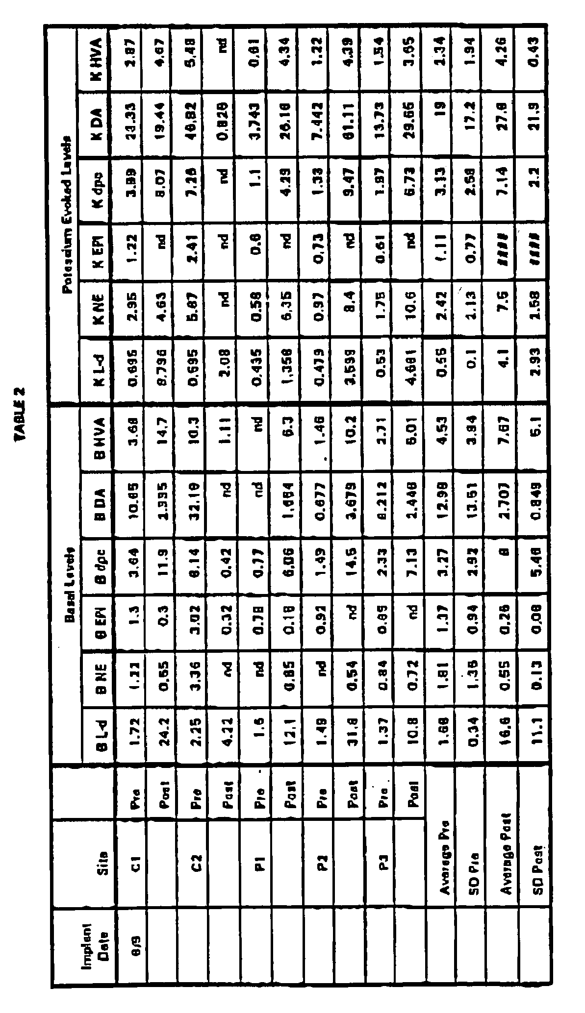


- PC12 cells are encapsulated and implanted in a recipient as described in Example 7. After 6 months, the capsules are explanted and assayed for the L-dopa to dopamine ratio. The cells are plated in multiwell dishes, and cultured under a range of O2 conditions in vitro. The cells are assayed periodically for L-dopa and dopamine output. Cells which release the target L-dopa to dopamine ratio are selected and implanted into human brain, using surgical procedures substantially as described in WO 92/00127.
- Capsules were prepared from hollow fibers with a phase inversion technique using PAN/PVC hollow fibers substantially as described in Example 7 (ID 450-500 µm, wall 50-65 µm,
MWCO 100 kD, hydraulic permeability 53 ml/min/m2/mmHg). - PC12 and PC12A cells were cultivated in 500 ml spinner cultures at 80 RPM in a serum-free defined medium HL-1 (Ventrex, Inc., Portland, Maine) at 37°C in a water-saturated, 7% CO2 ambient air atmosphere. Cells were harvested by collecting spinner culture supernatant and centrifuging at 800 g. Viability was assessed by exclusion of trypan blue and shown to be 90±5% prior to encapsulation.
- The cells were resuspended in HL-1, pH 7.3, at a concentration of 4 x 10⁷ cells/ml. An equal volume of a solution containing 2% (w/v) of pH 6.7 low viscosity chitosan (PROTOSAN®, chitosan chloride, Protan Biopolymer, Drammen, Norway), was added to the PC12 and PC12A cells resulting in a final cell concentration of 2.0 x 10⁷ cells/ml.
- Individual capsules were 7±0.5 mm in length. PC12 and FC12A cell-loaded capsules were fabricated in two separate batches and placed in a 24 multiwell tissue culture plate containing 1 ml of HL-1 medium.
- At 5-7 days following encapsulation, free-floating capsules were washed twice with 1 mL of HBSS HEPES buffered saline (containing 10 µM ascorbate) to remove residual culture medium. Sampling consisted of a 30 min incubation (basal release) in 250 µL HBSS. All samples were protected from oxidation by the rapid addition of a citrate-reducing acidified buffer (CRAB) and yielded a stable sample preparation in 10 mM citric acid, 20 µM sodium metabisulfite, and 0.1 N perchloric acid. Extended storage was at -80°C. standards were prepared by diluting stock solutions into HBSS and stabilizing them with CRAB and 0.1 N perchloric acid. Striatal Dopamine Depletions
- Rats were anesthetized with a 1.0 ml/kg intramuscular injection of a mixture of ketamine (33 mg/ml), xylazine (1.7 mg/ml), and acepromazine (10 mg/ml) and positioned in a Kopf stereotaxic instrument. A total of 12 µg of 6-OHDA (6 µl volume at a concentration of 2 µg/µl dissolved in 0.9% saline containing 0.2 µg/µl ascorbic acid) were infused at a rate of 1.0 µl/minute and allowed to diffuse for 5 minutes before the infusion cannula was slowly retracted. Infusion coordinates were 4.2 mm posterior to bregma, 1.0 mm lateral from midline, and 7.4 mm ventral to dura.
- Four months after being lesioned (2.75 months after the last apomorphine injection), capsules were implanted. Rats were anesthetized as before, and a sagittal incision was made in the scalp and a hole drilled for placement of the polymer capsule. Rats were implanted by placing the capsule within an 18-gauge Teflon catheter mounted to the stereotaxic frame. A stainless steel obturator was placed within the cannula, the device lowered into the brain, and the obturator held in place while the outer cannula was raised to passively place the capsule within the host striatum. The stereotaxic coordinates for implantation were: 0.5 mm anterior to bregma, 3.8 mm lateral to the sagittal suture, and 7.5 mm below the surface of the cortex. Rats not receiving implants were given sham surgeries (anesthetized, scalp lacerated, skull drilled, and dura punctured).
- After the completion of behavioral testing, rats with no capsules, FC12, PC12A, and half the rats with the empty capsules were anesthetized in a CO2 chamber and quickly decapitated. Their brains were removed from the skulls, the capsules were removed, both striata were dissected out, placed in Eppendorf tubes, and quick-frozen in liquid nitrogen. Following capsule retrieval from the host striatum, capsules were placed in 1 ml of phosphate HBSS for approximately 30 min. The phosphate HBSS was removed, and 1 ml of HEPES HBSS was added. Catecholamine analyses were performed as described in Example 4. After the catecholamine assay was completed the devices were placed in 4% paraformaldehyde and submitted for morphologic analyses.
- Figure 8 shows release of dopamine and L-dopa measured over a 30 minute period plotted for individual capsules, each assigned an identifier number. As shown in Figure 8, the 90 day post-implant production levels of L-dopa and dopamine were significantly different than the pre-implant levels.
- Bovine adrenal chromaffin cells were prepared as described in Example 4. Adrenal cell aggregates were immobilized in a 1.5% alginate matrix crosslinked with CaCl2 and encapsulated in double
skinned Type 4, immunoisolatory PAN/PVC hollow fiber membranes (ID 770 µm,wall 70 µm, HMCO 60-100 kD), substantially as described in WO92/19195. - The encapsulated cells were implanted in the subarachnoid space in two human patients, with terminal cancer, pain incompletely relieved by narcotic therapy, and no evidence of active infection or tumor in the meningeal space. Informed consent was granted by the patients and approval was received from the Ethical committee of the Faculty of Medicine of the University of Lausanne, Switzerland.
- Local infiltration with 1.0% lidocaine was used to establish anesthesia of the skin as well as the periosteum and other deep connective tissue structures down to and including the ligamentum flavum. A 3-5 cm skin incision was made in the parasagittal plane 1-2 cm to the right or left of the midline and was continued down to the lumbodorsal fascia. Using traditional bony landmarks including the iliac crests and the lumbar spinous processes, as well as fluoroscopic guidance, an 18 gauge Touhy needle was introduced into the subarachnoid space between L-3 and L-4 via an oblique paramedian approach. The needle was directed so that it entered the space at a shallow, superiorly directed angle that was no greater than 30-35° with respect to the spinal cord in either the sagittal or transverse plane.
- A guide wire was passed down the lumen of the Touhy needle hub until it extended 4-5 cm into the subarachnoid space (determined by promeasuring). The Touhy needle was removed from the wire.
- A 7 French dilator was then placed over the guide wire and the wire was used to direct the dilator as it was gently but firmly pushed through the fascia, paraspinous muscle, and ligamentum flavum, following the track of the wire toward the subarachnoid space.
- After the wire track was "overdilated" by the 7 French dilator, a 6 French dilator and cannula sheath were assembled and placed over the guide wire. The 6 French dilator and cannula were advanced carefully into the subarachnoid space until the opening tip of the cannula was positioned 7 cm within the space.
- When appropriate positioning of the cannula was assured, the guide wire and the 6 French dilator were gently removed from the lumen of the cannula in sequence.
- The encapsulated adrenal chromaffin cell was provided in a sterile, double envelope container, bathed in transport medium, and fully assembled including a tubular silicone tether. The membrane portion of the device was carefully introduced into the cannula. The capsule was advanced until the tip of the membrane reached a point that was 2-10 mm within the cranial tip of the cannula in the subarachnoid space. After the capsule was positioned using a pusher, the cannula and pusher were completely withdrawn. The final placement of the capsule was such that the 5 cm long membrane portion of the device lay entirely within the CSF containing subarachnoid space ventral to the cauda equina.
- The capsules were explanted after 84 days in
patient 1 and after 55 days inpatient 2. catecholamine output -- norepinephrine ("NE") and epinephrine ("EPI") -- after the exposure to in vivo restrictive conditions was compared with pre-implant levels (Table 3). Catecholamines were measured as described in example 4. The data in Table 3 are expressed as picomoles per capsule per 30 min (Basal) or picomoles per capsule per 15 min (nicotine stimulated: N-S). The catecholamine output was significantly different after exposure to the restrictive conditions than before implantation.TABLE 3: Capsule Catecholamine Output implant explant NE E NE E Patient 1: Implantation For 84 Days Basal 0 64 37 8 N-S 2932 2673 6366 3886 Patient 2: Implantation Por 55 Days Basal 48 120 4.1 69.3 N-S 5027 5063 10.6 65 - Encapsulated BHK-hNGF cells were prepared according to Example 1, implanted in the lateral ventricle of rat brain and acclimated by exposure to in vivo restrictive conditions for 14 months. After 14 months, the capsules were explanted and the acclimated cells from two capsules pooled and expanded by passage in 10% FES DMEM medium. A clonal line, designated BHK-
hNGF clone 23, was used for this experiment. These cells were aliquoted and portions were frozen in liquid nitrogen. Control BHK-hNGF clone 23 cells, not acclimated in vivo, were grown in vitro in 10% FBS DMEM (BHK-hNGF clone 23 cell bank). - The following six groups of cells and capsules were tested:
- Group 1: BHK-
hNGF 23 cells (acclimated cells from in vivo explanted capsules); re-encapsulated in Vitrogen® matrix; capsules implanted in lateral ventricles of rat brain; - Group 2: BHK-
hNGF 23 cells; (acclimated cells) resuspended in 10% FBS DMEM for encapsulation (no matrix); capsules implanted in lateral ventricles of rat brain; - Group 3: BHK-
hNGF 23 control cells (from cell bank; not previously implanted); encapsulated in vitrogen® matrix; capsules implanted in lateral ventricles of rat brain; - Group 4: BHK-
hNGF 23 control cells (from cell bank; not previously implanted); resuspended in 10% FBS DMEM for encapsulation (no matrix); capsules implanted in lateral ventricles of rat brain; - Group 5: BHK-control cells (from bank; not previously implanted); encapsulated in Vitrogen® matrix; capsules implanted in lateral ventricles of rat brain; and Group 6: BHK-control cells (from bank; not previously implanted); resuspended in 10% FBS DMEM (no matrix) for encapsulation; capsules implanted in lateral ventricles of rat brain.
- Acclimated BHK-
hNGF clone 23 cells were encapsulated essentially as described in Example 1. Most capsules were made from HF120794-6 fibers and had a final length of 7 mm. Several control capsules were made from 101-97-9 fibers which are comparable to the XP11 fibers used in Examples 7-9. - HF120794-6 single skinned fibers have, on average, a wall thickness of 86.4µ; an inner diameter (I.D.) of 453.2µ; an outer diameter (O.D.) of 625.1µ; a hydraulic permeability (HP) of 43.0 mm/min/m2/mmHg; and a molecular weight cut off (MWCO) based on a dextran diffusion test of 187 kD.
- The 101-97-9 single skinned fibers have, on average, a wall thickness of 59µ; an I.D. of 541µ; a tensile strength of 31g; a HP of 62 mm/min/m2/mmHg; and a MWCO based on a dextran diffusion test of 88 kD.
- Cells encapsulated in matrix (
Groups groups - Capsules containing BHK-hNGF cells previously acclimated in vivo and control capsules were randomly chosen and implanted bilaterally in the lateral ventricle of rats. Parallel samples of non-acclimated BHK-bNGF cells served as controls.
- Capsules from Groups 1-6 were assayed for NGF release as described in Example 1. NGF release from capsules containing acclimated or non-acclimated cells was measured before implantation and after implantation for a one month period. The NGF output from capsules of Groups 1-4 is shown in Figure 11. The data for non-NGF producing control cells is not shown. After NGF output assays, capsules were fixed in 4% paraformaldehyde and processed for histology, sectioning and morphological analyses.
- PC12 cells were cultured and encapsulated substantially as described in Example 9.
- 6-hydroxydopamine (6-OHDA) is selectively toxic to dopaminergic neurons in the substantia nigra. Rats given a 6-OHDA lesion in the substantia nigra develop Parkinson's disease-like characteristics associated with low dopamine levels. Rats were anesthetized as described in Example 9 and positioned in a Kopf stereotaxic instrument. A total of 10 µg of 6-OHDA (4 µg volume at 2.5 µg/µl dissolved in 0.9% saline containing 0.2 µg/µl ascorbic acid) were infused unilaterally and allowed to diffuse for 5 minutes before the infusion cannula was slowly retracted. Infusion coordinates were 3.2 mm posterior to bregma, 2.7 mm lateral from midline, and 8.0 mm ventral to dura with the incisor bar sat at +5.0 mm.
- Capsules were implanted bilaterally three to four weeks after rate were unilaterally lesioned. Rats ware anesthetized and capsules implanted using the procedures and stereotaxic coordinates described in Example 9.
- Devices were removed at 3, 10, 28, and 60 days post-implantation. Rats were anesthetized in a CO2 chamber and quickly decapitated. The brains were removed from the skulls and capsules were removed from lesioned and non-lesioned (control) sides of the brain. Both striata and nuclei accumbens were dissected out for catecholamine analysis by placing them in Eppendorf tubes and quick-freezing the tubes in liquid nitrogen. Capsules were placed in 1 ml phosphate HBSS for approx. 30 min. The phosphate HBSS was removed, and 1 ml HEPES HBSS added. Catecholamine analyses were performed as described in Example 4. After catecholamine assays were completed the capsules were placed in 4% paraformaldehyde for morphological analyses. Figure 9 (Panel A) shows the average release of L-dopa over a 30 min. period from 8 capsules pre-implantation ("Pre-"), and from capsules explanted at 5, 14, 28 or 60 days post-implantation from either lesioned (low dopamine) or non-lesioned (control) striata. Panel A shows that the relative amounts of catecholamine produced by encapsulated PC12 cells changes over time. Capsules explanted from both lesioned and control sides showed higher L-dopa output rates after acclimation in vivo for between two and three weeks.
- Figure 9 (Panel B) shows the ratio of L-dopa to dopamine released from the capsules of Panel A described above. Panel B shows that after acclimation in vivo in a low dopamine environment, encapsulated cells produced a lower L-dopa to dopamine ratio than the cells acclimated in a control environment. The L-dcpa to dopamine ratios became relatively stable after about 14 days in vivo. Capsules explanted from both the lesioned and control sides showed higher output L-dopa to dopamine ratios after being acclimated in vivo compared to the pre-implant output levels.
- catecholamine output values from a number of studies in which PC12 cells -- encapsulated in "XP11-equivalent" fibers and acclimated in primate brain as described in Example 7 -- are compared to the catecholamine output values from the same capsules before they were implanted. Encapsulated PC12 cells were assayed for basal L-dopa and dopamine output rates before implantation. The data reported in Table 4 represents the pre-implant catecholamine output. Capsules were then implanted into primate brains for in vivo acclimation, explanted at 1, 2.5, 3, 4, 5 and 6 months after implantation and assayed for basal L-dopa and dopamine output rates (Figure 10).
- Comparison of the summary data in Figure 10 and Table 4 show that pre-implant output and explant output differ markedly. The pre-implant data in
Row 1 of Table 4 corresponds to the explant data points at the 1 month interval in Figure 11. Likewise, the data in the other rows in Table 4 correspond to the other monthly data points plotted in Figure 11.TABLE 4: PPre-Implant Values Primate PC12 Capsule Catecholamine Output Explanted PC12 "XP-11-equivalent" Fibers Pre-Implant Corresponding to Time In Vivo Basal L-dope Basal Dopamire pm/15 min/ml avg. (S.D.) pm/15 min/ml avg. (S.D.) 1 (n=6) 2.02 (0.08) 2.95 (4.1) 2.5 (n=6) 0.78 (0.16) nd 3 (n=6) 0.79 (0.25) nd 4 (n=2) 10.6 (2.33) 2.39 (0.82) 5 (n=6) 2.06 (0.01) 4.26 (1.62) 6 (n=15) 3.09 (1.84) 21.3 (21.7) - The human CNTF (hCNTF) gene was inserted into a dihydrofolate reductase (DHFR) based expression vector designated pNUT, which contains the entire pUC 18 sequence including the polylinker. The transcription of the cDNA encoding the mutant form of DHFR is driven by the SV40 promoter. The 3' end is fused with the 3' end of the hepatitis B virus gene (HBV 3') to ensure efficient polyadenylation and maturation signals.
- The hCNTF gene was obtained by PCR amplification of human DNA. The primers used contained EcoRI site at the position of the natural hCNTF initiation codon. The hCNTF gene was fused at its 5' extremity to a 150 bp sequence from a mouse immunoglobulin (Ig) gene. The EcoRI site was used in such a way that the amino terminal part of the hCNTF protein corresponds to the first 18 amino acids of the Ig gene. A 325 bp AvaI fragment from the human growth hormone gene (hGH) containing the polyadenylation sequence and other sequences important for mRNA 3' end maturation was cloned at the 3' extremity of the hCNTF gene. Briefly, this fragment was introduced in the SpeI site of a Bluescript polylinker creating a BamHI and NotI site at the 5' and 3' ends, respectively. The BamHI site was ligated to the BglII site engineered at the 3' end of hCNTF.
- This construction was inserted at position +6 of the mouse MT-I promoter and the entire 3050 bp MT/Ig/hCNTF/hGH KpnI-NotI fragment was inserted into the KpnI-NotI site of the pNUT vector. Finally, the HSV-TK gene was cloned in the NotI site of the vector, thus separating it from the DHFR gene by the entire pUC-18 plasmid. This final construction is named RP3224E2.
- The RP3224E2 vector DNA was amplified in a standard E. coli strain (HB101) and purified using a Qiagen-Plasmid Kit (Kontron). DNA was transfected using a standard calcium/phosphate transfection procedure and selected with increasing concentrations of methotrexate. Cells are selected continuously in methotrexate while being maintained in PC-1 tissue culture medium. The PC-1 medium is a defined medium containing protein from human recombinant sources.
- Following drug selection (25 to 200 µm methrotrexate), the BHK cells (BHK-CNTF) were maintained in vitro without drug selection for several months and showed no loss of CNTF expression as assessed by Northern blot analysis, bioassays or ELISA. The level of CNTF production was about 1.0 ng/103 cells/hour as determined by bioassay and by ELISA.
- The CNTF-secreting BHK cells (BHK-CNTF) of Example 14 were encapsulated essentially as described in Example 1, except that a longer tether appropriate in length for implantation into human lumbar subarachnoid space was used. Devices were loaded at a density of 2x10⁵ transfected cells/µl of collagen solution (Zyderm). The CNTF released from each capsule was measured by immersing the capsule in 2 ml of fresh PC1 medium for 30 min. CNTF determination was then performed on the collected medium using the R&D Elisa system. The capsules were chosen to deliver a dosage of CNTF of 1µ/day intrathecally. Each human patient received one device.
- The encapsulated BHK-CNTF cells have been implanted for 1 month into the lumbar subarachnoid space of a human subject. Additional capsules will be acclimated in vivo for up to 6 months. Upon explant at one month, the rate of CNTF release per capsule was assayed as described in Example 12. After CNTF assays were performed, capsules were fixed in 4% paraformaldehyde for morphological analyses.
- The BHK-CNTF cells acclimated in vivo were removed from the capsules, pooled, and immediately cultured by growing in PC1 medium under low oxygen and glucose restrictive conditions selected to most closely match the environment of the subarachnoid space of the subject. BHK-CNTF cells were cultured under ambient conditions as parallel controls. The acclimated cells will be characterized, and can be further acclimated to other restrictive conditions.
- In one set of experiments, in vivo acclimated BHK-CNTF cells, cultured in vitro under ambient or restrictive conditions, will be re-encapsulated (following essentially the same procedures used for the first encapsulation) and implanted into a host subject.
- In another set of experiments, the acclimated cells implanted into the human host will first be acclimated to in vitro restrictive conditions in a non-human primate recipient.
- Subjects will be patients diagnosed with ALS as manifested by a combination of both upper motor neuron and lower motor neuron deficits at multiple levels; confirmatory electrophysiologic studies demonstrating active and chronic denervation in 3 limbs or 2 limbs and bulbar musculature; no neurological involvement outside the voluntary motor system; no evidence of primary disease that could cause neurologic deficit, particularly cervical spondylosis of plasma cell dyscrasia. The patient is relatively strong, i.e. can walk unassisted and is early in the course of the disease. The patent has forced vital capacity >75% of normal at the time of entry.
- The patients are monitored every day during the first week, once a week from the second to the fourth week and once a month thereafter, for, inter alia, side effects such as fever, stomatitis, cough and the reactivation to herpes. The following tests are performed once a month for efficacy evaluation: Tufts Quantitative Neurological Exam (TQNE); Bulbar coordination; Respiratory function--forced vital capacity, inspiratory flow. Blood is drawn once a week for the first four week and once a month thereafter for detection of plasma CNTF, potential antibodies to CNTF, C-reactive protein, fibrinogen.
- The surgical procedure for implantation of the encapsulated BHK-CNTF cells is as follows. After establishing IV access and administering prophylactic antibiotics (cefazolin sodium, 1 gram IV), the patient is positioned on the operating table, generally in either the lateral decubitus or genu-pectoral position, with the lumbar spine flexed anteriorly. The operative field is steriley prepared and draped exposing the midline dorsal lumbar region from the levels of S-1 to L-1 and allowing for intraoperative imaging of the lumbar spine with C-arm fluoroscopy. Local infiltration with 1.0% lidocaine is used to establish anesthesia of the skin as well as the periosteum and other deep connective tissue structures down to and including the ligamentum flavum.
- A 3-5 cm skin incision is made in the parasagittal plane 1-2 cm to the right or left of the midline and was continued down to the lumbodorsal fascia using electrocautery for hemostasis. Using traditional bony landmarks including the iliac crests and the lumbar spinous processes, as well as fluoroscopic guidance, an is gauge Touhy needle is introduced into the subarachnoid space between L-3 and L-4 via an oblique paramedian approach. The needle is directed so that it enters the space at a shallow, superiorly directed angle that is no greater than 30-35° with respect to the spinal cord in either the sagittal or transverse plane. Appropriate position of the tip of the needle is confirmed by withdrawal of several ml of cerebrospinal fluid (CSF) for preimplantation catecholamine, enkephalin, glucose, human CNTF, and protein levels and cell counts.
- The Touhy needle hub is reexamined to confirm that the opening at the tip is oriented superiorly (opening direction is marked by the indexing notch for the obturator on the needle hub), and the guide wire is passed down the lumen of the needle until it extends 4-5 cm into the subarachnoid space (determined by premeasuring). Care should be taken during passage of the wire that there is not resistance to advancement of the wire out of the needle and that the patient does not complain of significant neurogenic symptoms, either of which observations might indicate misdirection of the guide wire and possible impending nerve root or spinal cord injury.
- After the guide wire appears to be appropriately placed in the subarachnoid space, the Touhy needle is separately withdrawn and removed from the wire. The position of the wire in the midline of the spinal canal, anterior to the expected location of the cauda equina, and without kinks or unexplainable bends is then confirmed with fluoroscopy. After removal of the Touhy needle the guide wire should be able to be moved freely into and out of the space with only very slight resistance due to the rough surface of the wire running through the dense and fibrous ligamentum flavum.
- The 7 French dilator is then placed over the guide wire and the wire is used to direct the dilator as it is gently but firmly pushed through the fascia, paraspinous muscle, and ligamentum flavum, following the track of the wire toward the subarachnoid space. Advancement of the 7 French dilator is stopped and the dilator removed from the wire as soon as a loss of resistance is detected after passing the ligamentum flavum. This is done in order to avoid advancing and manipulating this relatively rigid dilator within the subarachnoid space to any significant degree.
- After the wire track is "overdilated" by the 7 French dilator, the 6 French dilator and cannula sheath are assembled and placed over the guide wire. The 6 French dilator and cannula are advanced carefully into the subarachnoid space until the opening tip of the cannula is positioned 7 cm within the space. As with the 7 French dilator, the assembled 6 French dilator and cannula are directed by the wire within the lumen of the dilator. Position within the subarachnoid space is determined by premeasuring the device and is grossly confirmed by fluoroscopy. Great care should be taken with manipulation of the dilators and cannula within the subarachnoid space to avoid misdirection and possible neurologic injury.
- When appropriate positioning of the cannula is assured, the guide wire and the 6 French dilator are gently removed from the lumen of the cannula in sequence. Depending on the patient's position on the operating table, CSF flow through the cannula at this point should be noticeable and may be very brisk, requiring capping the cannula or very prompt placement of the capsule implant in order to prevent excessive CSF loss.
- The encapsulated acclimated BHK-CNTF cells may be provided in a sterile, double envelope container, bathed in transport medium, and fully assembled including a tubular silicone tether. Prior to implantation through the cannula and into the subarachnoid space, the capsule may be transferred to the insertion Kit tray where it is positioned in a location that allows the capsule to be maintained in transport medium while it is grossly examined for damage or major defects.
- The tether portion of the capsule is mounted onto the stainless steel pusher by inserting the small diameter wire portion of the pusher on the membrane portion of the device and is carefully introduced into the cannula. The capsule is advanced until the tip of the membrane reaches a point that is 2-10 mm within the cranial tip of the cannula in the subarachnoid space. This placement is achieved by premeasuring the cannula and the capsule-tether-pusher assembly, and it assures that the membrane portion of the capsule is protected by the cannula for the entire time that it is being advanced into position.
- After the capsule is positioned within the cannula, the pusher is used to hold the capsule in position (without advancing or withdrawing) in the subarachnoid space while the cannula is completely withdrawn from over the capsule and pusher. The pusher is then removed from the capsule by sliding its wire portion out of the silicone tether. Using this method, the final placement of the capsule is such that the membrane portion of the device lies entirely within the CSF containing subarachnoid space ventral to the cauda equina. It may be anchored at its caudal end by a roughly 1-2 cm length of silicone tether that runs within the subarachnoid space before the tether exits through the dura and ligamentum flavum. The tether continues externally from this level through the paraspinous muscle and emerges from the lumbodorsal fascia leaving generally 10-12 cm of free tether material available for securing the device.
- CSF leakage is minimized by injecting fibrin glue (Tissel®) into the track occupied by the tether in the paraspinous muscle, and by firmly closing the superficial fascial opening of the track with a purse-string suture. The free end of the tether is then anchored with non-absorbable suture and completely covered with a 2 layer closure of the skin and subcutaneous tissue.
- The patient is then transferred to a neurosurgical recovery area and kept at strict bed rest, recumbent, for 24 hours postoperatively. Antibiotic prophylaxis is also continued for 24 hours following the implantation procedure.
-
- 1. A method for implanting cells in a host comprising:
- (a) encapsulating the cells in a biocompatible capsule,
- (b) exposing the cells to one or more restrictive conditions for a sufficient period of time to establish a desired cell property in response to the restrictive condition or conditions, and
- (c) implanting the encapsulated cells at an implantation site in a host, the cell property being substantially maintained following implantation.
- 2. The method of
statement 1, wherein the encapsulated cells are exposed to the restrictive condition or conditions in vitro. - 3. The method of
statement 1, wherein the encapsulated cells are exposed to the restrictive condition or conditions in vivo by implantation at an implantation site in a recipient. - 4. The method of
statement 1, wherein the cells are xenogeneic to the host and the capsule is immunoisolatory. - 5. The method of
statement 1, wherein at least one restrictive condition is a glucose concentration ranging from about 40 mg/deciliter to about 70 mg/deciliter. - 6. The method of
statement 1, wherein at least one restrictive condition is an oxygen concentration ranging from about 40 mmHg to about 65 mmHg. - 7. The method of
statement 1, wherein at least one restrictive condition is a dopamine concentration which is less than the physiological dopamine concentration in cerebrospinal fluid or brain parenchyma. - 8. The method of
statement 3, wherein the cells are selected from the group consisting of adrenal chromaffin cells, baby hamster kidney cells, and PC12 cells. - 9. The method of
statement 8, wherein the cells arePC 12 cells and are implanted in a recipient for 6 months or longer. - 10. The method of
statement 8, wherein the cells are adrenal chromaffin cells and are implanted in a recipient for 4 months or longer. - 11. The method of
statement 8, wherein the cells arePC 12 cells and are implanted in a recipient for 3 weeks or longer. - 12. The method of
statement 1, wherein the cells produce a biologically active molecule selected from the group consisting of neurotransmitters, analgesics and growth factors. - 13. The method of
statement 12, wherein the biologically active molecule is at least one catecholamine. - 14. The method of
statement 1, wherein the capsule is implanted in a host at an implantation site selected from the group consisting of cerebrospinal fluid and brain parenchyma. - 15. The method of
statement 1, wherein the host is a human. - 16. Cells that exhibit a desired cell property in response to a restrictive condition, said cells produced by:
- (a) encapsulating the cells in a biocompatible capsule, and
- (b) implanting the encapsulated cells at an implantation site in a primate recipient, the cells being xenogeneic to the recipient, for at least 6 months to establish the desired cell property in response to the restrictive conditions encountered at the implantation site.
- 17. The cells of statement 16, wherein the cells are selected from the group consisting of non-primate adrenal chromaffin cells, baby hamster kidney cells, and PC12 cells.
- 18. The cells of statement 17, wherein the cells are non-primate adrenal chromaffin cells.
- 19. The cells of statement 17, wherein the cells are PC12 cells.
- 20. The cells of statements 18 or 19, wherein the desired cell property is changed catecholamine output of the cells.
- 21. The cells of
statement 20, wherein the desired cell property is increased catecholamine output of the cells. - 22. The cells of statement 16, wherein the capsule is implanted at an implantation site selected from the group consisting of cerebrospinal fluid or brain parenchyma.
- 23. The cells of statement 16, wherein the host is a human.
- 24. A method for implanting cells in a xenogeneic host comprising:
- (a) culturing the cells under two or more restrictive conditions,
- (b) encapsulating the cells in a biocompatible capsule and further exposing the encapsulated cells to one or more restrictive conditions for a sufficient period of time to establish a desired cell property in response to the restrictive condition, and
- (c) implanting the encapsulated cells at an implantation site in the host, the cell property being substantially maintained following implantation.
Claims (4)
- An encapsulated cell system comprising: at least one capsule, each capsule comprising a core containing a cellular source of a biologically active molecule and a surrounding biocompatible jacket, the jacket permitting diffusion of the biologically active molecule, wherein the biologically active molecule is selected from the group consisting of hormones, cytokines, growth factors, angiogenesis factors, antibodies, lymphokines, enzymes, neurotransmitters, neurotrophic factors, antiinflammatory agents, and combinations thereof and wherein the system is adapted for implantation into the aqueous and vitreous humor of the eye.
- The system of claim 1, wherein the jacket comprises a permselective, immunoisolatory membrane.
- The system of claim 1, wherein the jacket comprises a semi-permeable membrane.
- Use of the system of any one of claims 1-3 in the manufacture of a medicament for delivery of said biologically active molecule into the aqueous or vitreous humor of the eye.
Applications Claiming Priority (2)
| Application Number | Priority Date | Filing Date | Title |
|---|---|---|---|
| US08/228,403 US5550050A (en) | 1994-04-15 | 1994-04-15 | Method for implanting encapsulated cells in a host |
| EP95916970A EP0756487A1 (en) | 1994-04-15 | 1995-04-13 | Method for implanting encapsulated cells in a host |
Related Parent Applications (1)
| Application Number | Title | Priority Date | Filing Date |
|---|---|---|---|
| EP95916970A Division EP0756487A1 (en) | 1994-04-15 | 1995-04-13 | Method for implanting encapsulated cells in a host |
Publications (1)
| Publication Number | Publication Date |
|---|---|
| EP1632238A1 true EP1632238A1 (en) | 2006-03-08 |
Family
ID=22857036
Family Applications (2)
| Application Number | Title | Priority Date | Filing Date |
|---|---|---|---|
| EP95916970A Ceased EP0756487A1 (en) | 1994-04-15 | 1995-04-13 | Method for implanting encapsulated cells in a host |
| EP05023484A Withdrawn EP1632238A1 (en) | 1994-04-15 | 1995-04-13 | Encapsulated cells adapted for implantation into the aqueous and vitreous humor of the eye |
Family Applications Before (1)
| Application Number | Title | Priority Date | Filing Date |
|---|---|---|---|
| EP95916970A Ceased EP0756487A1 (en) | 1994-04-15 | 1995-04-13 | Method for implanting encapsulated cells in a host |
Country Status (19)
| Country | Link |
|---|---|
| US (1) | US5550050A (en) |
| EP (2) | EP0756487A1 (en) |
| JP (2) | JPH10501792A (en) |
| CN (1) | CN1146155A (en) |
| AU (1) | AU697600B2 (en) |
| BR (1) | BR9507391A (en) |
| CA (1) | CA2187335A1 (en) |
| CZ (1) | CZ300496A3 (en) |
| EE (1) | EE9600147A (en) |
| FI (1) | FI964124A0 (en) |
| HU (1) | HUT75661A (en) |
| IL (1) | IL113390A0 (en) |
| IS (1) | IS4368A (en) |
| LV (1) | LV11730B (en) |
| NO (1) | NO964358L (en) |
| PL (1) | PL316845A1 (en) |
| SK (1) | SK131696A3 (en) |
| WO (1) | WO1995028166A2 (en) |
| ZA (1) | ZA953126B (en) |
Families Citing this family (92)
| Publication number | Priority date | Publication date | Assignee | Title |
|---|---|---|---|---|
| AU3124793A (en) * | 1991-10-29 | 1993-06-07 | Clover Consolidated, Limited | Crosslinkable polysaccharides, polycations and lipids useful for encapsulation and drug release |
| US5908623A (en) * | 1993-08-12 | 1999-06-01 | Cytotherapeutics, Inc. | Compositions and methods for the delivery of biologically active molecules using genetically altered cells contained in biocompatible immunoisolatory capsules |
| US5853717A (en) * | 1994-07-20 | 1998-12-29 | Cytotherapeutics, Inc. | Methods and compositions of growth control for cells encapsulated within bioartificial organs |
| US5935849A (en) * | 1994-07-20 | 1999-08-10 | Cytotherapeutics, Inc. | Methods and compositions of growth control for cells encapsulated within bioartificial organs |
| US5620013A (en) * | 1994-10-21 | 1997-04-15 | American Cyanamid Company | Method for destroying residual lens epithelial cells |
| US5762925A (en) * | 1994-11-03 | 1998-06-09 | Sagen; Jacqueline | Preventing opiate tolerance by cellular implantation |
| US6495364B2 (en) | 1995-05-23 | 2002-12-17 | Neurotech, S.A. | Mx-1 conditionally immortalized cells |
| US5855613A (en) | 1995-10-13 | 1999-01-05 | Islet Sheet Medical, Inc. | Retrievable bioartificial implants having dimensions allowing rapid diffusion of oxygen and rapid biological response to physiological change |
| WO1997033912A2 (en) | 1996-03-14 | 1997-09-18 | Genentech, Inc. | Uses of gdnf and gdnf receptor |
| US5904144A (en) * | 1996-03-22 | 1999-05-18 | Cytotherapeutics, Inc. | Method for treating ophthalmic diseases |
| US6299895B1 (en) | 1997-03-24 | 2001-10-09 | Neurotech S.A. | Device and method for treating ophthalmic diseases |
| US6027721A (en) * | 1996-05-20 | 2000-02-22 | Cytotherapeutics, Inc. | Device and method for encapsulated gene therapy |
| US6054142A (en) | 1996-08-01 | 2000-04-25 | Cyto Therapeutics, Inc. | Biocompatible devices with foam scaffolds |
| US6150164A (en) * | 1996-09-30 | 2000-11-21 | The Regents Of The University Of Michigan | Methods and compositions of a bioartificial kidney suitable for use in vivo or ex vivo |
| US6074825A (en) * | 1997-07-31 | 2000-06-13 | Maine Medical Center | Stable encapsulated reference nucleic acid and method of making |
| AU9198898A (en) * | 1997-09-17 | 1999-04-05 | Alcon Laboratories, Inc. | Methods for treating ocular diseases |
| WO1999013726A1 (en) * | 1997-09-19 | 1999-03-25 | University Of South Florida | Neuronal cells and an immunosuppressant and anti-inflammatory factor |
| US6638501B1 (en) | 1997-09-29 | 2003-10-28 | Neurospheres Holdings Ltd. | Use of multipotent neural stem cell progeny to augment non-neural tissues |
| US6303136B1 (en) | 1998-04-13 | 2001-10-16 | Neurotech S.A. | Cells or tissue attached to a non-degradable filamentous matrix encapsulated by a semi-permeable membrane |
| US6184035B1 (en) | 1998-11-18 | 2001-02-06 | California Institute Of Technology | Methods for isolation and activation of, and control of differentiation from, skeletal muscle stem or progenitor cells |
| US6610540B1 (en) * | 1998-11-18 | 2003-08-26 | California Institute Of Technology | Low oxygen culturing of central nervous system progenitor cells |
| US6713293B1 (en) | 1999-02-08 | 2004-03-30 | Friedrich Grummt | Encapsulated cells containing an amplified expression vector as a drug delivery device |
| JP4812167B2 (en) * | 1999-02-12 | 2011-11-09 | モレキュラー インサイト ファーマスーティカルズ インコーポレイテッド | Drug transport matrix and methods for making and using the same |
| US6303355B1 (en) | 1999-03-22 | 2001-10-16 | Duke University | Method of culturing, cryopreserving and encapsulating pancreatic islet cells |
| US6365385B1 (en) | 1999-03-22 | 2002-04-02 | Duke University | Methods of culturing and encapsulating pancreatic islet cells |
| US20070071734A1 (en) * | 1999-04-06 | 2007-03-29 | Weng Tao | ARPE-19 as platform cell line for encapsulated cell-based delivery |
| US6361771B1 (en) | 1999-04-06 | 2002-03-26 | Neurotech S.A. | ARPE-19 as a platform cell line for encapsulated cell-based delivery |
| US20040213768A1 (en) * | 1999-04-30 | 2004-10-28 | Elliott Robert Bartlett | Preparation for biotransplantation and xenotransplantion and uses thereof |
| US20090047325A1 (en) * | 1999-04-30 | 2009-02-19 | Neurotrophincell Pty. Limited | Xenotransplant for cns therapy |
| US20050265977A1 (en) * | 1999-04-30 | 2005-12-01 | Elliott Robert B | Xenotransplant for CNS therapy |
| EP1176970B1 (en) * | 1999-04-30 | 2010-05-19 | NeurotrophinCell Pty Ltd | Xenotransplant for cns therapy |
| EP1248640B1 (en) * | 2000-01-20 | 2006-10-04 | Diabcell Pty Limited | Preparation and xenotransplantation of porcine islets |
| WO2001054593A1 (en) * | 2000-01-31 | 2001-08-02 | The General Hospital Corporation | Neural regeneration conduit |
| US6719970B1 (en) | 2000-07-10 | 2004-04-13 | Alkermes Controlled Therapeutics, Inc. | Method of generating cartilage |
| DK1333846T3 (en) * | 2000-10-17 | 2012-08-06 | Diatranz Otsuka Ltd | Preparation and xenotransplantation of porcine islands |
| US6538010B1 (en) * | 2000-11-08 | 2003-03-25 | Research Triangle Institute | Compounds and methods for promoting smoking cessation |
| CA2452005C (en) * | 2001-07-10 | 2011-03-15 | Idec Pharmaceutical Corporation | Inhibition of apoptosis process and improvement of cell performance |
| EP1281767A3 (en) | 2001-07-31 | 2003-05-28 | Aladar A. Szalay | Light emitting microorganisms and cells for diagnosis and therapy of tumors |
| US7332330B2 (en) | 2001-09-11 | 2008-02-19 | Renamed Biologics, Inc. | Device for maintaining vascularization near an implant |
| ATE535601T1 (en) * | 2001-09-28 | 2011-12-15 | Diabcell Pty Ltd | BREEDING OF FOREIGN TRANSPLANTATION MATERIAL IN CULTURE |
| US20030068308A1 (en) * | 2001-10-09 | 2003-04-10 | Jacek Rozga | Intrasplenic encapsulated cell therapy |
| NZ515310A (en) * | 2001-11-07 | 2004-08-27 | Diabcell Pty Ltd | Methods of treatment and delivery modes |
| US7442546B2 (en) * | 2002-03-15 | 2008-10-28 | The Regents Of The University Of Michigan | Method of modulating inflammatory response |
| US20030228261A1 (en) * | 2002-06-05 | 2003-12-11 | Aladar Szalay | Light emitting microorganisms and cells for diagnosis and therapy of diseases associated with wounded or inflamed tissue |
| EP1369491A1 (en) * | 2002-06-05 | 2003-12-10 | Aladar A. Szalay | Light emitting microorganisms and cells for diagnosis and therapy of diseases associated with wounded or inflamed tissue |
| AU2002952993A0 (en) | 2002-11-29 | 2002-12-12 | The Corporation Of The Trustees Of The Order Of The Sisters Of Mercy In Queensland | Therapeutic and diagnostic agents |
| AU2003901325A0 (en) * | 2003-03-21 | 2003-04-03 | Stephen Locarnini | Therapeutic, prophylactic and diagnostic agents |
| AU2004224825A1 (en) * | 2003-03-26 | 2004-10-07 | Crc For Asthma Limited | Therapeutic and prophylactic compositions and uses therefor |
| AU2004245175C1 (en) | 2003-06-10 | 2010-03-18 | Biogen Ma Inc. | Improved secretion of neublastin |
| AU2004289953B2 (en) * | 2003-06-18 | 2008-09-25 | Genelux Corporation | Modified recombinant vaccina viruses and other microorganisms, uses thereof |
| CN1791670A (en) * | 2003-06-24 | 2006-06-21 | 戴伯塞尔有限公司 | Porcine pancreatic islet cultured with porcine sertoli cells for xenotransplantation |
| WO2005020677A1 (en) | 2003-08-29 | 2005-03-10 | The Walter And Eliza Hall Institute Of Medical Research | Method of selecting animal models from animals which have been subject to mutagenesis, and the use of myb transcription factors for screening |
| US8066973B2 (en) * | 2003-09-05 | 2011-11-29 | The Ohio State University Research Foundation | Nanoparticulate probe for in vivo monitoring of tissue oxygenation |
| EP1677676A4 (en) * | 2003-09-05 | 2011-02-23 | Univ Ohio State Res Found | Nanoparticulate probe for in vivo monitoring of tissue oxygenation |
| JP2007509109A (en) | 2003-10-20 | 2007-04-12 | エヌエスジーン・アクティーゼルスカブ | In vivo gene therapy for Parkinson's disease |
| AU2004292957C1 (en) * | 2003-11-13 | 2011-04-28 | Eyepoint Pharmaceuticals Us, Inc. | Injectable sustained release implant having a bioerodible matrix core and a bioerodible skin |
| DK1709161T3 (en) * | 2004-01-19 | 2009-01-26 | Nsgene As | Human therapeutic cells that secrete nerve growth factor |
| US20070281986A1 (en) * | 2004-02-03 | 2007-12-06 | Collier Gregory R | Methods and Compositions for Modulating Satiety |
| AU2005229434B2 (en) | 2004-03-30 | 2010-09-30 | Nsgene A/S | Therapeutic use of a growth factor, NsG33 |
| WO2005108981A1 (en) | 2004-05-12 | 2005-11-17 | The Walter And Eliza Hall Institute Of Medical Research | A method of cell isolation |
| US20090136552A1 (en) * | 2004-07-30 | 2009-05-28 | Mette Gronborg | Growth factors nsg28, nsg30, and nsg32 |
| MX2007012155A (en) * | 2005-04-01 | 2008-04-04 | Nsgene As | A human immortalised neural precursor cell line. |
| US9364427B2 (en) * | 2005-05-17 | 2016-06-14 | Nsgene A/S | Implantable therapy system for treating a living being with an active factor |
| NZ540597A (en) * | 2005-06-08 | 2007-02-23 | Neurotrophincell Pty Ltd | A method for preventing the onset of type I diabetes comprising administering an implantable composition comprising living choroid plexus cells |
| DK2407484T3 (en) | 2005-06-24 | 2016-09-05 | The Walter And Eliza Hall Inst Of Medical Res | Therapeutic pro-apoptotic BH3-like molecules, and methods of generating and / or selecting those |
| US20080286250A1 (en) * | 2005-10-28 | 2008-11-20 | Jens Tornoe | Implantable Biocompatible Immunoisolatory Vehicle for Delivery of Gdnf |
| DE102005061801A1 (en) * | 2005-12-23 | 2007-06-28 | BSH Bosch und Siemens Hausgeräte GmbH | Multiple product dispenser for domestic dish washing machines has a housing in the door which can house multiple doses of various washing agents in liquid or tablet form with multiple valves to dispense one dose at a time |
| ES2406716T3 (en) | 2005-12-30 | 2013-06-07 | Neurotech Usa, Inc. | Micronized device for the supply of biologically active molecules and method of use thereof |
| TW200819540A (en) | 2006-07-11 | 2008-05-01 | Genelux Corp | Methods and compositions for detection of microorganisms and cells and treatment of diseases and disorders |
| JP5558817B2 (en) | 2006-07-18 | 2014-07-23 | ユニバーシティ・オブ・ユタ・リサーチ・ファウンデイション | Pain treatment method and analgesic compound screening method |
| NZ584848A (en) | 2007-09-28 | 2012-09-28 | Intrexon Corp | Therapeutic gene-switch constructs and bioreactors for the expression of biotherapeutic molecules, and uses thereof |
| US20090090214A1 (en) * | 2007-10-04 | 2009-04-09 | Chung Yuan Christian University | Method for forming nano-scale metal particles |
| US20090196854A1 (en) * | 2008-02-04 | 2009-08-06 | Kytos Biosystems S.A. | Methods and compositions for use of crl 5803 cells for expression of biotherapeutics and encapsulated cell-based delivery |
| US20100021437A1 (en) * | 2008-04-07 | 2010-01-28 | The McLean Hospital Corporation Whitehead Institute for Biomedical Research | Neural stem cells derived from induced pluripotent stem cells |
| EP2389435B1 (en) | 2009-01-23 | 2015-11-04 | NsGene A/S | Improved cell lines and their use in encapsulated cell biodelivery |
| US20120021039A1 (en) | 2009-01-23 | 2012-01-26 | Nsgene A/S | Expression of neuropeptides in mammalian cells |
| EP2421981B1 (en) * | 2009-04-23 | 2013-08-14 | Neurotech USA, Inc. | Cell lines that produce prostaglandin f2 alpha (pgf2a) and uses thereof |
| FR2948125B1 (en) | 2009-07-17 | 2011-08-26 | Zoran Dermanovic | METHODS FOR ESTABLISHING SYMBIOSES |
| CA2776452A1 (en) * | 2009-10-02 | 2011-04-07 | Baxter International Inc. | Hematopoietic stem cells for use in the treatment of a kidney injury |
| US20110111008A1 (en) * | 2009-10-08 | 2011-05-12 | Weng Tao | Use of PEDF in an Encapsulated Cell-Based Delivery System |
| WO2012041328A1 (en) | 2010-10-01 | 2012-04-05 | Nsgene A/S | Use of meteorin for the treatment of allodynia, hyperalgesia, spontaneous pain and phantom pain |
| AU2011336592A1 (en) | 2010-12-02 | 2013-07-11 | Neurotech Usa, Inc. | Cell lines that secrete anti-angiogenic antibody-scaffolds and soluble receptors and uses thereof |
| JP2012215543A (en) * | 2011-03-31 | 2012-11-08 | Sony Corp | Individual information determination method, individual information determination device, electronic apparatus, and individual information determination program |
| CN104105501B (en) | 2011-09-05 | 2017-10-20 | 霍巴治疗公司 | Allodynia, hyperalgia, the treatment of spontaneous pain and phantom pain |
| WO2013126590A2 (en) | 2012-02-21 | 2013-08-29 | Baxter International Inc. | Pharmaceutical composition comprising cd34+ cells |
| KR20150016367A (en) | 2012-05-30 | 2015-02-11 | 뉴로테크 유에스에이, 인코포레이티드. | Cryopreserved implantable cell culture devices and uses thereof |
| EP2964147B1 (en) | 2013-03-07 | 2018-05-09 | Viacyte, Inc. | 3-dimensional large capacity cell encapsulation device assembly |
| CN105377286B (en) | 2013-05-31 | 2021-07-02 | 犹他大学研究基金会 | CONOTOXIN (CONOTOXIN) peptides, pharmaceutical compositions and uses thereof |
| CA2922483A1 (en) | 2013-09-11 | 2015-03-19 | Neurotech Usa, Inc. | Encapsulated cell therapy cartridge |
| WO2016191645A1 (en) | 2015-05-27 | 2016-12-01 | Neurotech Usa, Inc. | Use of encapsulated cell therapy for treatment of ophthalmic disorders |
| ES2959601T3 (en) | 2015-10-27 | 2024-02-27 | Univ Queensland | A modified EphA4 polypeptide |
| WO2022011081A1 (en) | 2020-07-09 | 2022-01-13 | The United States Of America, As Represented By The Secretary, Department Of Health And Human Services | Cell lines that produce human retinoschisin proteins and uses thereof |
Citations (15)
| Publication number | Priority date | Publication date | Assignee | Title |
|---|---|---|---|---|
| US4353888A (en) * | 1980-12-23 | 1982-10-12 | Sefton Michael V | Encapsulation of live animal cells |
| US4409331A (en) * | 1979-03-28 | 1983-10-11 | Damon Corporation | Preparation of substances with encapsulated cells |
| US4753635A (en) | 1986-05-23 | 1988-06-28 | Jacqueline Sagen | Inducing analgesia by implantation of cells releasing neuroactive substances |
| US4868121A (en) | 1985-02-07 | 1989-09-19 | Mcdonnell Douglas Corporation | Islet isolation process |
| WO1991010470A1 (en) * | 1990-01-08 | 1991-07-25 | Brown University Research Foundation | Devices and methods for enhanced delivery of active factors |
| US5084350A (en) | 1990-02-16 | 1992-01-28 | The Royal Institution For The Advance Of Learning (Mcgill University) | Method for encapsulating biologically active material including cells |
| WO1992003536A1 (en) | 1990-08-15 | 1992-03-05 | University Of Miami And Its School Of Medicine | Autotransplantation of schwann cells to promote nervous system repair |
| US5158881A (en) | 1987-11-17 | 1992-10-27 | Brown University Research Foundation | Method and system for encapsulating cells in a tubular extrudate in separate cell compartments |
| WO1992019195A1 (en) | 1991-04-25 | 1992-11-12 | Brown University Research Foundation | Implantable biocompatible immunoisolatory vehicle for delivery of selected therapeutic products |
| WO1993000127A1 (en) | 1991-06-28 | 1993-01-07 | Brown University Research Foundation | Neural implant system |
| WO1993022427A1 (en) | 1992-04-24 | 1993-11-11 | Somatix Therapy Corporation | Method of culturing viable cells and method of regulating the level of a compound in a body fluid |
| US5283187A (en) | 1987-11-17 | 1994-02-01 | Brown University Research Foundation | Cell culture-containing tubular capsule produced by co-extrusion |
| WO1994003154A1 (en) | 1992-07-29 | 1994-02-17 | Washington University | Use of pouch for implantation of living cells |
| WO1995001203A2 (en) | 1993-06-23 | 1995-01-12 | Cytotherapeutics, Inc. | Implantable membrane encapsulation apparatus |
| WO1995005452A2 (en) | 1993-08-12 | 1995-02-23 | Cytotherapeutics, Inc. | Improved compositions and methods for the delivery of biologically active molecules using genetically altered cells contained in biocompatible immunoisolatory capsules |
Family Cites Families (22)
| Publication number | Priority date | Publication date | Assignee | Title |
|---|---|---|---|---|
| US4352883A (en) * | 1979-03-28 | 1982-10-05 | Damon Corporation | Encapsulation of biological material |
| US4298002A (en) * | 1979-09-10 | 1981-11-03 | National Patent Development Corporation | Porous hydrophilic materials, chambers therefrom, and devices comprising such chambers and biologically active tissue and methods of preparation |
| NO163060C (en) * | 1981-03-13 | 1990-03-28 | Damon Biotech Inc | PROCEDURE FOR THE PREPARATION OF SUBSTANCES MADE BY LIVING CELLS PRODUCING INTERFERONES OR ANTIBODIES. |
| JPS5876086A (en) * | 1981-10-28 | 1983-05-09 | Ajinomoto Co Inc | Culture of suspensible animal cell |
| US4479796A (en) * | 1982-11-15 | 1984-10-30 | Medtronic, Inc. | Self-regenerating drug administration device |
| EP0127989A3 (en) * | 1983-06-01 | 1986-03-26 | Connaught Laboratories Limited | Microencapsulation of living tissue and cells |
| US4806355A (en) * | 1983-06-06 | 1989-02-21 | Connaught Laboratories Limited | Microencapsulation of living tissue and cells |
| US4689293A (en) * | 1983-06-06 | 1987-08-25 | Connaught Laboratories Limited | Microencapsulation of living tissue and cells |
| JPS60141286A (en) * | 1983-12-28 | 1985-07-26 | Ajinomoto Co Inc | Method and apparatus for culturing animal cell |
| US4749620A (en) * | 1984-02-15 | 1988-06-07 | Massachusetts Institute Of Technology | Encapsulated active material system |
| CA1215922A (en) * | 1984-05-25 | 1986-12-30 | Connaught Laboratories Limited | Microencapsulation of living tissue and cells |
| GB8500121D0 (en) * | 1985-01-03 | 1985-02-13 | Connaught Lab | Microencapsulation of living cells |
| US4902295A (en) * | 1985-08-26 | 1990-02-20 | Hana Biologics, Inc. | Transplantable artificial tissue |
| US5026365A (en) * | 1987-04-29 | 1991-06-25 | The University Of Massachusetts | Method and apparatus for therapeutically treating immunological disorders and disease states |
| DE3724798A1 (en) * | 1987-07-27 | 1989-02-09 | Mengele & Soehne Masch Karl | HARVEST HEADER ON FIELD CHOPPER |
| US4942129A (en) * | 1987-07-28 | 1990-07-17 | Queen's University At Kingston | Multiple membrane microencapsulation |
| US5182111A (en) * | 1987-11-17 | 1993-01-26 | Boston University Research Foundation | In vivo delivery of active factors by co-cultured cell implants |
| US5106627A (en) * | 1987-11-17 | 1992-04-21 | Brown University Research Foundation | Neurological therapy devices |
| US4892538A (en) * | 1987-11-17 | 1990-01-09 | Brown University Research Foundation | In vivo delivery of neurotransmitters by implanted, encapsulated cells |
| KR0132666B1 (en) * | 1989-03-14 | 1998-04-14 | Hitachi Kk | Method for controlling cultivation conditions for animal cells |
| US5002661A (en) * | 1989-08-25 | 1991-03-26 | W. R. Grace & Co.-Conn. | Artificial pancreatic perfusion device |
| WO1991009119A1 (en) * | 1989-12-13 | 1991-06-27 | Trancel Corporation | Improved alginate microcapsules, methods of making and using same |
-
1994
- 1994-04-15 US US08/228,403 patent/US5550050A/en not_active Expired - Lifetime
-
1995
- 1995-04-13 WO PCT/US1995/004564 patent/WO1995028166A2/en not_active Application Discontinuation
- 1995-04-13 BR BR9507391A patent/BR9507391A/en not_active Application Discontinuation
- 1995-04-13 SK SK1316-96A patent/SK131696A3/en unknown
- 1995-04-13 EP EP95916970A patent/EP0756487A1/en not_active Ceased
- 1995-04-13 CA CA002187335A patent/CA2187335A1/en not_active Abandoned
- 1995-04-13 CN CN95192579A patent/CN1146155A/en active Pending
- 1995-04-13 AU AU23829/95A patent/AU697600B2/en not_active Ceased
- 1995-04-13 CZ CZ963004A patent/CZ300496A3/en unknown
- 1995-04-13 JP JP7527082A patent/JPH10501792A/en active Pending
- 1995-04-13 HU HU9602850A patent/HUT75661A/en unknown
- 1995-04-13 EE EE9600147A patent/EE9600147A/en unknown
- 1995-04-13 EP EP05023484A patent/EP1632238A1/en not_active Withdrawn
- 1995-04-13 PL PL95316845A patent/PL316845A1/en unknown
- 1995-04-14 IL IL11339095A patent/IL113390A0/en unknown
- 1995-04-18 ZA ZA953126A patent/ZA953126B/en unknown
-
1996
- 1996-10-01 IS IS4368A patent/IS4368A/en unknown
- 1996-10-14 FI FI964124A patent/FI964124A0/en unknown
- 1996-10-14 NO NO964358A patent/NO964358L/en not_active Application Discontinuation
- 1996-11-15 LV LVP-96-434A patent/LV11730B/en unknown
-
2005
- 2005-03-16 JP JP2005076076A patent/JP2005239726A/en active Pending
Patent Citations (16)
| Publication number | Priority date | Publication date | Assignee | Title |
|---|---|---|---|---|
| US4409331A (en) * | 1979-03-28 | 1983-10-11 | Damon Corporation | Preparation of substances with encapsulated cells |
| US4353888A (en) * | 1980-12-23 | 1982-10-12 | Sefton Michael V | Encapsulation of live animal cells |
| US4868121A (en) | 1985-02-07 | 1989-09-19 | Mcdonnell Douglas Corporation | Islet isolation process |
| US4753635A (en) | 1986-05-23 | 1988-06-28 | Jacqueline Sagen | Inducing analgesia by implantation of cells releasing neuroactive substances |
| US5158881A (en) | 1987-11-17 | 1992-10-27 | Brown University Research Foundation | Method and system for encapsulating cells in a tubular extrudate in separate cell compartments |
| US5284761A (en) | 1987-11-17 | 1994-02-08 | Brown University Research Foundation | Method of encapsulating cells in a tubular extrudate |
| US5283187A (en) | 1987-11-17 | 1994-02-01 | Brown University Research Foundation | Cell culture-containing tubular capsule produced by co-extrusion |
| WO1991010470A1 (en) * | 1990-01-08 | 1991-07-25 | Brown University Research Foundation | Devices and methods for enhanced delivery of active factors |
| US5084350A (en) | 1990-02-16 | 1992-01-28 | The Royal Institution For The Advance Of Learning (Mcgill University) | Method for encapsulating biologically active material including cells |
| WO1992003536A1 (en) | 1990-08-15 | 1992-03-05 | University Of Miami And Its School Of Medicine | Autotransplantation of schwann cells to promote nervous system repair |
| WO1992019195A1 (en) | 1991-04-25 | 1992-11-12 | Brown University Research Foundation | Implantable biocompatible immunoisolatory vehicle for delivery of selected therapeutic products |
| WO1993000127A1 (en) | 1991-06-28 | 1993-01-07 | Brown University Research Foundation | Neural implant system |
| WO1993022427A1 (en) | 1992-04-24 | 1993-11-11 | Somatix Therapy Corporation | Method of culturing viable cells and method of regulating the level of a compound in a body fluid |
| WO1994003154A1 (en) | 1992-07-29 | 1994-02-17 | Washington University | Use of pouch for implantation of living cells |
| WO1995001203A2 (en) | 1993-06-23 | 1995-01-12 | Cytotherapeutics, Inc. | Implantable membrane encapsulation apparatus |
| WO1995005452A2 (en) | 1993-08-12 | 1995-02-23 | Cytotherapeutics, Inc. | Improved compositions and methods for the delivery of biologically active molecules using genetically altered cells contained in biocompatible immunoisolatory capsules |
Non-Patent Citations (37)
| Title |
|---|
| "ANIMAL CELL CULTURE", 1990, HUMANA PRESS INC., pages: 696 - 700 |
| "GEIGY SCIENTIFIC TABLES", vol. I, 1984, CIBA-GEIGY, article "Units of Measurements, Body Fluids, Composition of the Body, Nutrition" |
| "METHODS IN ENZYMOLOGY", 1993, ACADEMIC PRESS |
| AEBISCHER P. ET AL: "Functional recovery in Hemiparkinsonian Primates Transplanted with Polymer-Encapsulated PC12 Cells", EXP. NEUROL. (UNITED STATES), vol. 126, no. 2, 1994, pages 151 - 158, XP000562226 * |
| AEBISCHER P. ET AL: "Polymer Encapsulated PC12 Cells Transplanted in MPTP Lesioned Primates", SOC. NEUROSC. ABSTR., vol. 16, no. 2, 1990, pages 963, XP000764307 * |
| AEBISCHER, BIO-MATERIALS |
| AEBISCHER, BIOMATERIALS, vol. 12, 1981, pages 50 - 55 |
| AEBISCHER, TRANS. AM. SOC.ARTIF. INTERN. ORGANS, vol. 32, 1986, pages 134 - 7 |
| ALTMAN, DIABETES, vol. 35, 1986, pages G25 - 33 |
| BAETGE, PROC. NATL. ACAD. SCI., vol. 83, 1986, pages 5454 - 58 |
| CABASSO: "KIRK-OTHMER ENCYCLOPEDIA OF CHEMICAL TECHNOLOGY", vol. 12, 1980, WILEY, article "Hollow Fiber Membranes", pages: 492 - 517 |
| CEPKO, NEURON, vol. 1, 1988, pages 345 - 353 |
| CHICK, PROC. NATL. ACAD. SCI. USA, vol. 74, 1977, pages 628 - 632 |
| DARQUAY; REACH, DIABETOLOGIA, vol. 28, 1985, pages 776 - 80 |
| DWAINE F.E. ET AL: "Implants of Polymer-Encapsulated Human NGF-Secreting Cells in the Nonhuman Primate: Rescue and Sprouting of Degenerating Cholinergic Basal Forebrain Neurons", THE JOURNAL OF COMPARATIVE NEUROLOGY, vol. 349, 1994, pages 148 - 164, XP000562227 * |
| EXPERIMENTAL NEUROLOGY, vol. 111, 1981, pages 269 - 75 |
| H. GOLDBLATT, BIOCHEMICAL MEDICINE, vol. 7, 1973, pages 241 - 52 |
| HAMAGUCHI, DIABETES, vol. 40, 1991, pages 842 |
| HOYLE, NEURON, vol. 10, 1993, pages 1019 - 34 |
| HUGHES, PROC. NATL. ACAD. SCI. USA, vol. 89, 1992, pages 688 - 692 |
| LAND, NATURE, vol. 304, 1983, pages 596 - 602 |
| LINDNER M.D. ET AL: "Implantation of Encapsulated Catecholamine and GDNF-Producing Cells in Rats with Unilateral Dopamine Depletions and Parkinsonian Symptoms", EXP. NEUROL. (UNITED STATES), vol. 132, no. 1, 1995, pages 62 - 76, XP004537486 * |
| LIVETT, PHYSIOL. REV., vol. 64, 1984, pages 1103 - 62 |
| LIVETT, PHYSIOLOGY REVIEWS, vol. 64, 1984, pages 1103 - 61 |
| MATSUMOTO, PROC. NATL. ACAD. SCI. USA, vol. 87, 1990, pages 9133 - 37 |
| RAY, PROC. NATL. ACAD. SCI. USA, vol. 90, 1993, pages 3602 - 06 |
| REYNOLDS; WEISS, SCIENCE, vol. 255, 1992, pages 1707 - 10 |
| RICHARDS, PROC. NATL. ACAD. SCI. USA, vol. 89, 1992, pages 8591 - 95 |
| SCHARP, DIABETES, vol. 29, 1980, pages 19 - 30 |
| SEFTON, BIOTECHNOLOGY AND BIOENGINEERING, vol. 29, 1987, pages 1135 - 1143 |
| SPECTOR; JOHANSON, SCIENTIFIC AMERICAN, November 1989 (1989-11-01), pages 68 - 74 |
| SUGAMORI, TRANS. AM. SOC. ARTF. INTERN. ORGANS, vol. 35, 1989, pages 791 - 799 |
| SUGAMORI; SEFTON, TRANS. AM. SOC.ARTIF. INTERN. ORGANS, vol. 35, 1989, pages 791 - 9 |
| SUN., BIOMAT. ART. CELLS, ART. ORG., vol. 15, 1987, pages 483 - 496 |
| TAL, MOL. CELL BIOL., vol. 12, 1992, pages 422 - 32 |
| WAINER; HELLER: "NEURONAL CELL LINES", 1992, IRL PRESS, article "Neuronal Hybrid Cell Lines: Generation, Characterization and Utility", pages: 20 |
| WOLFFE; TATA, FEBS LETTERS, vol. 176, 1984, pages 8 - 15 |
Also Published As
| Publication number | Publication date |
|---|---|
| WO1995028166A2 (en) | 1995-10-26 |
| US5550050A (en) | 1996-08-27 |
| IL113390A0 (en) | 1995-07-31 |
| ZA953126B (en) | 1996-01-04 |
| NO964358D0 (en) | 1996-10-14 |
| MX9604832A (en) | 1998-06-28 |
| HU9602850D0 (en) | 1996-12-30 |
| JP2005239726A (en) | 2005-09-08 |
| CA2187335A1 (en) | 1995-10-26 |
| HUT75661A (en) | 1997-05-28 |
| NO964358L (en) | 1996-12-13 |
| WO1995028166A3 (en) | 1995-11-09 |
| SK131696A3 (en) | 1997-06-04 |
| AU2382995A (en) | 1995-11-10 |
| EE9600147A (en) | 1997-04-15 |
| FI964124A (en) | 1996-10-14 |
| CN1146155A (en) | 1997-03-26 |
| JPH10501792A (en) | 1998-02-17 |
| LV11730B (en) | 1998-02-20 |
| IS4368A (en) | 1996-10-01 |
| BR9507391A (en) | 1997-09-16 |
| LV11730A (en) | 1997-04-20 |
| PL316845A1 (en) | 1997-02-17 |
| CZ300496A3 (en) | 1997-03-12 |
| FI964124A0 (en) | 1996-10-14 |
| EP0756487A1 (en) | 1997-02-05 |
| AU697600B2 (en) | 1998-10-08 |
Similar Documents
| Publication | Publication Date | Title |
|---|---|---|
| AU697600B2 (en) | Method for implanting encapsulated cells in a host | |
| EP0802800B1 (en) | Biocompatible immunoisolatory capsules containing genetically altered cells for the delivery of biologically active molecules | |
| Emerich et al. | Implants of polymer‐encapsulated human NGF‐secreting cells in the nonhuman primate: rescue and sprouting of degenerating cholinergic basal forebrain neurons | |
| US5908623A (en) | Compositions and methods for the delivery of biologically active molecules using genetically altered cells contained in biocompatible immunoisolatory capsules | |
| US5869077A (en) | Methods for treating diabetes by delivering insulin from biocompatible cell-containing devices | |
| Sagot et al. | Polymer encapsulated cell lines genetically engineered to release ciliary neurotrophic factor can slow down progressive motor neuronopathy in the mouse | |
| US5800829A (en) | Methods for coextruding immunoisolatory implantable vehicles with a biocompatible jacket and a biocompatible matrix core | |
| EP1007636B1 (en) | Cultures of human cns neural stem cells | |
| Deglon et al. | Central nervous system delivery of recombinant ciliary neurotrophic factor by polymer encapsulated differentiated C2C12 myoblasts | |
| RU2234338C2 (en) | Using neuro-derivative embryionic cellular lines for transplantation therapy | |
| JP2003512333A (en) | Methods for inducing the in vivo proliferation and migration of transplanted precursor cells in the brain |
Legal Events
| Date | Code | Title | Description |
|---|---|---|---|
| PUAI | Public reference made under article 153(3) epc to a published international application that has entered the european phase |
Free format text: ORIGINAL CODE: 0009012 |
|
| 17P | Request for examination filed |
Effective date: 20051027 |
|
| AC | Divisional application: reference to earlier application |
Ref document number: 0756487 Country of ref document: EP Kind code of ref document: P |
|
| AK | Designated contracting states |
Kind code of ref document: A1 Designated state(s): AT BE CH DE DK ES FR GB GR IE IT LI LU MC NL PT SE |
|
| AX | Request for extension of the european patent |
Extension state: LT SI |
|
| RIN1 | Information on inventor provided before grant (corrected) |
Inventor name: DIONNE, KEITH E. Inventor name: LYSAGHT, MICHAEL J. Inventor name: RUDNICK, SETH A. Inventor name: HAMMANG, JOSEPH P. Inventor name: HOLLAND, LAURA M. |
|
| 17Q | First examination report despatched |
Effective date: 20060707 |
|
| AKX | Designation fees paid |
Designated state(s): AT BE CH DE DK ES FR GB GR IE IT LI LU MC NL PT SE |
|
| STAA | Information on the status of an ep patent application or granted ep patent |
Free format text: STATUS: THE APPLICATION IS DEEMED TO BE WITHDRAWN |
|
| 18D | Application deemed to be withdrawn |
Effective date: 20070118 |